Anterior Cord Syndrome (Archived)

Archived, for historical reference only

Introduction

Anterior cord syndrome is an incomplete spinal cord syndrome that predominantly affects the anterior two-thirds of the spinal cord, resulting in motor deficits and loss of sensory function in pain and temperature. The patient presentation varies depending on the portion of the spinal cord affected and the time to treatment. Other findings include back pain or autonomic dysfunction, such as hypotension, neurogenic bowel or bladder, and sexual dysfunction.[1] The severity of motor dysfunction can vary, typically resulting in paraplegia or quadriplegia depending on how rostral the level of the spinal cord involved is.

The syndrome is caused by ischemia within the anterior spinal artery (ASA), which supplies blood to the anterior two-thirds of the spinal cord. The ASA forms from the bilateral vertebral arteries at the foramen magnum. The ASA runs as an uninterrupted artery within the anterior median sulcus of the spinal cord to the conus medullaris. Radicular arteries enter the spinal canal through the intervertebral foramen and primarily supply the nerve roots; however, some anastomoses contribute to the ASA. The largest of these radicular arteries is the artery of Adamkiewicz, which most commonly arises off of a left intercostal artery between segments T9 to T12 but can vary anatomically. The ASA branches into small sulcal and penetrating arteries that enter the body of the spinal cord.[2]

The ASA supplies blood to the spinal cord's bilateral anterior and lateral horns and the bilateral spinothalamic tracts and corticospinal tracts. The anterior horns and corticospinal tracts control the somatic motor system from the neck to the feet. The lateral horns span levels T1 to L2 of the spinal cord and comprise the neuronal cell bodies of the sympathetic nervous system. The spinothalamic tracts relay pain, temperature, and sensory information. The anterior spinal artery, with a few radicular artery contributions, is the sole source of blood supply to these areas of the spinal cord via its sulcal and penetrating arteries.

Ischemia of the ASA causes symptoms consistent with the dysfunction of these tracts. Since the ASA is formed from the more cephalad vertebral arteries and runs caudally, the more rostral the location of ischemia, the more widespread or severe the symptoms are. Since the lateral horns are located only between T1 to L2 of the spinal cord, autonomic symptoms are not always present if ischemia does not involve this region.[1] Proprioception, vibratory sense, two-point discrimination, and fine touch are not affected in anterior cord syndrome and are carried by the dorsal column of the spinal cord, which is perfused by two posterior spinal arteries running in the posterior lateral sulci.[3]

Etiology

The root cause of anterior cord syndrome is occlusion of the ASA with resultant spinal cord ischemia in its vascular distribution. Thus, the etiology of anterior cord syndrome is wide in that it includes any procedure or disease process that can limit blood flow to this region. The most common cause of anterior cord syndrome is iatrogenic, namely aortic aneurysm repair. Several factors during surgery can contribute to cord ischemia, including hypotension, cross-clamping of the aorta, increased spinal canal pressure, and occlusion of arteries perfusing the spinal cord.[4][5][6] Aortic dissection is a rare cause of spinal cord ischemia due to occlusion of branch vessels of the aorta, which include radicular arteries that feed the ASA.[7] Anterior cord syndrome has been rarely reported to occur following transient intraoperative hypotension during elective anterior cervical spine surgery.[8]

Other causes include hypotension secondary to disease processes such as cardiac arrest, atherothrombotic disease, emboli, vasculitis, disorders affecting blood vessels, sickle cell, hypercoagulable diseases, arteriovenous malformation, intervertebral disc herniation, and cocaine use.[9] Transverse myelitis, a rare condition in patients with myelin oligodendrocyte antibody-associated disease, has a similar presentation to anterior cord syndrome.[10] Furthermore, anterior cord syndrome can occur iatrogenically as a complication of endovascular embolization.[11]

Epidemiology

Epidemiologic data on the subject of anterior cord syndrome is sparse. A limited number of studies on spinal cord infarction exist as a whole; estimates reveal that spinal cord infarction accounts for approximately 1.2% of all strokes.[12] The incidence of spinal cord infarction has been estimated to be 3.1 per 100,000 persons or 7,600 people per year in the United States.[13] Anterior cord syndrome is the most common of spinal cord infarction.[9] Given the gap between available data and the prevalence of this syndrome, further studies are needed. 

History and Physical

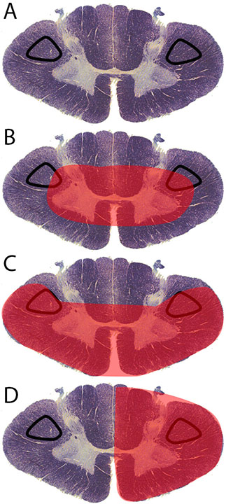
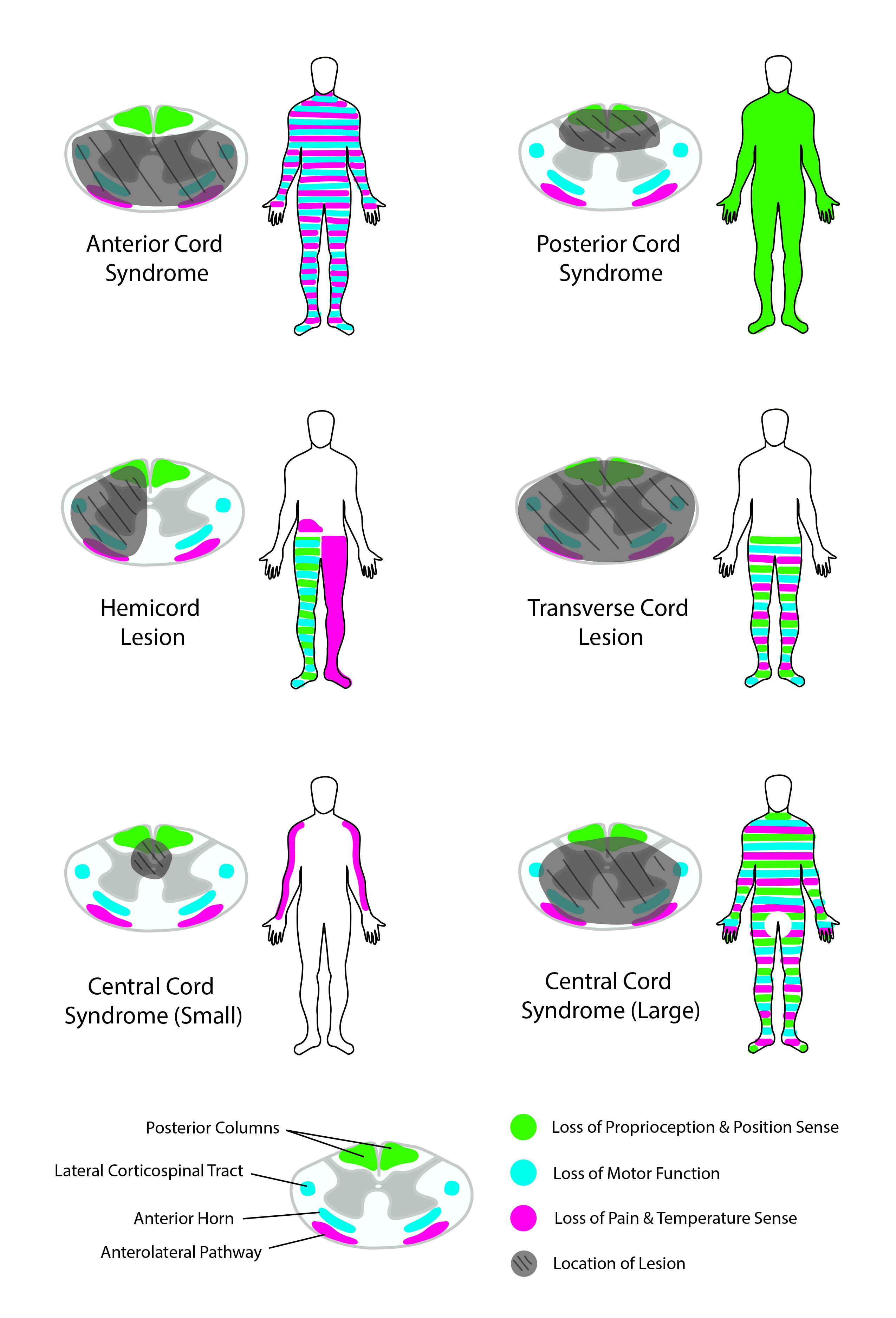
Due to the ischemic etiology of anterior cord syndrome, symptoms are typically acute in onset. Patients present with acute motor dysfunction and the loss of pain and temperature sensation below the infarction level (see Image. Corticospinal Tract Lesion Locations). These symptoms are almost always bilateral because both halves of the anterior spinal cord receive vascular supply from one midline anterior spinal artery. Motor dysfunction severity varies from paraplegia to quadriplegia, depending on the infarction location (see Image. Comparison of Spinal Cord Lesions and Syndromes).

Often, the location of infarction includes the lateral horns within levels T1 to L2 of the spinal cord, causing autonomic dysfunction, the most common manifestations being neurogenic bowel or bladder requiring bladder catheterization, hypotension, and sexual dysfunction. Acute onset of back pain is also a common symptom of cord infarctions in general and usually localizes at the infarction level.[14][15][16] Reported cases of anterior cord syndrome with unilateral symptomatology are rare; this may be due to occlusion of unilateral sulcal arteries or collateralization from one posterior spinal artery.[16]

Evaluation

Magnetic resonance imaging (MRI) is the primary imaging modality in diagnosing anterior cord syndrome. T2 hyperintensities within the region of the anterior horns are the hallmark finding. These hyperintensities on the sagittal view appear as thin “pencil-like” lesions extending vertically across several spinal levels. On the axial view, these hyperintensities appear as two bright dots, one within each anterior horn, resembling “owl’s eyes” in appearance. If imaging is obtained in the early stages of development, edema is present and can cause the spinal cord to appear expanded in the infarction region. Sometimes, signs of vertebral body infarction can be identified near the area in question, increasing the specificity of imaging for cord infarction.[16][17][18] MRI is also used to rule out compressive pathologies causing myelopathy.

Patients with positive MRI findings, clinical history, and consistent findings of anterior cord syndrome likely do not require further testing. Other tests can rule out alternative diagnoses and determine the underlying cause of cord infarction. Additional imaging tests can rule out aortic or vertebral artery dissection, multiple sclerosis, or look for sources of embolism. Lumbar puncture and cerebrospinal fluid (CSF) testing can help to evaluate for infectious or inflammatory diseases. Blood and urine tests can rule out infectious, hypercoagulable, atherosclerotic, inflammatory/rheumatic, or drug-related etiologies.

It is vital to differentiate between a non-surgical condition, such as anterior spinal artery occlusion, and a surgical one, such as a compressive anterior bone fragment or a herniated intervertebral disc.[19] If an MRI cannot be obtained, computed tomography (CT) or CT myelography are options. While CT imaging has less resolution of the spinal cord parenchyma, it can rule out a compressive surgical etiology. 

Treatment / Management

The most common etiology of anterior cord syndrome is aortic surgery. A protocol exists to reduce ischemia affecting the spinal cord after aortic surgery, which involves creating incremental increases in the patient’s mean arterial pressure by using intravenous (IV) fluids to increase intravascular volume in addition to vasopressor medications. A lumbar drain is also placed to drain cerebrospinal fluid. These interventions should increase blood flow to the affected spine region by optimizing vasculature and reducing overall pressure within the spinal canal.[20][21]

For atherothrombotic or embolic etiologies of anterior cord syndrome, thrombolysis is not yet considered a standard of care. However, throughout the literature, several acute cord ischemia patients were treated with intravenous thrombolysis within 4.5 hours of symptom onset with rapid improvement in neurologic symptoms and no hemorrhagic complications. Thrombolysis for this condition can be effective if administered within a similar time frame as stroke treatment, but safety and efficacy require further study.[22] If the cause of anterior cord syndrome is due to ongoing compression of the spinal cord, such as from intervertebral disc or osteophyte, surgical intervention may be necessary.

Ultimately, the underlying cause of anterior cord syndrome should be the focus of treatment; this may be surgery to correct an aortic dissection or immunosuppression therapy to treat vasculitis. After a cord infarction, focusing on managing the symptoms is crucial. This management could include fluid or vasopressor support for neurogenic hypotension, intubation, mechanical ventilation for high cervical spinal cord infarctions that affect the diaphragm, and bladder catheterization for neurogenic bladder dysfunction.

Long-term sequelae of hospitalization must be addressed as well, including the risk of deep vein thrombosis and pressure sores in an immobilized patient and gastrointestinal stress ulcers, which can sometimes occur as a result of cervical spinal cord infarctions. Physical and occupational therapy are also crucial in regaining physical function in this patient population.

Differential Diagnosis

The following should be included in the differential diagnosis:

  • Central cord syndrome
  • Dorsal cord syndrome
  • Brown-Séquard syndrome
  • Conus medullaris syndrome
  • Cauda equina syndrome
  • Transverse myelitis
  • Guillain-Barré syndrome
  • Multiple sclerosis
  • Spinal epidural abscess
  • Epidural hematoma
  • Disk herniation
  • Spinal cord neoplasm
  • Meningitis/encephalitis

Prognosis

The overall mortality rate for spinal cord infarction has reportedly been between 9% and 23%, and the majority of deaths occur shortly after the initial injury.[5] Etiology is also important in that patients with aortic dissection or rupture or with high cervical lesions are at greater risk of death. Survivors will have varying degrees of functional, motor, and sensory dysfunction. Two crucial prognostic factors are the severity of symptoms at initial presentation and the improvement in the first 24 hours.

More severe presenting symptoms and lack of significant improvement in the first 24 hours lead to a much poorer prognosis. Two additional poor prognostic factors identified are female sex and old age. Studies have shown favorable outcomes with improved neurologic function are possible in less severe cases, and fewer patients regain their full walking ability. Functional improvements can slowly develop over several years following injury.[5][23]

Complications

The majority of life-threatening complications occur shortly after the initial spinal cord infarct or during the hospitalization period. Hypotension can occur due to the involvement of the lateral horns containing sympathetic neuronal cell bodies. High cervical infarctions affecting C1 to C4 may cause respiratory failure due to phrenic nerve involvement, which innervates the diaphragm.

High cervical infarctions can also cause bradycardia, requiring treatment with atropine or electrical pacing. Since patients typically have a significant motor impairment, prolonged immobilization can result in deep vein thrombosis or pulmonary embolism.

Other complications during the hospitalization period include neurogenic bowel and bladder, pressure sores, and gastrointestinal stress ulcers. Survivors face long-term complications such as functional, motor, and sensory dysfunction, chronic pain, and spasticity, as well as bowel, bladder, and sexual dysfunction.

Deterrence and Patient Education

Many of the etiologies of anterior cord syndrome result from preventable disease processes. Educating patients about primary disease prevention is crucial to avoid developing anterior cord syndrome. Aortic surgery is the most common cause of anterior cord syndrome, which is the result of many pathologies, including aortic aneurysm or dissection. Atherosclerosis leading to thrombosis, as well as an embolus, are also common causes of anterior cord syndrome. Preventable risk factors for these diseases include hypertension, dyslipidemia, diabetes, and tobacco smoking.[24][25][26] 

Patient education on smoking cessation and developing a healthy lifestyle through exercise and proper nutritional habits can help minimize contributing risk factors. If patients develop the aforementioned risk factors despite primary prevention, secondary prevention is necessary through lifestyle guidance and medical management to maintain these conditions in a well-controlled state. In the case of spinal cord infarction, strict management of underlying disease processes can help prevent recurrence or continued functional decline. Ultimately, physical and occupational therapy and psychiatry play a significant role in the patient’s return to functional independence regarding physical function, social health, and overall quality of life.[27]

Enhancing Healthcare Team Outcomes

Anterior cord syndrome is a severe, life-changing disease. It affects multiple organ systems throughout the body and thus requires a multidisciplinary team approach to optimally care for the patient. Emergency department providers must be able to recognize the clinical signs quickly and subsequently move to confirmatory imaging and treatment.

Neurologists play a vital role in evaluating and treating spinal cord injury, as do vascular surgeons if an aortic etiology is suspected, as do neurosurgeons and orthopedic spine surgeons if a structural etiology is suspected or present. Nurses can identify and prevent complications such as pressure sores, urinary retention, and/or deep venous thromboses. Pharmacists can help manage multiple medications for patients with spinal cord infarction, preventing complications.

Physical and occupational therapy plays an important prognostic role in helping patients with rehabilitation and improving their functional status. Patients with spinal cord injury have experienced improvements in lifespan and functional independence due to advancements in multidisciplinary patient care.[28] 


(Click Image to Enlarge)
<p>Corticospinal Tract Lesion Locations

Corticospinal Tract Lesion Locations. Corticospinal tract lesion locations: (A) normal spinal cord cross-section, (B) area affected by central cord syndrome, (C) area affected by anterior cord syndrome, (D) area affected by Brown-Sequard syndrome.


Contributed by D Peterson, PhD


(Click Image to Enlarge)
<p>Spinal Cord Syndromes. This image shows&nbsp;the spinal cord syndromes and the neurological deficits they may cause.</p>

Spinal Cord Syndromes. This image shows the spinal cord syndromes and the neurological deficits they may cause.


Contributed by R Kabir, MD

Details

Updated:

2/24/2024 1:58:46 PM

References


[1]

Klakeel M, Thompson J, Srinivasan R, McDonald F. Anterior spinal cord syndrome of unknown etiology. Proceedings (Baylor University. Medical Center). 2015 Jan:28(1):85-7     [PubMed PMID: 25552812]


[2]

Yogendranathan N, Herath HMMTB, Jayamali WD, Matthias AT, Pallewatte A, Kulatunga A. A case of anterior spinal cord syndrome in a patient with unruptured thoracic aortic aneurysm with a mural thrombus. BMC cardiovascular disorders. 2018 Mar 5:18(1):48. doi: 10.1186/s12872-018-0786-4. Epub 2018 Mar 5     [PubMed PMID: 29506472]

Level 3 (low-level) evidence

[3]

McKinley W, Hills A, Sima A. Posterior cord syndrome: Demographics and rehabilitation outcomes. The journal of spinal cord medicine. 2021 Mar:44(2):241-246. doi: 10.1080/10790268.2019.1585135. Epub 2019 Apr 2     [PubMed PMID: 30939076]


[4]

Piffaretti G, Bonardelli S, Bellosta R, Mariscalco G, Lomazzi C, Tolenaar JL, Zanotti C, Guadrini C, Sarcina A, Castelli P, Trimarchi S. Spinal cord ischemia after simultaneous and sequential treatment of multilevel aortic disease. The Journal of thoracic and cardiovascular surgery. 2014 Oct:148(4):1435-1442.e1. doi: 10.1016/j.jtcvs.2014.02.062. Epub 2014 Feb 26     [PubMed PMID: 24698563]


[5]

Robertson CE, Brown RD Jr, Wijdicks EF, Rabinstein AA. Recovery after spinal cord infarcts: long-term outcome in 115 patients. Neurology. 2012 Jan 10:78(2):114-21. doi: 10.1212/WNL.0b013e31823efc93. Epub 2011 Dec 28     [PubMed PMID: 22205760]


[6]

Messé SR, Bavaria JE, Mullen M, Cheung AT, Davis R, Augoustides JG, Gutsche J, Woo EY, Szeto WY, Pochettino A, Woo YJ, Kasner SE, McGarvey M. Neurologic outcomes from high risk descending thoracic and thoracoabdominal aortic operations in the era of endovascular repair. Neurocritical care. 2008:9(3):344-51. doi: 10.1007/s12028-008-9104-9. Epub     [PubMed PMID: 18483880]


[7]

Joo JB, Cummings AJ. Acute thoracoabdominal aortic dissection presenting as painless, transient paralysis of the lower extremities: a case report. The Journal of emergency medicine. 2000 Nov:19(4):333-7     [PubMed PMID: 11074326]

Level 3 (low-level) evidence

[8]

Greig D, Zoller S, Sheppard WL, Park DY. Intermittent and Transient Hypotension-related Anterior Cord Syndrome following Elective Cervical Spine Surgery: A Case Report. Journal of orthopaedic case reports. 2021 Mar:11(3):21-24. doi: 10.13107/jocr.2021.v11.i03.2070. Epub     [PubMed PMID: 34239823]

Level 3 (low-level) evidence

[9]

Schneider GS. Anterior spinal cord syndrome after initiation of treatment with atenolol. The Journal of emergency medicine. 2010 Jun:38(5):e49-52. doi: 10.1016/j.jemermed.2007.08.061. Epub 2008 Jul 2     [PubMed PMID: 18597977]


[10]

Datta AK, Mukherjee A, Ray BK, Biswas A. Anti-MOG antibody associated long segment myelitis presenting as anterior cord syndrome. BMJ case reports. 2021 May 19:14(5):. doi: 10.1136/bcr-2020-240055. Epub 2021 May 19     [PubMed PMID: 34011662]

Level 3 (low-level) evidence

[11]

Padgett M, Abi-Jaoudeh N, Benn BS, Rahimian R, Nelson K. Anterior Cord Syndrome after Embolization for Malignant Hemoptysis. Seminars in interventional radiology. 2019 Jun:36(2):111-116. doi: 10.1055/s-0039-1688424. Epub 2019 May 22     [PubMed PMID: 31123382]


[12]

Sandson TA, Friedman JH. Spinal cord infarction. Report of 8 cases and review of the literature. Medicine. 1989 Sep:68(5):282-92     [PubMed PMID: 2677596]

Level 3 (low-level) evidence

[13]

Qureshi AI, Afzal MR, Suri MFK. A Population-Based Study of the Incidence of Acute Spinal Cord Infarction. Journal of vascular and interventional neurology. 2017 Jun:9(4):44-48     [PubMed PMID: 28702119]


[14]

Weidauer S, Nichtweiss M, Lanfermann H, Zanella FE. Spinal cord infarction: MR imaging and clinical features in 16 cases. Neuroradiology. 2002 Oct:44(10):851-7     [PubMed PMID: 12389137]

Level 3 (low-level) evidence

[15]

Foo D, Rossier AB. Anterior spinal artery syndrome and its natural history. Paraplegia. 1983 Feb:21(1):1-10     [PubMed PMID: 6835686]


[16]

Novy J, Carruzzo A, Maeder P, Bogousslavsky J. Spinal cord ischemia: clinical and imaging patterns, pathogenesis, and outcomes in 27 patients. Archives of neurology. 2006 Aug:63(8):1113-20     [PubMed PMID: 16908737]


[17]

Vargas MI, Gariani J, Sztajzel R, Barnaure-Nachbar I, Delattre BM, Lovblad KO, Dietemann JL. Spinal cord ischemia: practical imaging tips, pearls, and pitfalls. AJNR. American journal of neuroradiology. 2015 May:36(5):825-30. doi: 10.3174/ajnr.A4118. Epub 2014 Oct 16     [PubMed PMID: 25324492]


[18]

Masson C, Pruvo JP, Meder JF, Cordonnier C, Touzé E, De La Sayette V, Giroud M, Mas JL, Leys D, Study Group on Spinal Cord Infarction of the French Neurovascular Society. Spinal cord infarction: clinical and magnetic resonance imaging findings and short term outcome. Journal of neurology, neurosurgery, and psychiatry. 2004 Oct:75(10):1431-5     [PubMed PMID: 15377691]


[19]

Santillan A, Goldberg JL, Carnevale JA, Kirnaz S, Hartl R, Knopman J. Anterior spinal artery syndrome caused by thoracic disc herniation. Journal of clinical neuroscience : official journal of the Neurosurgical Society of Australasia. 2020 Jul:77():211-212. doi: 10.1016/j.jocn.2020.05.040. Epub 2020 May 11     [PubMed PMID: 32409217]


[20]

Cheung AT, Pochettino A, McGarvey ML, Appoo JJ, Fairman RM, Carpenter JP, Moser WG, Woo EY, Bavaria JE. Strategies to manage paraplegia risk after endovascular stent repair of descending thoracic aortic aneurysms. The Annals of thoracic surgery. 2005 Oct:80(4):1280-8; discussion 1288-9     [PubMed PMID: 16181855]


[21]

McGarvey ML, Cheung AT, Szeto W, Messe SR. Management of neurologic complications of thoracic aortic surgery. Journal of clinical neurophysiology : official publication of the American Electroencephalographic Society. 2007 Aug:24(4):336-43     [PubMed PMID: 17938602]


[22]

Jankovic J, Rey Bataillard V, Mercier N, Bonvin C, Michel P. Acute ischemic myelopathy treated with intravenous thrombolysis: Four new cases and literature review. International journal of stroke : official journal of the International Stroke Society. 2019 Dec:14(9):893-897. doi: 10.1177/1747493019851289. Epub 2019 May 15     [PubMed PMID: 31092154]

Level 3 (low-level) evidence

[23]

Nedeltchev K, Loher TJ, Stepper F, Arnold M, Schroth G, Mattle HP, Sturzenegger M. Long-term outcome of acute spinal cord ischemia syndrome. Stroke. 2004 Feb:35(2):560-5     [PubMed PMID: 14726546]


[24]

Tang W, Yao L, Roetker NS, Alonso A, Lutsey PL, Steenson CC, Lederle FA, Hunter DW, Bengtson LG, Guan W, Missov E, Folsom AR. Lifetime Risk and Risk Factors for Abdominal Aortic Aneurysm in a 24-Year Prospective Study: The ARIC Study (Atherosclerosis Risk in Communities). Arteriosclerosis, thrombosis, and vascular biology. 2016 Dec:36(12):2468-2477     [PubMed PMID: 27834688]


[25]

Gawinecka J, Schönrath F, von Eckardstein A. Acute aortic dissection: pathogenesis, risk factors and diagnosis. Swiss medical weekly. 2017:147():w14489. doi: 10.4414/smw.2017.14489. Epub 2017 Aug 25     [PubMed PMID: 28871571]


[26]

Elsharawy MA, Alkhadra AH, Ibrahim MF, Selim F, Hassan K, Elsaid AS, Bahnassy A. Impact of atherosclerosis risk factors on the clinical presentation of arterial occlusive disease in Arabic patients. The International journal of angiology : official publication of the International College of Angiology, Inc. 2008 Winter:17(4):203-6     [PubMed PMID: 22477450]


[27]

Gómara-Toldrà N, Sliwinski M, Dijkers MP. Physical therapy after spinal cord injury: a systematic review of treatments focused on participation. The journal of spinal cord medicine. 2014 Jul:37(4):371-9. doi: 10.1179/2045772314Y.0000000194. Epub 2014 Jan 21     [PubMed PMID: 24621042]

Level 1 (high-level) evidence

[28]

Ragnarsson KT. Medical rehabilitation of people with spinal cord injury during 40 years of academic physiatric practice. American journal of physical medicine & rehabilitation. 2012 Mar:91(3):231-42. doi: 10.1097/PHM.0b013e3182489f5e. Epub     [PubMed PMID: 22317933]