Continuing Education Activity
Hallux varus is a clinical condition characterized by medial deviation of the great toe at the metatarsophalangeal joint, often resulting in discomfort, functional impairment, and cosmetic concerns for patients. This condition has multiple etiologies, and patients may present with various symptoms and varying severity of hallux varus. Adult-acquired hallux varus deformity is commonly iatrogenic, occurring as a result of surgery for hallux valgus. Mild hallux varus can be managed with stretching exercises and splints. However, surgery should be considered if symptoms become significant and affect daily activities.
This course aims to provide healthcare professionals with a comprehensive understanding of hallux varus, including its etiology, diagnostic approaches, and evidence-based treatment modalities. Participants will gain the knowledge and skills necessary to evaluate and manage patients with hallux varus effectively, thereby enhancing clinical outcomes and improving patient satisfaction. This activity also highlights the role of an interprofessional team in caring for affected patients.
Objectives:
Differentiate between various etiologies of hallux varus to accurately diagnose underlying causes in clinical practice.
Screen patients presenting with foot deformities to identify hallux varus.
Select optimal evidence-based treatment strategies based on individual patient factors, including age, activity level, and comorbidities.
Evaluate interprofessional team strategies for improving care coordination and communication to advance the treatment of hallux varus and improve outcomes.
Introduction
Hallux varus is a clinical condition characterized by medial deviation of the great toe at the metatarsophalangeal (MTP) joint. It represents a significant challenge in orthopedic and podiatric practice, often necessitating a multifaceted approach for effective management. This condition may present varying severity, causes, and symptoms.
Adult-acquired hallux varus deformity is commonly iatrogenic, resulting from surgery for hallux valgus. Whether acquired through iatrogenic factors or congenital anomalies, hallux varus can lead to functional impairment, discomfort, and cosmetic concerns for patients. A patient may have difficulty in walking and wearing shoes. Mild hallux varus can be managed with stretching exercises and splints. However, if the symptoms become significant and affect daily activities, then surgery should be considered.[1][2]
Etiology
Rarely is hallux varus congenital, with no apparent genetic component identified.[3] Often, the condition is associated with a combination of structural or biomechanical abnormalities such as a short first metatarsal, accessory bones, or a fibrous band along the first MTP joint.[3] Flexible hallux varus may be found in newborns and reflects their intrauterine positioning. It corrects to valgus in early childhood when walking begins.[4]
More frequently, this deformity develops after a surgical procedure for hallux valgus because of overcorrection, excessive lateral release, over-resection of the medial eminence, over-plication of the medial capsule, a 0° or negative intermetatarsal angle, or immobilization of the toe in excessive varus after surgery. Iatrogenic hallux varus from hallux valgus correction has been reported to have an incidence of 2% to 13%.[5] Other causes include trauma and certain systemic inflammatory diseases such as psoriasis and rheumatoid arthritis.[6]
Noniatrogenic causes of hallux varus include the following:
- Congenital hallux varus which is divided into 3 classifications as follows:
- Primary hallux varus is rare and related to an overactive abductor hallucis.
- Secondary hallux varus is associated with congenital abnormalities such as great toe polydactyly, a delta phalanx longitudinal epiphyseal bracket syndrome, and metatarsus adductus.
- Tertiary hallux varus is associated with severe anomalies such as dwarfism.[7][8]
- Medial insertion of the abductor tendon causes primary dynamic infantile hallux varus.
- Acquired adult hallux varus is an inflammatory arthropathy that includes psoriatic and rheumatoid arthritis. The mechanism of arthropathies combines destruction of the articular surfaces by distention of the joint capsule with subsequent laxity of the collateral ligaments, intrinsic muscular contracture, and pannus.
- Traumatic hallux varus occurs with sports injuries secondary to rupture of the lateral collateral ligament and conjoined tendon.
Congenital hallux varus may be due to connective tissue disorders (ie, Marfan syndrome and Ehlers-Danlos syndrome) or is associated with Down syndrome and neuromuscular disorders (ie, cerebral palsy).[9] Spontaneous idiopathic hallux varus is noted incidentally, and the cause is not usually demonstrable.
Epidemiology
The incidence of iatrogenic postoperative hallux varus varies from 2% to 14% after corrective surgery for hallux valgus deformity. Crescentic osteotomies have an overall varus rate of 10%. The incidence of idiopathic, congenital/infantile, traumatic, and otherwise acquired hallux varus is unknown.[10]
Pathophysiology
Hallux varus occurs due to musculotendinous and bony imbalances. The first MTP joint is stabilized by the flexor halluces brevis (FHB), extensor halluces brevis (EHB), abductor hallucis brevis, adductor hallucis, flexor hallucis longus (FHL), and extensor hallucis longus (EHL).[11] In a typical anatomic foot, the average position of the first MTP joint is 15° valgus.[12] Muscular imbalances may result from the overcorrection of a hallux valgus deformity.[11]
Juliano et al evaluated the tendinous restraints acting on the first MTP joint that influence the acquisition of a hallux varus deformity. Their study identified that the release of the lateral capsule decreased the load across the first MTP by 42.2% to displace the hallux 4 mm. Transection of the adductor tendon further reduced the force by 67.4%, and the addition of the FHB reduced the force by 81.6%.[13]
With a chevron osteotomy, a hallux varus deformity can develop if the capital fragment is excessively displaced laterally. Likewise, with a proximal osteotomy, the distal segment can be translated too far laterally. For the classical McBride procedure, the fibular sesamoid is excised, which causes MTP joint hyperextension, interphalangeal (IP) joint flexion, and medial deviation of the hallux.[11] The incidence of hallux varus following a classical McBride procedure has been reported between 5.1% and 15.4%.[14]
With time, the deformity becomes fixed, making it difficult for the patient to obtain comfortable footwear. The deformity typically manifests as a medial deviation of the great toe, supination of the phalanx, and claw toe deformity.[5]
History and Physical
The hallux varus deformity is often asymptomatic. Some patients complain of deformity and have difficulty in wearing shoes, instability, and weakness with push-off. Other patients may present with chronic pain, difficulty walking and standing for extended time periods, foot weakness, ingrown toenails, limited MTP joint range of motion, foot swelling, and occasionally redness or ulceration of the big toe. The symptoms become worse when patients wear closed-toe shoes that crowd the toes. The most common cause of pain in hallux varus is irritation of the deformed toe due to a poorly fitting shoe. Pain indicates an underlying arthritic process.
Information regarding previous surgery is a crucial aspect of the patient's history that should be obtained. Primary procedure selection, fixation, and time since the initial surgery will influence prognostic and treatment options.
A thorough physical exam focuses on bone and joint deformity, joint flexibility and integrity, and soft-tissue balance. Assessment may demonstrate the following:
- Bone deformity: Varus orientation of the great toe and/or medial displacement of the medial sesamoid.
- Joint flexibility and integrity: Dorsal contracture of the MTP joint with or without IP joint contracture. The degree of extension of the first MTP joint should be analyzed, considering whether it is weight-bearing and whether the dynamics of ambulation increase the deformity or not.
- Soft tissue balance: Medial displacement of the EHL with a bowstring deformity. The plantar surface should be examined for any callosities.[11]
Evaluation
Evaluation with weight-bearing radiographs can provide significant insight into the degree of deformity and potential treatment options. Useful radiographic measurements to assess a structural deformity include the metatarsal protrusion distance, first intermetatarsal angle, and hallux valgus angle.[15] Specifically, a hallux varus deformity of 16° to 24° was found to be clinically significant and defined as a higher degree of deformity.[16] Additional evaluation of the IP joint can assess for rotational deformity, arthritic changes, or osseous deformities.
Understanding the type of deformity aids in treatment selection. Hallux varus deformities are classified into 3 types according to Akhtar et al as follows:
- Type 1: bone deformity
- Type 2: myoligamentous deformity
- Type 3: combined deformity [17]
Blood tests are only needed if an infectious or inflammatory process is considered.
Treatment / Management
Nonoperative treatment includes shoe stretching and modification. Shoes with wide toe boxes and padding over bony prominences should be recommended. Tapping or splinting the toes can be effective for early postoperative varus deformities after hallux valgus correction surgery. This should be continued for 12 weeks until soft tissue healing. If there is persistent pain or inability to wear shoes, surgery is indicated.[18][19][20][21]
The aims of surgery include maintaining and/or restoring a normal gait pattern and weight-bearing mechanism, realigning the sesamoids, correcting deformity in the sagittal and transverse plane, and preserving the first MTP joint range of motion, if possible.
Operative treatment
Operative treatment may involve soft tissues and/or bones. Surgical planning should consider the following:
- Type of deformity- flexible or rigid
- Degree of deformity
- Presence of arthritis in the first MTP joint
As a rule, a flexible deformity can be corrected with a soft tissue procedure. Lengthening the medial capsular structures may be sufficient if the deformity is not too advanced. For advanced but flexible deformities, procedures have been described either alone or in combination as follows:
- Adductor hallucis tendon reattachment with medial release
- Abductor hallucis tendon transfer on the base of the lateral base of the proximal phalanx in combination with reattachment or reefing of the conjoined tendon in the webspace or transfer of a part of EHB or EHL under the transverse intermetatarsal ligament to the distal metatarsal neck
- Abductor hallucis tendon transfer to the lateral aspect of the proximal phalanx in combination with medial capsule release and medial sesamoid mobilization [14]
- Medial release without or with IP joint arthrodesis
- Split EHB transfer and reverse Akin procedure
- Tenodesis of the EHB tendon in combination with the release of the medial soft tissues [22][13]
- EHL transfer to the proximal phalanx to act as a dynamic stabilizer.[23] This is done in combination with IP arthrodesis.
- Keller resection arthroplasty
- Implant arthroplasty
Contraindications for tendon transfer procedures include the following:
- Arthropathies (degenerative and inflammatory)
- Active infection
- Neurovascular compromise (eg, peripheral neuropathy, peripheral vascular diseases)
- Excessive resection of the medial eminence
- Fixed deformity of the MTP joint
In cases with rigid deformity, deformity with limited first MTP joint motion, or arthritic changes in the first MTP joint, arthrodesis of the first MTP joint is considered. Lateral closing wedge osteotomy with or without repair of the lateral ligaments can be offered in cases of iatrogenic hallux varus due to overcorrection of a hallux valgus deformity with either metatarsal or proximal phalangeal osteotomy.[24] A reverse scarf osteotomy has also shown improvement in the hallux valgus angle and clinical outcomes following the treatment of iatrogenic hallux varus.[25]
Differential Diagnosis
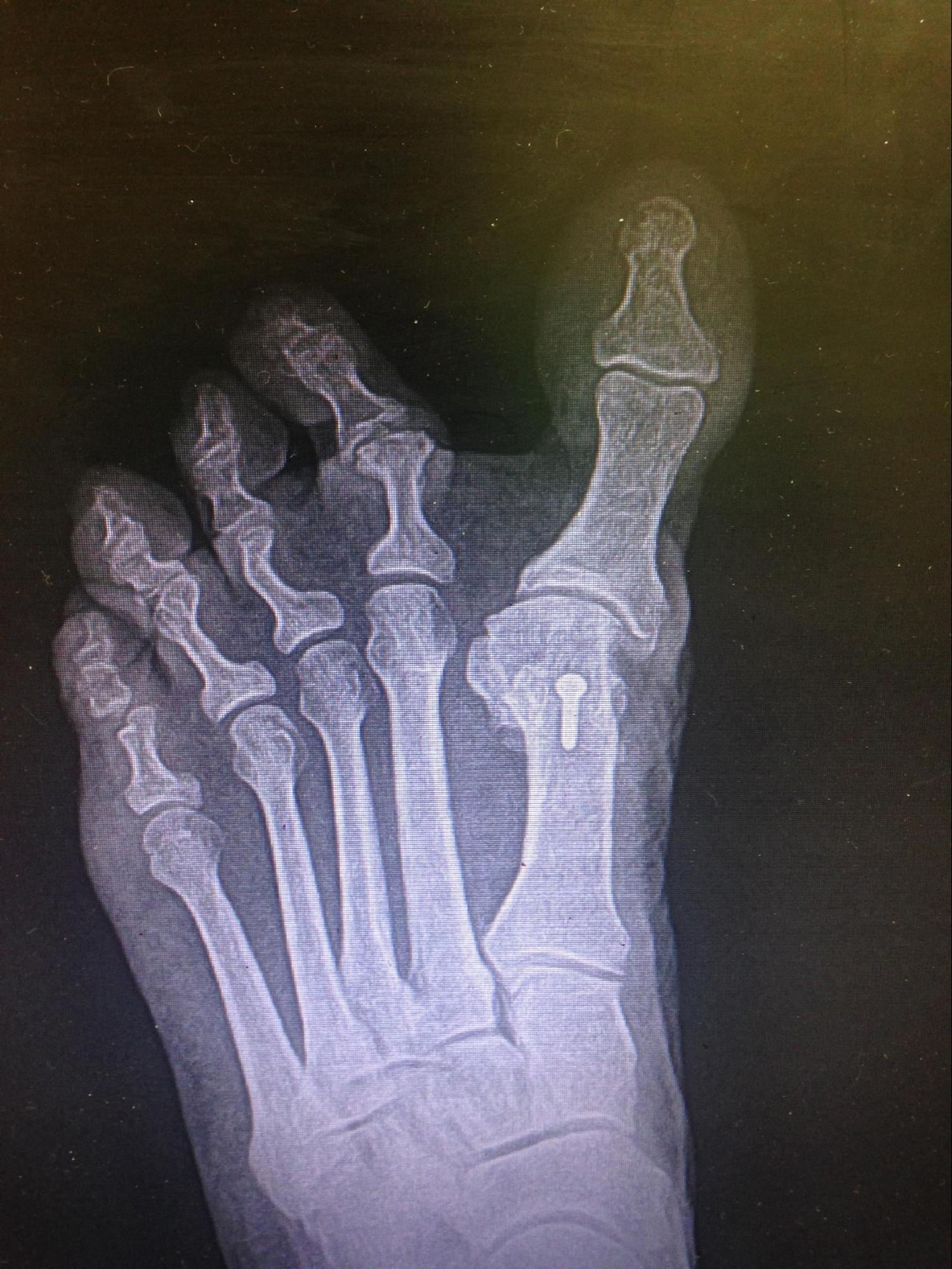
The presentation of hallux varus may be clear clinically and radiographically (see Image. Iatrogenic Hallux Varus). However, underlying disease processes contributing to the deformity are essential to consider and include the following:
- Osteoarthritis: Cortical degeneration with subchondral changes may result in medial deviation of the hallux in individuals affected by osteoarthritis. Radiographic evaluation will differentiate osteoarthritis from iatrogenic or congenital hallux varus.
- Rheumatoid arthritis: Rheumatoid arthritis causes dysregulation of chondrocytes and synovial fibroblasts in the musculoskeletal system, manifesting in articular and non-articular presentations. This often depreciates the capsular integrity of the first MTP joint, shifting the hallux in valgus; however, hallux varus has been reported in 0.2% to 10% of cases. Laboratory evaluation can confirm the diagnosis of rheumatoid arthritis.[26]
- Charcot-Marie-Tooth (CMT) disease: CMT is a neuromuscular disorder that initially presents motor and sensory dysfunction in the foot. The disease progresses proximally and gradually affects the lower extremity.[27] The musculotendinous imbalances often present as hallux malleus.[28]
- Avascular necrosis (AVN) of the metatarsal head: Historically, AVN following a distal chevron osteotomy for hallux valgus correction has been cited as a 20% occurrence. More recent literature has shown a 0% occurrence. However, the clinical presentation of AVN can range from asymptomatic to significant pain, deformity, and bone collapse.[29]
Prognosis
Surgery improves the overall position of the hallux but not necessarily its motion. In cases of flexible iatrogenic hallux varus, tendon transfer with soft tissue release has been reported to result in sustainable correction.[30] Patient satisfaction rates of 94% following an arthrodesis procedure for fixed deformity have also been reported.[5]
Complications
Patients undergoing conservative treatments may experience limited efficacy or adverse reactions to orthotic devices or splints, necessitating adjustments or alternative approaches. Moreover, any delay in seeking treatment or failure to adhere to postoperative rehabilitation protocols can compromise treatment outcomes and exacerbate existing complications.
Complications of surgical treatment in hallux varus can arise despite meticulous preoperative planning and execution. Potential complications of hallux varus surgery include the following:
- Soft tissue complications, including infection, impaired wound healing, and nerve damage or irritation
- MTP joint instability
- Recurrence of hallux varus
- Under-correction or over-correction
- Avascular necrosis of the metatarsal head
- Stiffness
- Hardware failure
- Progression of degenerative changes in the MTP joint
- Shortening of the medial column
- Transfer metatarsalgia
Comprehensive preoperative assessment, patient counseling, and vigilant post-treatment monitoring are crucial to minimize the risk of complications and optimize the success of interventions for hallux varus.
Postoperative and Rehabilitation Care
Postoperative care following hallux varus correction is dependent on the procedure. In cases of soft tissue procedures, including tendon transfers, tenodesis, and soft tissue releases, weight-bearing as tolerated is permitted in a stiff-soled shoe.[22] For osseous correction, similar postoperative protocols are followed for correction of hallux valgus procedures, including weight-bearing in a postoperative shoe for distal metatarsal osteotomies and non-weight bearing for arthrodesis procedures.[31][32]
Deterrence and Patient Education
Deterrence and patient education play pivotal roles in the management of hallux varus, aiming to prevent progression and optimize treatment outcomes. Through patient education initiatives, clinicians can empower individuals with knowledge about the condition's etiology, risk factors, and potential consequences. By highlighting the importance of early intervention and lifestyle modifications, such as appropriate footwear choices and toe exercises, patients can actively engage in self-care practices to mitigate symptoms and slow the deformity's progression. Additionally, emphasizing the potential risks associated with delaying treatment or neglecting conservative measures can serve as a deterrent, motivating patients to seek timely medical attention and adhere to recommended management strategies. Effective deterrence and patient education efforts enhance treatment compliance and foster proactive engagement in self-management, ultimately improving overall outcomes for individuals with hallux varus.
Pearls and Other Issues
Hallux varus can be a debilitating condition requiring specialized care. The etiology of the deformity will drive treatment options.
Conservative options
- Orthotics
- Wide shoe gear
- Activity modification
- Ice
Conservative treatment is useful for symptom management, though it does not correct the deformity. If pain continues, difficulty fitting into shoe gear, or ulceration occurs, surgical intervention may be needed.
Surgical options
- Metatarsal osteotomy
- First MTP joint arthrodesis
- Soft tissue rebalancing
Surgical intervention varies according to the etiology, prior procedures, and degree of deformity. A comprehensive history and physical are needed to address the underlying cause of the deformity.
Enhancing Healthcare Team Outcomes
Hallux varus is a relatively common foot deformity seen in clinics. Given somewhat limited treatment, early diagnosis and changes in shoe wear are key. Although changing shoe wear is beneficial, many patients have a significant deformity that requires surgery. Surgery improves the overall position of the hallux but not necessarily its motion. Salvage procedures may be necessary, and corrective iatrogenic hallux varus procedures are 60% to 80% effective.[33][34]
The condition is best managed by an interprofessional team that includes podiatrists, orthopedic surgeons, physical therapists, advanced care practitioners, nurses, pharmacists, and other healthcare professionals. Skills encompass a broad range, from clinicians adept in differentiating between hallux varus etiologies to specialists skilled in conservative treatments or surgical interventions. A strategic approach involves early screening, implementing evidence-based interventions, and ensuring ongoing patient education and follow-up. Ethically, practitioners must prioritize patient autonomy, informed decision-making, and transparency regarding treatment options and potential risks. Responsibilities include advocating for patient needs, ensuring informed consent, and adhering to best practices in care delivery.
Physical therapists play a crucial role in the management of hallux varus by designing customized rehabilitation programs to improve foot strength, flexibility, and alignment. Through targeted exercises and manual therapy techniques, they help patients regain mobility, reduce pain, and enhance overall function, complementing both conservative and surgical interventions.
Interprofessional communication is paramount, facilitating seamless collaboration, exchange of information, and shared decision-making among team members. Effective care coordination involves streamlining patient pathways, ensuring continuity of care, and addressing potential gaps or barriers to treatment. By harnessing these skills, strategies, ethical principles, responsibilities, interprofessional communication, and care coordination, healthcare professionals can collectively optimize patient-centered care, outcomes, safety, and team performance in managing hallux varus.