Continuing Education Activity
Mycoplasma pneumoniae is a common respiratory pathogen responsible for approximately 10% of all cases of community-acquired pneumonia. This bacterium is more prevalent in children and young adults. Aside from respiratory manifestations, M pneumoniae causes extrapulmonary syndromes, affecting approximately 25% of patients. These syndromes include cold-agglutinin hemolytic anemia, arthritis, pericarditis, thrombosis, and mucocutaneous manifestations.
Mucocutaneous eruptions, characterized by rashes affecting both mucous membrane and skin, can manifest as urticaria, erythema multiforme, Stevens-Johnson syndrome, toxic epidermal necrolysis, or drug reaction with eosinophilia and systemic symptoms, stemming from varied causes. However, accurately classifying these rashes in association with M pneumoniae infections has proven challenging. Recently, a distinct pattern of mucocutaneous rash associated with M pneumoniae respiratory infections has proposed an additional mucocutaneous eruption entity, which is Mycoplasma pneumoniae–induced rash and mucositis (MIRM). Diagnosing MIRM typically entails assessing recent or concurrent respiratory symptoms, with laboratory tests confirming Mycoplasma involvement. This activity reviews the etiology and pathophysiology of Mycoplasma mucositis while emphasizing the crucial role of the interprofessional healthcare team in achieving optimal patient outcomes through effective management.
Objectives:
Identify the clinical features and characteristic manifestations of Mycoplasma pneumoniae–induced rash and mucositis to facilitate accurate diagnosis.
Implement evidence-based management strategies for Mycoplasma pneumoniae–induced rash and mucositis, including antibiotic therapy and supportive care, to optimize patient outcomes.
Select appropriate pharmacological interventions for the treatment of Mycoplasma pneumoniae–induced rash and mucositis, considering factors such as antibiotic resistance patterns and patient comorbidities.
Collaborate with interprofessional healthcare team members, including infectious disease specialists, dermatologists, and intensivists, to provide comprehensive care for patients with Mycoplasma pneumoniae–induced rash and mucositis.
Introduction
Mycoplasma pneumoniae is a common respiratory pathogen responsible for approximately 10% of all cases of community-acquired pneumonia, with rates rising as high as 37% among children in certain studies and geographic areas.[1][2] This bacterium is more prevalent in children and young adults. Aside from respiratory manifestations, M pneumoniae causes extrapulmonary syndromes, affecting approximately 25% of patients.[2][3] These syndromes include cold-agglutinin hemolytic anemia, arthritis, pericarditis, thrombosis, and mucocutaneous manifestations. Mucocutaneous eruptions, characterized by rashes affecting both mucous membrane and skin, have varied inciting etiologies, including infectious, frequently viral, drug-induced reactions, and autoimmune factors.[4][5]
Previously, M pneumoniae infections have been associated with rashes and mucocutaneous manifestations such as urticaria, erythema multiforme, Stevens-Johnson syndrome (SJS), toxic epidermal necrolysis (TEN), and drug reaction with eosinophilia and systemic symptoms (DRESS).[6][7] However, accurately classifying these rashes associated with M pneumoniae infections has proven challenging, leading to considerable controversy in the literature. Recently, a distinct pattern of mucocutaneous rash linked to M pneumoniae respiratory infections has spurred the proposal for a novel mucocutaneous entity, coined Mycoplasma pneumoniae–Induced rash and mucositis (or MIRM) by Canavan et al in 2015 in a systematic review.[8][9][10]
Etiology
M pneumoniae infections can lead to both pulmonary and extra-pulmonary diseases.[11] These extra-pulmonary conditions encompass vasculitides, neurological complications, immunological disorders, thrombotic events, and mucocutaneous manifestations. Notably, mucocutaneous manifestations occur in approximately 25% of patients with M pneumonia infections.[12]
MIRM, a recently identified mucocutaneous entity, was introduced and coined in a 2015 systematic review by Canavan et al.[13] This review, comprising 202 cases of M pneumoniae-associated erythema multiforme, SJS, and mucositis without rash, delineated the distinct features of MIRM.[13] MIRM is characterized by prominent mucositis with comparatively less cutaneous involvement than other mucocutaneous syndromes associated with M pneumoniae, such as urticaria, erythema nodosum, erythema multiforme, SJS, TEN, and DRESS.[6][7][14]
Epidemiology
M pneumoniae is a prevalent respiratory pathogen, accounting for about 10% of all cases of community-acquired pneumonia. This bacterium can lead to extrapulmonary syndromes in roughly 25% of patients, which include cold-agglutinin hemolytic anemia, arthritis, pericarditis, thrombosis, and mucocutaneous manifestations.[2][3] The hallmark of MIRM is the involvement of the mucosa, which typically manifests in the urogenital and oral regions with ulcerations, vesicles, bullae, and ocular involvement. These symptoms often include conjunctivitis and, in severe instances, may lead to conjunctival ulceration and pseudomembranous formation.[15]
M pneumoniae most frequently causes community-acquired pneumonia, particularly in children aged 5 or older, with some regions reporting it as the cause in up to 37% of pediatric cases.[1][2] Both M pneumoniae and MIRM occur mostly during the winter months.[16] A systematic review by Canavan et al, using their novel proposed definition, found that MIRM was mostly reported in children and young adolescents with a mean age of 12.[13] However, a more recent systematic review by Lofgren and Leinkeit found that MIRM cases ranged between the ages of 4 and 46, with a mean age of 16.[16] However, MIRM has also been reported in young adults.[17][18] In the systematic review by Canavan et al, 60% of the identified cases of MIRM occurred in males, with 47% of patients experiencing mucositis without significant skin involvement and 34% presenting with mucositis alone without any skin involvement.[13]
The exact incidence of MIRM is not known for various reasons. Importantly, until now, a distinct definition distinguishing it from other causes of mucocutaneous syndromes associated with M pneumoniae infection does not exist, leading to misclassification of the syndrome. Other contributing factors include potential underreporting, often due to M pneumoniae not being considered in the initial diagnosis, limited availability of resources for testing, failure to identify a definitive cause, and possible lack of reporting. Until 2015, the lack of a discreet definition for MIRM led to inconsistent naming conventions in publications describing cases of M pneumoniae infection. Examples include M pneumoniae–associated SJS, Fuchs syndrome, and SJS without skin lesions.[17][19][20]
Pathophysiology
The exact pathogenesis of MIRM is not completely elucidated. Proposed mechanisms include immune activation, resulting in polyclonal B-cell and antibody production, leading to immune complex deposition and complement activation, subsequently causing skin lesions. Other causes could be molecular mimicry between Mycoplasma P1-adhesion molecules and the host's keratinocytes, potentially inducing injury via antibodies or cytotoxic T cells.[12][21] These processes differ from the mechanisms underlying erythema multiforme and SJS/TEN, which are mediated by delayed hypersensitivity reaction and Fas ligand–mediated toxicity.[22] These differences can aid in the differentiation of MIRM from other cutaneous reactions.[13]
Histopathology
The presence of histopathological features unique to MIRM that differentiate it from erythema multiforme, SJS, and TEN remains uncertain. Erythema multiforme, SJS, and MIRM exhibit similar and overlapping histopathological characteristics, including apoptotic keratinocytes and sparse perivascular dermal infiltrates. The existence of distinct biopsy features enabling histopathological differentiation between these diseases remains controversial.
Rzany et al investigated specimens from erythema multiforme, SJS, and TEN and did not observe significant, consistent histologic distinctions. However, Wetter and Camilleri identified histopathologic features in drug-induced SJS that were absent in MIRM or immunization-induced SJS in some examined specimens.[23][24] Specifically, SJS may exhibit more necrotic keratinocytes, denser dermal infiltrates, microscopic red blood cell extravasation, pigment incontinence, and parakeratosis compared to MIRM.[23]
History and Physical
Obtaining a thorough and detailed history of the present illness is particularly crucial. Such information can provide valuable clues aiding in the differentiation of MIRM from other mucocutaneous eruptions, such as erythema multiforme and SJS/TEN. In the case of MIRM, nearly all patients typically experience prodromal symptoms, such as fever, cough, and malaise, approximately 1 week before the onset of the rash.[13] SJS/TEN, viewed as different spectra of the same underlying process, are often preceded by a prodrome of fever and upper respiratory infection symptoms. Additionally, patients with SJS/TEN usually have a history of recent exposure to new medications, such as antibiotics, nonsteroidal anti-inflammatory drugs, allopurinol, antiepileptics, and nevirapine.[25] This medication history may sometimes lead to a lower index of suspicion for infection during differential diagnosis evaluation.
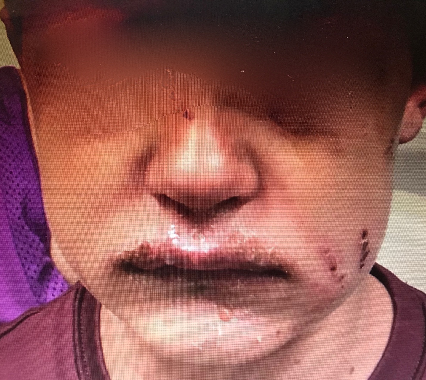
Physical examination findings in MIRM commonly reveal a predominance of mucosal rashes, with involvement of the oral mucosa (94%), ocular region (82%), and urogenital area (63%). Other mucosal sites, such as the nares and anus, may also be affected.[26] Mucosal lesions are typically characterized as ulcerative or hemorrhagic and may cause discomfort. Nasal involvement may present as dense hemorrhagic crusts, sometimes manifesting as blood on the tissue. Lesions in the anus, part of the gastrointestinal mucosa, can lead to pain during defecation (see Images 1 and 2. Mycoplasma pneumoniae–Induced Rash and Mucositis).[26]
Cutaneous non-mucosal rashes have been reported in 47% of MIRM cases, and if absent, the condition is classified as MIRM sine rash. When present, the cutaneous rash in MIRM displays distinct features compared to other mucocutaneous eruptions. MIRM rashes are typically sparse in distribution and are more commonly located in the acral regions (46%) than in the trunk (23%). The predominant morphology of the cutaneous rash in MIRM is described as vesiculobullous in 77% of cases. Additionally, both typical target lesions, characterized by 3 circumferential demarcation zones, and atypical target lesions, featuring 2 color zones, are observed in 48% of cases. Less commonly, rashes are described as papules (14%), macules (12%), or morbilliform (9%). The extent of detached skin typically involves less than 10% of the body surface area.[13]
In contrast, erythema multiforme presents initially as a cutaneous acral rash with macules that evolve into papules, plaques, and typical target lesions. These target lesions spread centripetally to the trunk and face. Erythema multiforme minor has little or no mucous membrane involvement, and erythema multiforme major has a rash on one or more mucous membranes. SJS/TEN manifests as a rash characterized by macules, purpura, diffuse erythema, atypical target lesions, and numerous flaccid blisters. These lesions are extensive in number and initially more concentrated centrally and gradually coalesce, spreading to involve the face and limbs. Extensive mucous membrane involvement affecting 2 or more mucosal sites is common. The amount of skin detachment determines the extent of SJS/TEN—SJS involves less than 10% skin detachment. Skin detachment ranging from 10% to 30% overlaps between SJS and TEN, whereas skin detachment exceeding 30% meets the criteria for TEN.[27]
Evaluation
Diagnosing MIRM involves the presence or recent occurrence of pulmonary symptoms, often leading to clinical findings suggestive of pneumonia, which may be confirmed through clinical examination and/or chest radiography. Laboratory assessment for the cause of pneumonia should include testing for elevated M pneumoniae IgM antibodies, detecting M pneumoniae from oropharyngeal or polymerase chain reaction (PCR) or bullae cultures, or obtaining serum cold agglutinins.[16]
The proposed definition, as outlined by Canavan et al,[13] and the classic diagnostic criteria for MIRM are mentioned below.[11]
- Evidence of atypical M pneumoniae pneumonia includes:
- Signs: Fever, cough, and auscultatory or radiographic findings.
- Laboratory findings: Increased M pneumoniae IgM antibodies, detection of M pneumoniae in oropharyngeal or bullae cultures or PCR, and/or serial cold agglutinins.
- Involvement of at least 2 mucosal sites.
- Few vesiculobullous lesions or scattered atypical targets may be observed.
Within MIRM, 3 types differ in their cutaneous, non-mucosal rash patterns [13][22][16]
Classic MIRM: This subtype meets the classic criteria outlined above and additionally presents with a non-mucosal rash, characterized by vesiculobullous lesions (77%), scattered target lesions (48%), papules (14%), macules (12%), and morbilliform eruptions (9%).
MIRM sine rash: This subtype fulfills the classic criteria mentioned above but lacks significant cutaneous, non-mucosal rash presence, although a few fleeting morbilliform lesions or a few vesicles may be present.
Severe MIRM: This subtype meets the above classic criteria, with the involvement of more than 2 mucosal sites reported. Additionally, the cutaneous rash is extensive, featuring widespread non-mucosal blisters or flat atypical target lesions.
Treatment / Management
In the acute care setting, distinguishing MIRM from other mucocutaneous eruptions can be challenging for clinicians. Therefore, consultation with an infectious disease physician or dermatologist, or transfer to a facility with appropriate resources, may be warranted. Patients with MIRM require supportive care, including pain management for skin lesions and oral ulcerations (such as "magic mouthwash" solution or sucralfate), mucosal care, and correction of any fluid and nutritional deficiencies resulting from reduced oral intake. Severe cases of MIRM with extensive skin detachment may necessitate early transfer to a burn center.[16] In addition, lesions in particular mucosal areas may warrant specialty consultation with ophthalmology, otolaryngology, gastroenterology, and urology to mitigate long-term complications.
Although specific treatment guidelines are not tailored for MIRM, patients diagnosed with the condition often exhibit evidence of atypical pneumonia and may, therefore, benefit from antibiotic treatment.[28] Common oral antibiotic options for atypical pneumonia, such as macrolides, tetracyclines, and fluoroquinolones, are typically recommended, with macrolides being the preferred choice.[28] Reports of macrolide resistance have been increasing globally, with varying prevalence rates across different regions. The highest resistance to the lowest prevalence of resistance is observed in the following regions—the Western Pacific, South East Asia, the United States of America, Europe, and the East Mediterranean.[29] Therefore, clinicians must remain vigilant, particularly in cases of refractory Mycoplasma pneumoniae pneumonia, as alternative antibiotic regimens may need to be considered.
Empirical administration of corticosteroids and other immunosuppressive agents has been documented, particularly in patients with severe MIRM. Intravenous immunoglobulin (IVIG) has also been utilized in cases of MIRM with severe mucositis. In a study by Canavan et al, 35% of patients received systemic corticosteroids, and 8% received IVIG.[13] In a more recent systematic review by Lofgren and Lenkeit, 77% of patients received antibiotics, 37% were treated with corticosteroids, and 11% received IVIG.[16]
Differential Diagnosis
Common differential considerations for MIRM include other conditions that can cause similar skin findings and/or mucosal manifestations[30][31]:
- Erythema multiforme major
- SJS/TEN
- DRESS
- Staphylococcal scalded skin syndrome
- Hand-foot-and-mouth disease
- Kawasaki disease
- Herpetic gingivostomatitis
- Severe cutaneous adverse reactions (eg, drug hypersensitivity syndrome)
- Bullous systemic lupus erythematosus
- Plasma cell stomatitis
- Coxsackie virus and other enteroviruses
- SARS-CoV-2 infection
- Autoimmune diseases, such as Behçet disease and systemic lupus erythematosus
Prognosis
Overall, the prognosis of MIRM is generally favorable. Only 4% of patients required admission to intensive care, with 81% experiencing complete recovery.[13] The recurrence rate of MIRM is 8%.[32] In contrast, patients afflicted with SJS/TEN often necessitate intensive care more frequently and face higher mortality rates.[33][24]
Complications
Although 81% of patients with MIRM experience a full recovery,[13] there are potential long-term sequelae, mostly involving the mucosa. Ocular mucosal damage occurs in 8.9% of patients with MIRM, resulting in conjunctival shrinkage, corneal ulcerations, blindness, ocular synechiae, and loss of eyelashes.[13] Postinflammatory pigmentary changes occurred in 5.6% of patients, while oral and genital mucosal synechiae were reported in 0.8% and 0.8% of patients, respectively. [13] Rare complications include persistent cutaneous lesions, B-cell lymphopenia, restrictive lung disease, and chronic obliterative bronchitis. However, while the mortality of MIRM is low, experts suspect that the vast majority of mild cases are underreported in the literature, suggesting that the true rate of morbidity and mortality is likely far lower.
Consultations
When considering the diagnosis of MIRM, consultation with infectious diseases specialists or dermatologists may be beneficial. Additionally, obtaining consultations with otolaryngologists, ophthalmologists, gastroenterologists, and urologists may be helpful for site-dependent mucosal lesions to mitigate long-term sequelae. In cases with extensive cutaneous lesions and detachment, consulting burn specialists is advisable.
Deterrence and Patient Education
Patients diagnosed with MIRM should be informed about the potential for painful mucosal lesions, which can lead to dehydration and reduced nutrient intake. In addition, it is important to keep cutaneous lesions clean to minimize the risk of secondary bacterial infection. Although the exact transmission patterns of MIRM are not well established, it is advisable for patients and close contacts of suspected M pneumoniae pulmonary infection to implement basic infection control measures, including droplet transmission precautions.
Pearls and Other Issues
- MIRM is considered a clinically distinct entity from other mucocutaneous eruptions.
- Historical features suggesting MIRM include a pulmonary infection approximately 1 week before the onset of the rash.
- Other mucocutaneous reactions, such as erythema multiforme, often develop after a herpes simplex infection.
- Although the rash of SJS/TEN may occur in the context of new medication use, it is crucial to include MIRM in the differential diagnosis.
- The rash pattern of MIRM may differ from that of other conditions, often characterized by mucous membrane involvement. In MIRM, the cutaneous rash often tends to be sparse rather than consolidated, frequently appearing in acral areas. Typically, the rash in MIRM is usually vesiculobullous or targets lesions.
- Although MIRM may result in some long-term sequelae, such as mucous membrane damage and cutaneous skin changes, it generally follows a more benign course than other conditions, including SJS/TEN.
- Currently, no specific guidelines exist for treating MIRM, but patients may benefit from antibiotics targeting atypical pneumonia and immunosuppressive therapy with corticosteroids. Additionally, IVIG administration should be considered as part of the treatment approach.
- Consulting with an infectious disease specialist or dermatologist can provide valuable guidance in directing patient care.
Enhancing Healthcare Team Outcomes
MIRM management is optimally conducted by an interprofessional team comprising infectious disease specialists, internists, primary care providers, and dermatologists. Collaborative efforts involving diagnosis, education, and patient support are crucial components of the interprofessional approach. The interprofessional team can achieve optimal outcomes by coordinating care and offering comprehensive patient education and support.
Patients diagnosed with MIRM should be informed about the potential for painful mucosal lesions, which can lead to dehydration and limit oral nutrient intake. In addition, it is important to keep cutaneous lesions as clean as possible to prevent secondary bacterial infection. Although the transmission patterns of MIRM are not well-established, it is advisable for patients and close contacts of suspected M pneumoniae pulmonary infection to implement basic infection control measures, including droplet transmission precautions.