Continuing Education Activity
The oculocardiac reflex (OCR), also known as the Aschner reflex or trigeminovagal reflex, was first described in 1908 as a heart rate reduction from direct pressure placed on the eyeball or periorbital structures. Currently, this reflex is defined by a heart rate decrease greater than 20% following globe pressure or extraocular muscle traction. The OCR most commonly results in sinus bradycardia, though associations with reduced arterial pressure, arrhythmia, asystole, and even cardiac arrest have been reported. Ophthalmologic procedures, specifically strabismus surgery, consistently trigger the OCR. However, facial trauma, regional anesthetic nerve blocks, and mechanical stimulation may also activate this reflex.
This activity for healthcare professionals is designed to enhance learners' proficiency in recognizing the OCR and managing its potential complications. Learners gain a deeper insight into the reflex's relevant anatomy, mechanisms, symptomatology, and appropriate interventions. Participants become prepared to collaborate within an interprofessional team caring for patients at risk of developing the OCR during surgery.
Objectives:
Identify the anatomical structures involved in the oculocardiac reflex.
Determine the risk factors for oculocardiac reflex occurrence.
Create a management strategy for a patient with oculocardiac reflex.
Implement effective collaboration and communication among interprofessional team members to improve outcomes and treatment efficacy for patients with oculocardiac reflex.
Introduction
The oculocardiac reflex (OCR), also known as the Aschner reflex or trigeminovagal reflex, was first described in 1908 as a heart rate diminishment when direct pressure is placed on the eyeball. The heart rate is characteristically decreased by over 20% following globe pressure or extraocular muscle traction. The OCR typically leads to sinus bradycardia, though arterial pressure reduction, arrhythmia, asystole, and even cardiac arrest have also been documented arising from this reflex. Ophthalmic procedures, particularly strabismus surgery, often induce the OCR. Facial trauma, regional anesthetic nerve blocks, and mechanical stimulation may also trigger this reflex.[1][2][3]
The OCR's incidence is reported to be anywhere from 14% to 90% and decreases with age. Pediatric populations face the highest risk of OCR development due to their increased susceptibility. Young individuals are also particularly vulnerable to OCR's complications due to their heavy dependence on the heart rate for maintaining cardiac output. The OCR's varying occurrence and severity are linked to factors like hypoxia, hypercarbia, acidosis, and the anesthetic agent type employed during surgery.[4][5]
Anatomy and Physiology
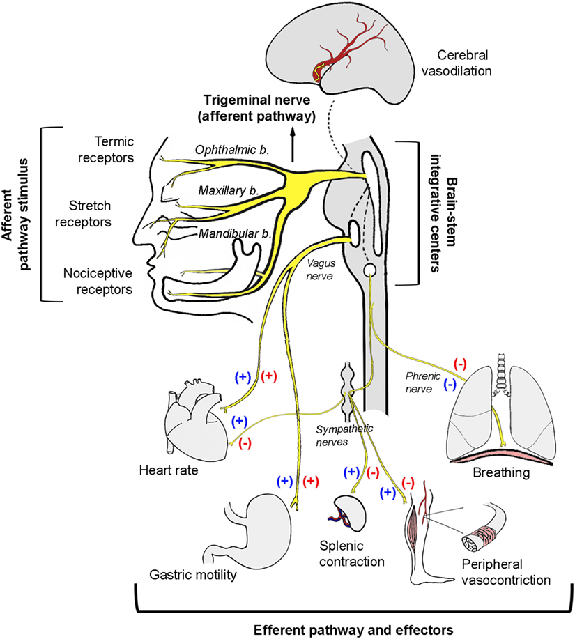
The OCR arc consists of an afferent and efferent limb (see Image. Schematic Diagram of Trigeminal Nerve Stimulation Effects). The trigeminal or 5th cranial nerve (CN V) comprises the reflex's afferent or sensory limb. The vagus or 10th cranial nerve (CN X) constitutes the efferent OCR limb. Ocular and periorbital stretch receptors activate this pathway.
The short and long ciliary nerves conduct impulses that carry the sensory message to the ciliary ganglion. The impulses are then transported via CN V's ophthalmic division to the Gasserian ganglion, followed by the trigeminal nucleus, where the afferent limb terminates in the central nervous system (CNS). The CNS processes the sensory input, facilitating communication between the trigeminal sensory nucleus and the vagus nerve's visceral motor nucleus. The outflow triggers the efferent limb, sending impulses from the brainstem to the myocardium, specifically the sinoatrial node, activating the vagal motor response. Negative chronotropy consequently arises, causing bradycardia.
Risk Factors
The OCR is often triggered by extraocular muscle traction, with the medial rectus muscle most commonly implicated. However, some studies show no particular muscle significantly associated with increased OCR occurrence.
Additional stimuli linked to OCR activation include direct globe pressure, ocular manipulation, and ocular pain. Retrobulbar blocks, ocular hematomas, and facial or orbital trauma may also activate the reflex due to increased pressure. However, the OCR is fatigable, decreasing in intensity with multiple, repeated stimuli.[6][7][8]
Complications
The OCR's complications arise from vagal responses and may include the following:
- Sinus bradycardia
- Arrhythmia
- Reduced atrial pressure
- Ventricular tachycardia
- Ventricular fibrillation
- Multifocal premature ventricular contractions
- Ventricular bigeminy
- Asystole
- Cardiac arrest
- Dizziness
- Lightheadedness
- Nausea
- Weakness
Prompt recognition and management are essential to avert significant morbidity and mortality.
Clinical Significance
Bradycardia is the OCR's most common manifestation, though it may deteriorate to potentially fatal arrhythmias, asystole, and cardiac arrest. Apt et al (1973) and Espahbodi et al (2015) reported OCR occurrence rates in patients undergoing ophthalmologic surgeries to be 67.9% and 63%, respectively. Most patients do not experience detrimental outcomes following OCR activation, but its effects can be potentially injurious.[9][10][11]
OCR activation has also been linked to noncardiac manifestations, including hypotension, syncope, and gastrointestinal symptoms like nausea and vomiting due to the vagal motor response. Vagal effects may influence postoperative nausea and vomiting severity. Nausea and vomiting rates in pediatric patients can reach 85% poststrabismus surgery, making it a leading cause of inpatient admission after outpatient procedures.
Emphasis must be given to the immediate cessation of the triggering stimulus as the only definitive treatment when addressing OCR prevention, treatment, and complications. Relieving eye or orbital pressure can deactivate the reflex. Proceeding cautiously is essential after removing the source. However, stimulus withdrawal may be more challenging in uncontrolled situations like trauma. Pharmacological management with cardiac monitoring may be necessary in instances where the OCR may develop.
The occurrence of the reflex may be reduced by choosing the appropriate anesthetic, as some agents are associated with a greater risk of OCR activation than others. Studies indicate that pretreatment with intravenous anticholinergics like atropine or glycopyrrolate decreases OCR incidence. Atropine blocks peripheral cardiac muscarinic receptors, increasing sinoatrial node firing and atrioventricular node conduction and countering OCR's vagal outflow.
Ketamine may also counteract vagal stimulation by enhancing sympathetic activity. Choi et al. reported lower OCR incidence with ketamine infusion than sevoflurane, halothane, and propofol. Espahbodi et al. (2015) found ketamine superior to atropine in OCR reduction. Other studies associated ketamine with a lower incidence of postoperative nausea, vomiting, and agitation.
Blunting the afferent OCR limb may also decrease its occurrence. Retro- or peribulbar blocks with xylocaine hydrochloride can block the ciliary ganglion. Combining with other agents known to decrease OCR incidence, such as atropine, can further prevent OCR activation.
Fast-acting opioids like fentanyl, sufentanil, and remifentanil can precipitate OCR and induce bradycardia. Many anesthetic agents' effects on the OCR have been studied, but several require further investigation. Preanesthetic medications like atropine and retrobulbar blocks should be routinely used during ocular procedures or when managing eye trauma to mitigate OCR effects and safeguard patients.
Enhancing Healthcare Team Outcomes
Manipulating the orbit poses a potential challenge in the operating room due to various stimuli activating the OCR. Therefore, anesthesiologists, ophthalmologists, maxillofacial surgeons, trauma teams, anesthesia nurses, and emergency medicine physicians working with orbital or facial structures must be aware of this reflex, its potential consequences, and strategies for managing or preventing its occurrence.
Vigilance and comprehensive monitoring are paramount to early detection and intervention in patient care. Clinicians must take advantage of diagnostic modalities like electrocardiography and blood pressure monitoring when caring for at-risk patients. The healthcare team must also be prepared to seamlessly administer drugs like atropine and glycopyrrolate and initiate Advanced Cardiac Life Support when necessary. Preparedness and vigilance are vital for promptly identifying and addressing potential complications, especially if symptomatic bradycardia or cardiac arrest arises from OCR activation. Continuous monitoring allows healthcare providers to intervene swiftly, potentially saving lives and enhancing patient outcomes.