Silica-Induced Pneumoconiosis (Archived)

Archived, for historical reference only

Introduction

Silicosis is occupational pneumoconiosis caused by inhalation of crystalline silicon dioxide. This disease is one of several well-described pulmonary complications associated with toxic exposures in the workplace, along with asbestosis, berylliosis, coal miner's lung, hard metal pneumoconiosis, among others.[1]

Etiology

Two forms of silicon dioxide exist in nature: amorphous and crystalline. The crystalline form of silicon dioxide is an abundant natural mineral, commonly found in substances such as sandstone, quartz, and granite. While inhalation of the amorphous form does not appear to cause clinically significant complications, inhalation of the crystalline compound can lead to pulmonary disease.

As silica deposits in the airways, contact with the alveolar and endobronchial surfaces generates reactive oxygen species. Smaller particles are taken up through phagocytosis into macrophages, which generate additional free radicals. Oxidative damage by activated macrophages and silica particles result in the release of inflammatory cytokines, increase in cell signaling, and apoptosis of parenchymal cells and macrophages. Fibroblast infiltration occurs in a nodular pattern as the disease progresses.[2]

Epidemiology

Numerous occupations may place workers at risk for exposure to silica dust across a wide variety of industries. [3] High-risk occupations include road repair, concrete manufacturing, coal mining, brick working, and rock excavation. Also at risk are workers employed in stone cutting, petroleum extraction, steel working, sandblasting, and others. According to the Occupational Safety and Health Administration, there are over two million workers exposed to some form of respirable silicon dioxide on a regular basis. Improved awareness and safety measures in the workplace have led to a substantial decrease in this disease over the last several decades; however, safeguards remain imperfect and there continue to be new cases of silicosis that arise.[3]

Pathophysiology

Inhalation of crystalline silicon dioxide causes mineral deposits to form at the level of terminal bronchioles and alveoli. The presence of foreign material results in the activation of alveolar macrophages and also exerts direct toxic effects on the surrounding lung parenchyma. Cellular damage results in the release of inflammatory cytokines (such as IL-1 and TNF-alpha), the generation of free radicals, and augmentation of cell-signaling pathways. The different cytokines result in the promotion of fibrosis. A role of tissue mast cells has also been reported. There is also evidence that silica interferes with the ability of macrophages to inhibit the growth of mycobacteria and this effect explains the common association silicosis/tuberculosis. In the acute form of silicosis, a direct toxic effect of alveolar type 2 cells, as well as on macrophages may occur. A role of immunologic factors has long been postulated but not proven in silicosis.[4][5]

Histopathology

Nodular silicosis is characterized by the presence of silicotic nodules. Grossly, silicotic nodules are firm, discrete, rounded lesions that contain a variable amount of black pigment. The nodules tend to occur around respiratory bronchioles and small pulmonary arteries and in the subpleural and paraseptal areas. Progressive expansion causes obliteration of small airways and pulmonary vessels. The latter are characterized by concentrically arranged hyalinized collagen bundles surrounded by variable numbers of dust-filled histiocytes. In early nodules, fibroblasts and histiocytes are prominent with acellular collagen lamellae. Small, polarizable, doubly refractile, round or oval particles are often found within nodules. Their presence helps to confirm the diagnosis but they may be absent in some cases and they are not specific of silicosis. Granulomatous reaction may be observed in the capsule of silicotic nodules. Its presence raises the possibility of the coexistence of mycobacterial infection. Central necrosis is unusual. The surrounding lung may be unremarkable although dust macules and pigment-laden macrophages are usually seen around small airways. A non specific type of interstitial fibrosis has been reported occasionally in patients with chronic silicosis.

In acute silicosis, microscopic findings mimic pulmonary alveolar proteinosis. Unlike ordinary alveolar proteinosis, there are usually also interstitial inflammation and fibrosis or irregular hyaline scars as well as a variable amount of pigment. Silicotic nodules are poorly formed or absent in most cases. 

Microscopic exam may be helpful in revealing some complications including progressive massive fibrosis or tuberculosis and non tuberculous infections. Progressive massive fibrosis is characterized by the presence of a nodular fibrosis that is greater than 1 cm in dimension. Usually, there is a large amorphous mass of fibrous tissue that is composed of conglomerated nodules causing obliteration and contracture of lung parenchyma. this lesion also occurs in other pneumoconioses including asbestosis, coal workers' pneumoconiosis or mixed dust fibrosis. Tuberculosis and non tuberculous mycobacteriosis can be suspected when facing a granulomatous infection with necrosis.[6][7]

History and Physical

Three clinical entities exist along the spectrum of disease including chronic forms, accelerated forms, and acute silicosis. Nodular or pure silicosis can occur after 20 or more years of exposure in occupations. An accelerated form of the disease occurs after a heavier exposure for 5 to 10 years. [8] Acute silicosis is an unusual reaction caused by heavy exposure over a short period to high levels of silica of small particle size.[8]

Evaluation

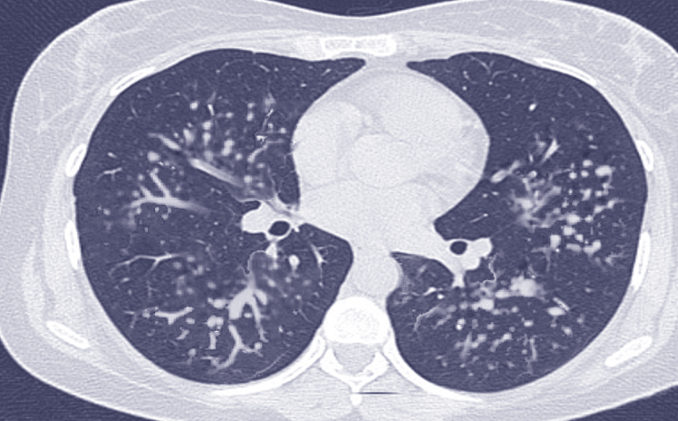
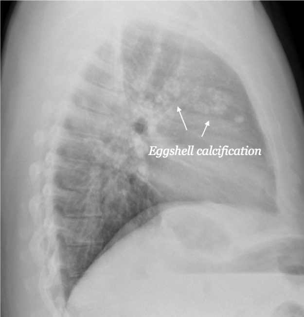
Radiologic findings depend on the form of silicosis. In simple silicosis, chest X-ray shows variable features consisting mainly in well-defined opacities ranging from 1 to 10 mm in diameter, located in the upper lobe and posterior portion of the lung. CT-scan findings consist of multiple small nodules with hilar and mediastinal lymphadenopathy. Some nodules may be calcified. 

In acute silicosis, chest X-ray shows bilateral consolidation associated with ground-glass opacities. CT-scan findings consist of numerous centrilobular ground-glass opacities with consolidation.[9][10]

Treatment / Management

There are no specific therapeutic modalities. The primary treatment procedure consists of removing the source of exposure.[6]

Differential Diagnosis

The principal differential diagnosis is rheumatoid arthritis. The silicotic nodules may show features mimicking a rheumatoid nodule. These nodules are characterized by central, acellular areas of necrosis surrounded by palisading histiocytes and a peripheral rim of lymphocytes and plasma cells. 

Toxicity and Adverse Effect Management

Complications of silicosis are represented by:

  • Progressive massive fibrosis
  • Tuberculosis and nontuberculous mycobacteriosis: they should be suspected clinically when there is rapid enlargement of small nodules or when nodules undergo cavitation. 

There are reports of an increased incidence of scleroderma in patients who suffer chronic exposure to silica but this phenomenon remains unexplained. Reports of acute glomerulonephritis are also in the literature.[2]

Enhancing Healthcare Team Outcomes

Silicosis is mainly a professional disease necessitating an eviction of the source of exposure.

The clinical symptoms and radiologic findings are nonspecific.

The microscopic exam is necessary to assess the diagnosis and to rule out the major diagnostic pitfalls consisting of progressive massive fibrosis and tuberculosis and nontuberculous mycobacteriosis.


(Click Image to Enlarge)
Silicosis, Computed Tomography
Silicosis, Computed Tomography. This CT of a patient with silicosis reveals the characteristic nodules in the middle lobes of both lungs accompanied by peri-hilar lymphadenopathy.
Contributed by S Bhimji, MD

(Click Image to Enlarge)
Eggshell calcification of hilar lymph nodes highly suggestive of silicosis
Contributed by Howard Sachs, MD
Details

Author

Mouna Mlika

Author

Rotimi Adigun

Updated:

8/17/2023 8:47:51 AM

References


[1]

Gottesfeld P, Tirima S, Anka SM, Fotso A, Nota MM. Reducing Lead and Silica Dust Exposures in Small-Scale Mining in Northern Nigeria. Annals of work exposures and health. 2019 Jan 7:63(1):1-8. doi: 10.1093/annweh/wxy095. Epub     [PubMed PMID: 30535234]


[2]

Skowroński M, Halicka A, Barinow-Wojewódzki A. Pulmonary tuberculosis in a male with silicosis. Advances in respiratory medicine. 2018:86(3):. doi: 10.5603/ARM.2018.0019. Epub     [PubMed PMID: 29960278]

Level 3 (low-level) evidence

[3]

Hoy R, Yates DH. Artificial stone-associated silicosis in Belgium: response. Occupational and environmental medicine. 2019 Feb:76(2):134. doi: 10.1136/oemed-2018-105563. Epub 2018 Dec 11     [PubMed PMID: 30538143]


[4]

Peruzzi C, Nascimento S, Gauer B, Nardi J, Sauer E, Göethel G, Cestonaro L, Fão N, Cattani S, Paim C, Souza J, Gnoatto D, Garcia SC. Inflammatory and oxidative stress biomarkers at protein and molecular levels in workers occupationally exposed to crystalline silica. Environmental science and pollution research international. 2019 Jan:26(2):1394-1405. doi: 10.1007/s11356-018-3693-4. Epub 2018 Nov 13     [PubMed PMID: 30426371]


[5]

Wang XX, Zhang HD, Wang XM. [The analysis of the epidemiological characteristics of pneumoconiosis notified in Chongqing from 2011 to 2015]. Zhonghua lao dong wei sheng zhi ye bing za zhi = Zhonghua laodong weisheng zhiyebing zazhi = Chinese journal of industrial hygiene and occupational diseases. 2018 Mar 20:36(3):194-197. doi: 10.3760/cma.j.issn.1001-9391.2018.03.008. Epub     [PubMed PMID: 29996220]

Level 2 (mid-level) evidence

[6]

Joubert KD, Awori Hayanga J, Strollo DC, Lendermon EA, Yousem SA, Luketich JD, Ensor CR, Shigemura N. Outcomes after lung transplantation for patients with occupational lung diseases. Clinical transplantation. 2019 Jan:33(1):e13460. doi: 10.1111/ctr.13460. Epub 2018 Dec 30     [PubMed PMID: 30506808]


[7]

Li J, Yao W, Hou JY, Zhang L, Bao L, Chen HT, Wang D, Yue ZZ, Li YP, Zhang M, Yu XH, Zhang JH, Qu YQ, Hao CF. The Role of Fibrocyte in the Pathogenesis of Silicosis. Biomedical and environmental sciences : BES. 2018 Apr:31(4):311-316. doi: 10.3967/bes2018.040. Epub     [PubMed PMID: 29773095]


[8]

Guarnieri G,Bizzotto R,Gottardo O,Velo E,Cassaro M,Vio S,Putzu MG,Rossi F,Zuliani P,Liviero F,Mason P,Maestrelli P, Multiorgan accelerated silicosis misdiagnosed as sarcoidosis in two workers exposed to quartz conglomerate dust. Occupational and environmental medicine. 2019 Mar     [PubMed PMID: 30514749]


[9]

Takahashi M, Nitta N, Kishimoto T, Ohtsuka Y, Honda S, Ashizawa K. Computed tomography findings of arc-welders' pneumoconiosis: Comparison with silicosis. European journal of radiology. 2018 Oct:107():98-104. doi: 10.1016/j.ejrad.2018.08.020. Epub 2018 Aug 24     [PubMed PMID: 30292280]


[10]

Zhang H,Li L,Xiao H,Sun XW,Wang Z,Zhang CL, Silicotuberculosis with oesophagobronchial fistulas and broncholithiasis: a case report. The Journal of international medical research. 2018 Feb     [PubMed PMID: 28703631]

Level 3 (low-level) evidence

[11]

Yang J,Xue J,Hu W,Zhang L,Xu R,Wu S,Wang J,Ma J,Wei J,Wang Y,Wang S,Liu X, Human embryonic stem cell-derived mesenchymal stem cell secretome reverts silica-induced airway epithelial cell injury by regulating Bmi1 signaling. Environmental toxicology. 2023 Sep;     [PubMed PMID: 37227716]


[12]

Zhang Y,Liu F,Jia Q,Zheng L,Tang Q,Sai L,Zhang W,Du Z,Peng C,Bo C,Zhang F, Baicalin alleviates silica-induced lung inflammation and fibrosis by inhibiting TLR4/NF-?B pathway in rats. Physiological research. 2023 Apr 30;     [PubMed PMID: 37159856]


[13]

Wang Y,Cheng D,Li Z,Sun W,Zhou S,Peng L,Xiong H,Jia X,Li W,Han L,Liu Y,Ni C, IL33-mediated NPM1 promotes fibroblast-to-myofibroblast transition via ERK/AP-1 signaling in silica-induced pulmonary fibrosis. Toxicological sciences : an official journal of the Society of Toxicology. 2023 Aug 29;     [PubMed PMID: 37399107]