Posterior Myocardial Ischemia (Archived)

Archived, for historical reference only

Introduction

Acute myocardial infarction is a common cause of chest pain in adult patients, responsible for more than 8 million emergency department presentations every year. Ischemic heart disease accounts for more than 500,000 deaths in the United States annually. Posterior myocardial infarction (PMI) accounts for a substantial number of cases of acute coronary syndrome (ACS); however, true PMI is often under-recognized, potentially leading to delays in treatment. PMI, like other acute myocardial infarction, is a true medical emergency with significant morbidity and mortality and requires urgent reperfusion therapy.

Etiology

PMI typically results from a reduction or cessation of blood flow to one or several of the coronary arteries. Normally, oxygenated blood is supplied to the coronary arteries after leaving the left ventricle. This oxygenated blood then supplies oxygen to the myocardial tissue and removes waste. There are two main coronary arteries: the right coronary artery (RCA) and the left main coronary artery, which divides into the circumflex artery (LCx) and the left anterior descending artery (LAD). The coronary artery that supplies the posterior third of the interventricular septum determines circulatory “dominance.” The dominant artery is responsible for flow to the posterior descending artery (PDA). Approximately 70% of the population is “right-dominant” (i.e., the RCA supplies the PDA), while 10% are “left-dominant (i.e., the LCx supplies the PDA), and 20% are “co-dominant” (i.e., both the RCA and LCx jointly supply the PDA).

Epidemiology

Posterior wall involvement is reported to occur in 15% to 21% of acute MI.[1] An “isolated PMI” occurs when the exclusively posterior wall is affected, most commonly due to occlusion of the LCx.[2] The rate of “isolated PMI” has been reported to be approximately 3.3%[1]; however, some experts contend that this number is likely higher as it is often under-diagnosed electrocardiographically.[3]

Pathophysiology

Ischemia is the result of a reduction of blood supply to a particular tissue. Reduction of blood supply prevents oxygen delivery and waste removal from the afflicted tissue. In myocardial infarction, this is most commonly due to atherosclerotic plaques, which are aggregates of macrophages, smooth muscle, and lipids that form on the injured vascular wall. A fixed atherosclerotic lesion in a vessel supplying cardiac tissue that experiences increased oxygen demand (e.g., tachycardia, hypotension, hypoxemia, among others) may produce myocardial ischemia. Type 1 acute myocardial infarction (AMI) occurs when a fixed atherosclerotic plaque ruptures, causing platelet aggregation and, ultimately, either critically low blood flow or no blood flow to cardiac myocytes distal to the lesion, resulting in cardiac myocytes necrosis. “Occlusion MI” (OMI) occurs when type 1 AMI is caused by near or total occlusion with insufficient collateral circulation, whereas non-occlusion MI (NOMI) describes any AMI without such occlusion or near-occlusion. Posterior myocardial infarction refers specifically to necrosis of cardiac myocytes located in the dorsal, infra-atrial portion of the left ventricle located beneath the atrioventricular sulcus.[1]

History and Physical

The most common complaint associated with any AMI including PMI is chest pain or discomfort, though it is not always present. The chest discomfort is classically described as a squeezing, heaviness, or tightness. Other manifestations of PMI include shortness of breath, epigastric pain or discomfort, back pain, or shoulder pain. The pain may radiate to the arms, back, neck, or jaw. Associated symptoms often include nausea, diaphoresis, and/or lightheadedness. Symptoms brought on by physical exertion and improved with rest are highly concerning for ACS. Women, diabetics, and the elderly often have “atypical” presentations of ACS.[4][5] Risk factors for coronary artery disease (CAD) include smoking, family history of early CAD, hypertension, hyperlipidemia, diabetes mellitus, and obesity.[6]

The physical exam is not always a reliable indicator of the presence of ACS. Patients with actively infarcted myocardium may range from mild discomfort to obtundation. Frequently, the episode of discomfort that prompts an encounter for ACS may have resolved by the time of presentation. Vital signs may range from hypertension to hypotension in the case of AMI induced cardiogenic shock with a compensatory tachycardia. However, bradycardia is also a common vital sign abnormality if the sinoatrial (SA) node is involved (most commonly in the setting of a proximal occlusion of a dominant RCA). The presence of an S3 gallop, crackles, and cold extremities may indicate acute heart failure and cardiogenic shock as a result of AMI. A new murmur may indicate papillary muscle rupture or new valve dysfunction. The patient may have a clenched fist or open hand pressed against the sternum, termed Levine sign and Cossio sign respectively.[7]

Finally, cardiac arrest, especially with ventricular tachycardia or ventricular fibrillation as the initial aberrant rhythms is a common presentation for AMI.

Evaluation

The diagnosis of PMI is established by the ECG in conjunction with history and physical.

PMI most commonly manifests on ECG in conjunction with other vascular territories (e.g., inferoposterior) or in isolation.

Isolated or “True” Posterior Myocardial Infarction

Unlike the anterior, lateral, and inferior walls of the left ventricle, there is no lead on the standard 12-lead ECG that overlies the posterior wall. While transmural infarction (the result of STEMI or OMI) of other walls registers as “positive” or elevated changes on the corresponding leads of the standard 12 leads (e.g., inferior STEMI has ST elevations in the inferior leads II, III, and aVF), posterior MI registers only reciprocal changes on the standard anterior leads because there is no standard lead overlying the posterior wall.

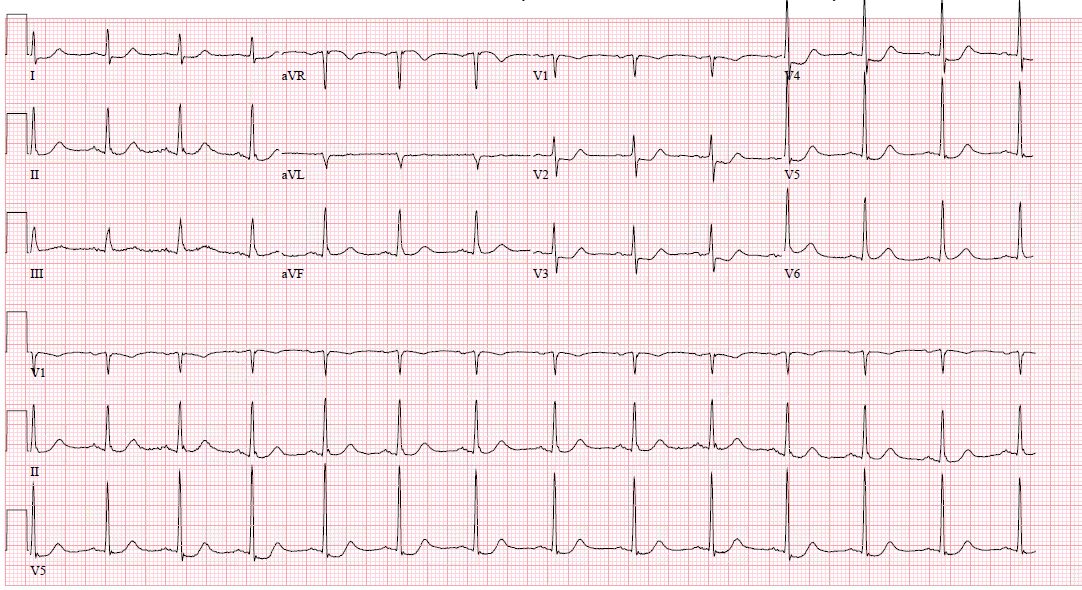
Therefore, posterior infarction appears on the standard 12-lead ECG primarily as ST depressions in leads V1-V4,[8][9] usually deepest in leads V2-V4.[10][11] This is contrary to subendocardial ischemia (the result of demand ischemia or non-total occlusion or sufficient collateral circulation) that manifests as diffuse ST depressions, usually deepest in V4-V6 and lead II (leads that are generally in line with the overall average vector of depolarization of the entire left ventricle). Diagnosis of isolated PMI is defined by the European Society of Cardiology (ESC) 2017 guidelines as isolated ST-segment depression greater than or equal to 0.5 mm in leads V1-V3 or ST elevation of 0.5 mm in leads V7-V9 with the use of posterior leads.[2] Other documents, guidelines, and societies have differing criteria or no specific criteria for the definition of posterior MI:

  • The National Cardiovascular Data Registry (NCDR) defines isolated PMI as “ST elevations in leads V7-V9 or ST depressions maximal in V1-V3, without ST elevation in other leads is considered a STEMI equivalent and qualifies for emergent reperfusion.”
  • The 2009 Standardization of ECG Interpretation Guidelines defines isolated PMI as 0.05 mV of abnormal J-point elevation in leads V7-V9 or “abnormal J-point depression of -0.05 mV in V1-V3 or -0.1 mV in all other leads” without qualification of what indicates abnormal J-point depression.[12]
  • The 2013 ACCF/AHA STEMI guidelines suggest that “ST depression in 2 or more leads of V1-4 may indicate transmural posterior injury”[13] and the AHA/ACC 2017 Clinical Performance and Quality Measures for Adults With ST-Elevation and Non–ST-Elevation Myocardial Infarction indicate that isolated PMI is a “STEMI equivalent” and should be treated with emergent reperfusion, though no definition for isolated PMI was provided.[14]
  • The Fourth Universal Definition of MI in 2018 suggests that the pattern of “new horizontal or downsloping ST-depression 0.5 mm or greater in two contiguous leads and/or T inversion >1 mm in 2 contiguous leads with prominent R wave or R/S ratio >1” is abnormal and likely ischemic; however, no specific recommendations for management are made.[15]

The distinction between STEMI and OMI is demonstrated in many cases of true isolated PMI. It is clear that ST elevations meeting STEMI criteria are a medical emergency requiring emergent reperfusion; however, in many true PMI (as in the example below) the ECG manifests only ST depression and therefore does not meet current ACC/AHA STEMI criteria. Persistent symptomatic occlusion of the coronary artery supplying the posterior wall (OMI) requires emergent reperfusion and should therefore be treated as a STEMI, because persistent total occlusion is the typical cause of classic STEMI, and there has never been any evidence demonstrating a difference in the benefit of reperfusion between STEMI(+) occlusion and STEMI(-) Occlusion MI. As expected based on this knowledge, failure to recognize PMI is common and results in delays in time to reperfusion and worsened outcomes.[16] A subanalysis of the TRITON-TIMI 38 trial revealed that 314 of 1198 (26%) of patients with isolated ST depressions in V1-V6 had an acute occlusion (TIMI 0-1) demonstrated with coronary angiography. Furthermore, only 14 of the 314 (4.5%) with confirmed posterior OMI were interpreted as a STEMI equivalent by the study investigators. There were delays of 6 hours or more from presenting ECG to PCI in all of these patients. This data does not take into account the number of patients that had reperfusion of the completely occluded artery with medical therapy before undergoing PCI.[3]

Posterior Leads

Performing a posterior ECG involves taking leads V4-V6 and applying them to the left side of the patient’s back. Specifically, V4 is applied to the posterior axillary line at the same level as V6 and is then referred to as V7. V5 is placed at the mid-scapular line at the same level as V6 and is referred to as V8. Finally, V6 is placed midway between V8 and the left paraspinal muscles at the level of V8 and is then referred to as V9.[17] The application of posterior leads has been demonstrated to increase sensitivity and specificity when diagnosing acute posterior MI.[16]

Air is a poor conductor of electricity, and therefore, the ST elevations in posterior leads are much less profound since they must first go through the air of the lungs before reaching the leads. This may create a scenario in which the depressions in V1-V4 are obvious, but the posterior leads do not show much if any, elevation. ST elevation in the posterior leads of up to 0.5 mm may be normal. [17] Conversely, ST elevation of 0.5 mm or more in posterior leads (V7-V9) without ST depression in V1-V3 has been demonstrated in 22% to 39% of PMI. [16][18][19] Using the criteria of ST elevations of 0.5 mm or greater rather than 1 mm in posterior leads when evaluating for PMI improves the sensitivity from 49% to between 94% to 100%.[16][18][19] It is essential to maintain a high level of suspicion for PMI in ECGs with ST depressions that are maximal in V1-V4 and with a suspicious clinical picture. Repeat ECGs also increase the sensitivity and specificity of the ECG. If clinical uncertainty remains, a STAT echocardiogram can be performed to look for wall motion abnormalities.

Biomarkers

Troponin elevation greater than the 99th percentile of the upper limit of normal for the particular assay plus one of the following is criteria for acute MI:

  • Symptoms of myocardial ischemia
  • New ischemic ECG changes
  • Development of pathological Q waves
  • Imaging evidence of new loss of viable myocardium or new regional wall motion abnormalities in a pattern consistent with an ischemia etiology
  • Identification of a coronary thrombus by angiography or autopsy[15]

Importantly, the current contemporary troponin assays widely used in the United States will not be positive even in true AMI until up to 4 to 6 hours after the onset of acute coronary occlusion. Over-reliance on troponin in the diagnosis of AMI will result in delays of care and worse outcomes. The decision to perform emergent reperfusion is a bedside clinical decision based on history, physical, and ECG findings, and should not be delayed for any laboratory test.

Treatment / Management

When there is a concern for PMI, the patient should have continuous cardiac monitoring with defibrillator capacity and pulse oximetry, and IV access must be established. Patients with hypoxemia or in respiratory distress should receive supplemental oxygen.[20] Aspirin reduces mortality in the setting of AMI and should be administered to all patients with suspected ischemia without an aspirin allergy.[21] Heparin infusion should be initiated to all patients without contraindication who are clinically diagnosed with active, ongoing acute coronary syndrome, as the combination of heparin and aspirin reduces the short-term risk of death or progression to AMI by 56% in patients with ACS.[22] Additional agents such as PGY12 inhibitors, GIIb/IIIa antagonists, and other antithrombin agents should be considered in consultation with cardiology. Nitrates should be used to treat ongoing ischemic chest pain unless there is evidence of right ventricular infarction, hypotension is present, significant bradycardia or tachycardia, allergy to nitrates, or phosphodiesterase inhibitor (e.g., sildenafil, tadalafil) use within the last 24 to 48 hours.[13]

Emergent reperfusion therapy is formally recommended in PMI that meets STEMI criteria in the standard 12-leads or posterior leads V7-V9, as well as any patient with ischemia refractory to maximal medical management or ACS with hemodynamic or electrical instability.[2][12][13] Mechanical treatment with percutaneous intervention (PCI) is superior and to have fewer complications compared to medical reperfusion therapy with fibrinolytics.[23] PCI should be performed within 90 minutes of arrival to PCI center or within 120 minutes if nearby PCI-capable center is available. If PCI is unavailable, fibrinolytics can be administered up to 12 hours from symptom onset.[14] The authors do not recommend using opioid pain medication to treat chest pain suspicious for ACS unless the decision to perform emergent reperfusion is already made and maximal medical therapy has already been initiated, as it may mask, and therefore, delay definitive treatment which is required for ischemia refractory of maximal medical management.[2][12]

Differential Diagnosis

  • Acute coronary syndrome 
  • Pulmonary embolism 
  • Pneumothorax
  • Aortic dissection
  • Boerhaave Syndrome
  • Pneumonia
  • Gastroesophageal reflux disease 
  • Esophageal dysfunction
  • Pancreatitis
  • Musculoskeletal pain

Prognosis

Patients with inferior or lateral MI with posterior extension are at an increased risk of complications including death, likely due to the size of the infarct.[16][24] Further, isolated PMI is less likely to be detected on ECG, owing to the absence of traditional or familiar “red-flag” ECG findings of AMI (for example, ST elevations). [3][11] Patients who present with STEMI and undergo PCI in under 2 hours have 30-day mortality of 3% to 5%.[25][26] NSTEMI with proven coronary occlusion (OMI) on coronary angiography has double the mortality rate at 30 days (2% to 5%) compared to NSTEMI without a demonstrated coronary occlusion (NOMI).[27][28][29][30] There are no specific data to support a significant difference in mortality of posterior STEMI compared to STEMI of other anatomical location.

Complications

Dysrhythmia, free wall rupture, mitral regurgitation, heart failure, Dressler syndrome, and death.[16][31] The rate of complication increases with increasing time to reperfusion.[24]

Consultations

When concern exists for PMI early consultation with an interventional cardiologist and a PCI-capable facility are essential.

Pearls and Other Issues

  • Isolated PMI is difficult to diagnose owing to the absence of traditional STEMI criteria and standard 12-lead positioning
  • ST depressions in the precordial leads, deepest in V1-V4, are concerning for PMI and require further immediate attention
  • Posterior ECGs should be performed when investigating PMI; however, posterior leads may be falsely negative and should not dissuade against emergent reperfusion if there are concerning findings such as ST depression in the anterior leads
  • Occlusion MI affecting the posterior wall frequently does not meet STEMI criteria, requiring the practitioner to develop ECG interpretation skills beyond the current STEMI vs. NSTEMI paradigm to deliver optimal care to patients with ACS.

Enhancing Healthcare Team Outcomes

Paying attention to changes in the patient’s status, particularly in terms of recurrence of chest discomfort or intractable pain is essential. Serial ECGs should be obtained in patients with chest pain. Furthermore, deliberate reassessment of these patients by physicians, nurses, and other staff members is essential to optimizing outcomes and providing expedient care.

Patients should be counseled on lifestyle modifications including weight loss, smoking/illicit drug secession, and the importance of medication adherence. This is particularly important after an AMI as cessation or non-compliance with antiplatelet agents in particular following PCI can lead to in-stent restenosis and additional AMI events.

The outcomes after a PMI depend on the time to treatment, type of treatment, patient age, the extent of infarct, hemodynamic changes,  and other comorbidities.


(Click Image to Enlarge)
Isolated posterior MI (OMI), cath proven 100% proximal circumflex with TIMI 0 flow (example 2)
Contributed by Alexander Bracey, MD
Details

Updated:

4/7/2023 3:04:21 PM

References


[1]

van Gorselen EO, Verheugt FW, Meursing BT, Oude Ophuis AJ. Posterior myocardial infarction: the dark side of the moon. Netherlands heart journal : monthly journal of the Netherlands Society of Cardiology and the Netherlands Heart Foundation. 2007 Jan:15(1):16-21     [PubMed PMID: 17612703]


[2]

Ibanez B, James S, Agewall S, Antunes MJ, Bucciarelli-Ducci C, Bueno H, Caforio ALP, Crea F, Goudevenos JA, Halvorsen S, Hindricks G, Kastrati A, Lenzen MJ, Prescott E, Roffi M, Valgimigli M, Varenhorst C, Vranckx P, Widimský P. [2017 ESC Guidelines for the management of acute myocardial infarction in patients presenting with ST-segment elevation.]. Kardiologia polska. 2018:76(2):229-313. doi: 10.5603/KP.2018.0041. Epub     [PubMed PMID: 29457615]


[3]

Pride YB, Tung P, Mohanavelu S, Zorkun C, Wiviott SD, Antman EM, Giugliano R, Braunwald E, Gibson CM, TIMI Study Group. Angiographic and clinical outcomes among patients with acute coronary syndromes presenting with isolated anterior ST-segment depression: a TRITON-TIMI 38 (Trial to Assess Improvement in Therapeutic Outcomes by Optimizing Platelet Inhibition With Prasugrel-Thrombolysis In Myocardial Infarction 38) substudy. JACC. Cardiovascular interventions. 2010 Aug:3(8):806-11. doi: 10.1016/j.jcin.2010.05.012. Epub     [PubMed PMID: 20723851]

Level 2 (mid-level) evidence

[4]

Canto JG, Goldberg RJ, Hand MM, Bonow RO, Sopko G, Pepine CJ, Long T. Symptom presentation of women with acute coronary syndromes: myth vs reality. Archives of internal medicine. 2007 Dec 10:167(22):2405-13     [PubMed PMID: 18071161]


[5]

Arora G, Bittner V. Chest pain characteristics and gender in the early diagnosis of acute myocardial infarction. Current cardiology reports. 2015 Feb:17(2):5. doi: 10.1007/s11886-014-0557-5. Epub     [PubMed PMID: 25618302]


[6]

Han JH, Lindsell CJ, Storrow AB, Luber S, Hoekstra JW, Hollander JE, Peacock WF 4th, Pollack CV, Gibler WB, EMCREG i*trACS Investigators. The role of cardiac risk factor burden in diagnosing acute coronary syndromes in the emergency department setting. Annals of emergency medicine. 2007 Feb:49(2):145-52, 152.e1     [PubMed PMID: 17145112]


[7]

Edmondstone WM. Cardiac chest pain: does body language help the diagnosis? BMJ (Clinical research ed.). 1995 Dec 23-30:311(7021):1660-1     [PubMed PMID: 8541748]


[8]

Rich MW, Imburgia M, King TR, Fischer KC, Kovach KL. Electrocardiographic diagnosis of remote posterior wall myocardial infarction using unipolar posterior lead V9. Chest. 1989 Sep:96(3):489-93     [PubMed PMID: 2788559]


[9]

Levis JT. ECG Diagnosis: Isolated Posterior Wall Myocardial Infarction. The Permanente journal. 2015 Fall:19(4):e143-4     [PubMed PMID: 26828074]


[10]

Bough EW, Korr KS. Prevalence and severity of circumflex coronary artery disease in electrocardiographic posterior myocardial infarction. Journal of the American College of Cardiology. 1986 May:7(5):990-6     [PubMed PMID: 3958381]


[11]

Boden WE, Kleiger RE, Gibson RS, Schwartz DJ, Schechtman KB, Capone RJ, Roberts R. Electrocardiographic evolution of posterior acute myocardial infarction: importance of early precordial ST-segment depression. The American journal of cardiology. 1987 Apr 1:59(8):782-7     [PubMed PMID: 3825938]


[12]

Wagner GS, Macfarlane P, Wellens H, Josephson M, Gorgels A, Mirvis DM, Pahlm O, Surawicz B, Kligfield P, Childers R, Gettes LS, Bailey JJ, Deal BJ, Gorgels A, Hancock EW, Kors JA, Mason JW, Okin P, Rautaharju PM, van Herpen G, American Heart Association Electrocardiography and Arrhythmias Committee, Council on Clinical Cardiology, American College of Cardiology Foundation, Heart Rhythm Society. AHA/ACCF/HRS recommendations for the standardization and interpretation of the electrocardiogram: part VI: acute ischemia/infarction: a scientific statement from the American Heart Association Electrocardiography and Arrhythmias Committee, Council on Clinical Cardiology; the American College of Cardiology Foundation; and the Heart Rhythm Society. Endorsed by the International Society for Computerized Electrocardiology. Journal of the American College of Cardiology. 2009 Mar 17:53(11):1003-11. doi: 10.1016/j.jacc.2008.12.016. Epub     [PubMed PMID: 19281933]


[13]

O'Gara PT, Kushner FG, Ascheim DD, Casey DE Jr, Chung MK, de Lemos JA, Ettinger SM, Fang JC, Fesmire FM, Franklin BA, Granger CB, Krumholz HM, Linderbaum JA, Morrow DA, Newby LK, Ornato JP, Ou N, Radford MJ, Tamis-Holland JE, Tommaso CL, Tracy CM, Woo YJ, Zhao DX, Anderson JL, Jacobs AK, Halperin JL, Albert NM, Brindis RG, Creager MA, DeMets D, Guyton RA, Hochman JS, Kovacs RJ, Kushner FG, Ohman EM, Stevenson WG, Yancy CW, American College of Cardiology Foundation/American Heart Association Task Force on Practice Guidelines. 2013 ACCF/AHA guideline for the management of ST-elevation myocardial infarction: a report of the American College of Cardiology Foundation/American Heart Association Task Force on Practice Guidelines. Circulation. 2013 Jan 29:127(4):e362-425. doi: 10.1161/CIR.0b013e3182742cf6. Epub 2012 Dec 17     [PubMed PMID: 23247304]

Level 3 (low-level) evidence

[14]

Jneid H, Addison D, Bhatt DL, Fonarow GC, Gokak S, Grady KL, Green LA, Heidenreich PA, Ho PM, Jurgens CY, King ML, Kumbhani DJ, Pancholy S. 2017 AHA/ACC Clinical Performance and Quality Measures for Adults With ST-Elevation and Non-ST-Elevation Myocardial Infarction: A Report of the American College of Cardiology/American Heart Association Task Force on Performance Measures. Journal of the American College of Cardiology. 2017 Oct 17:70(16):2048-2090. doi: 10.1016/j.jacc.2017.06.032. Epub 2017 Sep 21     [PubMed PMID: 28943066]

Level 2 (mid-level) evidence

[15]

Thygesen K, Alpert JS, Jaffe AS, Chaitman BR, Bax JJ, Morrow DA, White HD, Executive Group on behalf of the Joint European Society of Cardiology (ESC)/American College of Cardiology (ACC)/American Heart Association (AHA)/World Heart Federation (WHF) Task Force for the Universal Definition of Myocardial Infarction. Fourth Universal Definition of Myocardial Infarction (2018). Circulation. 2018 Nov 13:138(20):e618-e651. doi: 10.1161/CIR.0000000000000617. Epub     [PubMed PMID: 30571511]


[16]

Matetzky S, Freimark D, Feinberg MS, Novikov I, Rath S, Rabinowitz B, Kaplinsky E, Hod H. Acute myocardial infarction with isolated ST-segment elevation in posterior chest leads V7-9: "hidden" ST-segment elevations revealing acute posterior infarction. Journal of the American College of Cardiology. 1999 Sep:34(3):748-53     [PubMed PMID: 10483956]


[17]

Taha B, Reddy S, Agarwal J, Khaw K. Normal limits of ST segment measurements in posterior ECG leads. Journal of electrocardiology. 1998:31 Suppl():178-9     [PubMed PMID: 9988025]


[18]

Wung SF, Drew BJ. New electrocardiographic criteria for posterior wall acute myocardial ischemia validated by a percutaneous transluminal coronary angioplasty model of acute myocardial infarction. The American journal of cardiology. 2001 Apr 15:87(8):970-4; A4     [PubMed PMID: 11305988]


[19]

Matetzky S, Freimark D, Chouraqui P, Rabinowitz B, Rath S, Kaplinsky E, Hod H. Significance of ST segment elevations in posterior chest leads (V7 to V9) in patients with acute inferior myocardial infarction: application for thrombolytic therapy. Journal of the American College of Cardiology. 1998 Mar 1:31(3):506-11     [PubMed PMID: 9502627]


[20]

Lellouche F, Simon M, L’Her E. Oxygen Therapy in Suspected Acute Myocardial Infarction. The New England journal of medicine. 2018 Jan 11:378(2):201. doi: 10.1056/NEJMc1714937. Epub     [PubMed PMID: 29322760]


[21]

Antman EM, Anbe DT, Armstrong PW, Bates ER, Green LA, Hand M, Hochman JS, Krumholz HM, Kushner FG, Lamas GA, Mullany CJ, Ornato JP, Pearle DL, Sloan MA, Smith SC Jr, American College of Cardiology, American Heart Association, Canadian Cardiovascular Society. ACC/AHA guidelines for the management of patients with ST-elevation myocardial infarction--executive summary. A report of the American College of Cardiology/American Heart Association Task Force on Practice Guidelines (Writing Committee to revise the 1999 guidelines for the management of patients with acute myocardial infarction). Journal of the American College of Cardiology. 2004 Aug 4:44(3):671-719     [PubMed PMID: 15358045]

Level 1 (high-level) evidence

[22]

Théroux P, Waters D, Qiu S, McCans J, de Guise P, Juneau M. Aspirin versus heparin to prevent myocardial infarction during the acute phase of unstable angina. Circulation. 1993 Nov:88(5 Pt 1):2045-8     [PubMed PMID: 8222097]

Level 2 (mid-level) evidence

[23]

Keeley EC, Boura JA, Grines CL. Primary angioplasty versus intravenous thrombolytic therapy for acute myocardial infarction: a quantitative review of 23 randomised trials. Lancet (London, England). 2003 Jan 4:361(9351):13-20     [PubMed PMID: 12517460]

Level 1 (high-level) evidence

[24]

Oraii S, Maleki M, Tavakolian AA, Eftekharzadeh M, Kamangar F, Mirhaji P. Prevalence and outcome of ST-segment elevation in posterior electrocardiographic leads during acute myocardial infarction. Journal of electrocardiology. 1999 Jul:32(3):275-8     [PubMed PMID: 10465571]


[25]

Cannon CP, Gibson CM, Lambrew CT, Shoultz DA, Levy D, French WJ, Gore JM, Weaver WD, Rogers WJ, Tiefenbrunn AJ. Relationship of symptom-onset-to-balloon time and door-to-balloon time with mortality in patients undergoing angioplasty for acute myocardial infarction. JAMA. 2000 Jun 14:283(22):2941-7     [PubMed PMID: 10865271]


[26]

Stone GW, Grines CL, Cox DA, Garcia E, Tcheng JE, Griffin JJ, Guagliumi G, Stuckey T, Turco M, Carroll JD, Rutherford BD, Lansky AJ, Controlled Abciximab and Device Investigation to Lower Late Angioplasty Complications (CADILLAC) Investigators. Comparison of angioplasty with stenting, with or without abciximab, in acute myocardial infarction. The New England journal of medicine. 2002 Mar 28:346(13):957-66     [PubMed PMID: 11919304]


[27]

Dixon WC 4th, Wang TY, Dai D, Shunk KA, Peterson ED, Roe MT, National Cardiovascular Data Registry. Anatomic distribution of the culprit lesion in patients with non-ST-segment elevation myocardial infarction undergoing percutaneous coronary intervention: findings from the National Cardiovascular Data Registry. Journal of the American College of Cardiology. 2008 Oct 14:52(16):1347-8. doi: 10.1016/j.jacc.2008.07.029. Epub     [PubMed PMID: 18929247]


[28]

Bahrmann P, Rach J, Desch S, Schuler GC, Thiele H. Incidence and distribution of occluded culprit arteries and impact of coronary collaterals on outcome in patients with non-ST-segment elevation myocardial infarction and early invasive treatment strategy. Clinical research in cardiology : official journal of the German Cardiac Society. 2011 May:100(5):457-67. doi: 10.1007/s00392-010-0269-9. Epub 2010 Dec 17     [PubMed PMID: 21165625]


[29]

Kim MC, Ahn Y, Rhew SH, Jeong MH, Kim JH, Hong YJ, Chae SC, Kim YJ, Hur SH, Seong IW, Chae JK, KAMIR Investigators. Impact of total occlusion of an infarct-related artery on long-term mortality in acute non-ST-elevation myocardial infarction patients who underwent early percutaneous coronary intervention. International heart journal. 2012:53(3):160-4     [PubMed PMID: 22790683]


[30]

Karwowski J, Poloński L, Gierlotka M, Ciszewski A, Hawranek M, Bęćkowski M, Gąsior M, Kowalik I, Szwed H. Total coronary occlusion of infarct-related arteries in patients with non-ST-elevation myocardial infarction undergoing percutaneous coronary revascularisation. Kardiologia polska. 2017:75(2):108-116. doi: 10.5603/KP.a2016.0130. Epub 2016 Oct 7     [PubMed PMID: 27714715]


[31]

Aufderheide TP. Arrhythmias associated with acute myocardial infarction and thrombolysis. Emergency medicine clinics of North America. 1998 Aug:16(3):583-600, viii     [PubMed PMID: 9739776]