
Introduction
Frontal fibrosing alopecia is a type of hair loss identified by Kossarden in 1994. The condition is characterized by band-like cicatricial alopecia of the frontotemporal zone of the scalp. Signs of perifollicular inflammation are apparent at the edge of the hairy region, with associated eyebrow alopecia and involvement of axillary, pubic, facial, and hairy lesions of the limbs. Histopathological examination shows a lymphocytic infiltrate around the isthmus and infundibulum, associated with a decrease in the number of follicles replaced by fibrosis.[1] Frontal fibrosing alopecia is a special form of lichen planopilaris affecting the frontal area in women, most often after menopause. The description in humans is recent, and the recognition of cases in families is newly described (see Image. Frontal Fibrosing Alopecia).
Etiology
Register For Free And Read The Full Article
 Search engine and full access to all medical articles
- 10 free questions in your specialty
- Free CME/CE Activities
- Free daily question in your email
- Save favorite articles to your dashboard
- Emails offering discounts
Learn more about a Subscription to StatPearls Point-of-Care
Etiology
The mechanism of the disease is still unclear. Family cases are increasingly reported, which suggests a genetic factor. Environmental factors, including dioxin-like substances in the food of animal origin, drugs, sun exposure, sunscreens, and viral infections, could be the trigger in genetically predisposed patients.[2]
Epidemiology
The incidence of frontal fibrosing alopecia is increasing in Europe, the United States, and Japan. Frontal fibrosing alopecia mainly affects women after menopause, typically around the age of 60.[3] However, instances have been documented in premenopausal women, with the youngest reported case aged 21, and rarely in men.[4] Men are mainly affected when genetic factors are involved. Among African women, frontal fibrosing alopecia is often associated with traction alopecia that worsens the progression of the disease. Japanese women have less severe forms compared to European women.[5]
Pathophysiology
Frontal fibrosing alopecia is a special form of lichen planopilaris. Affected follicles may express a specific antigen to induce a T-lymphocyte immune response. Drug-induced lichen planopilaris has been documented with the use of certain medications, such as β-blockers and nonsteroidal anti-inflammatory drugs. In contrast, some drugs, such as angiotensin-converting enzyme inhibitors, have a protective role.[6] No drug-induced cases of frontal fibrosing alopecia have been described to date.
Many cases of frontal fibrosing alopecia have been reported after surgery, such as hair transplantation or facial lifting.[7] A Koebner phenomenon or the loss of the immune privilege of the follicle could explain this. The occurrence after menopause is in favor of a hormonal factor. The efficacy of anti-androgens, reported by some authors, favors this hypothesis.[8][9] However, the condition affects men and premenopausal women with normal hormonal balances. Hormone replacement therapy does not affect the course of the disease.[10]
Histopathology
In a series of 20 cases of frontal fibrosing alopecia, Vaisse et al confirmed the stereotypical clinical aspects of this disease.[11] Therefore, the diagnosis is exclusively clinical. In complex cases, a histological examination is useful. The examination demonstrates perifollicular lymphocytic infiltrate, apoptotic cells in the outer epithelial sheath, and perifollicular fibrosis followed by the destruction of the follicle, replaced by a nonspecific vertical fibrous band. More apoptosis and fewer inflammatory infiltrates occur in frontal fibrosing alopecia compared to lichen planopilaris.[12] The inflammatory infiltrates simultaneously affect the terminal, intermediate, and vellus hair, regardless of the hair cycle phase.
In the advanced stages of the disease, the histological aspect is not specific, showing cicatricial alopecia in all the affected areas.[13] Direct cutaneous immunofluorescence is often negative in frontal fibrosing alopecia. In contrast, the testing may reveal immunoglobulin M (IgM)-positive colloidal antibodies more rarely in IgA or C3 in 40% of lichen planopilaris cases.
History and Physical
Frontal fibrosing alopecia results in a retraction of the frontal implantation line of the scalp. This cicatricial alopecia involves the frontal line and can extend to the preauricular and retroauricular regions of the scalp. In the advanced stages of the disease, the contrast is evident between the alopecic area, where the skin is pale and devoid of follicular openings, and the rest of the forehead, which is hyperpigmented with signs of solar elastosis. The implantation line has an unusual appearance due to the disappearance of all hair follicles. Isolated hair, however, is observed in the affected areas. Erythema or perifollicular papules are present in the active stages of the disease and on the edges of the alopecic zone.
In some cases, alopecia may extend to the occipital or parietal implantation line and may form circular patterns.[14] The hair pull test is positive on the edges of affected areas during the active stages. The hair pull test is positive without clinical evidence of inflammation. Pruritus is rare in frontal fibrosing alopecia, unlike lichen planopilaris. Burns and pains are typically non-existent. Total or partial eyebrow alopecia affects 81% of patients with Kossard and 100% of the more recent series.[13][15] This eyebrow injury may precede frontal alopecia. The involvement of axillary, pubic, and limb regions has been documented, along with eyelashes.[16] Therefore, frontal fibrosing alopecia is a generalized hair condition close to the Graham Little-Piccardi-Lassueur syndrome, which combines cicatricial alopecia of the scalp and axillary and pubic alopecia.[17]
Evaluation
In a patient with frontal fibrosing alopecia, dermoscopy reveals the following signs:
- The disappearance of follicular orifices and discrete peri-follicular desquamation
- Marked perifollicular hyperkeratosis, more visible without immersion
- Perifollicular erythema is more visible with non-contact dermatoscopy
- The disappearance of the follicular openings, which is not easy to highlight [18]
Special examination of the frontal line is required, especially during the initial stages of the disease.
Treatment / Management
No treatment is fully effective in frontal fibrosing alopecia. The absence of controlled trials and the potential for spontaneous resolution in frontal fibrosing alopecia necessitate caution before confirming treatment efficacy. No standardized criteria are available for measuring treatment effectiveness. Objective criteria include the hair count, a photographic evaluation, or measuring the distance between the glabella and the frontotemporal line. Price developed a severity score of the lichen planopilaris (LPPAI: Lichen Planopilaris Activity Index) but evaluates pain, burns, and pruritus, often absent in the frontal fibrosing alopecia. The LPP develops slowly; this is necessary to affirm the effectiveness of treatment 2 years after follow-up with standardized photographs and accounts of hair on marked areas.[19][20][21](B2)
Potent local corticosteroids are insufficient to halt the progression of alopecia in 93% of cases, in some isolated stabilization cases, and sometimes in combination with minoxidil (2% or 5%). Intralesional corticosteroids provide an improvement in almost 60% of patients using intralesional injections of triamcinolone acetonide (10 mg/mL). The risk is to worsen atrophy. The efficiency is much better on the eyebrows, with 80% of patients experiencing partial or total regrowth after intralesional triamcinolone injections in the early stages.[22] Minoxidil is ineffective, except in cases with associated androgenic alopecia. Synthetic antimalarials, such as hydroxychloroquine or chloroquine, are ineffective in several series. Price reports an improvement in LPPAI in 73% of cases after 6 months of hydroxychloroquine in 16 patients with frontal fibrosing alopecia, but this improvement is considered crucial in only 36% of patients. Price, in the same series, treated 4 patients with doxycycline, with improvement in half of the patients after 6 months of treatment.[11] (B2)
The most promising outcomes are achieved with 5-α-reductase inhibitors, finasteride, and dutasteride. Stabilization was observed in 4 out of 8 patients treated with finasteride at a dosage of 2.5 mg per day and minoxidil 2% for 18 months. Similarly, stabilization was noted in a patient treated with dutasteride at a dosage of 0.5 mg per day for 6 months with topical pimecrolimus for 3 months.[23] Frontal fibrosing alopecia is often associated with androgenic alopecia. 5ARI and minoxidil can improve androgenic alopecia, but they falsely suggest an improvement.
Topical tacrolimus, used alone, is ineffective and difficult on the scalp. Oral ciclosporin and mycophenolate mofetil have been proposed in lichen planopilaris with some efficiency, but a high recurrence rate is reported.[24][25] The number of cases of frontal fibrosing alopecia treated with ciclosporin or mycophenolate mofetil reported in the literature is too low to conclude, and adverse effects limit the use of these treatments. However, some consider ciclosporin as a future treatment. No indication of the use of hormone replacement therapy is indicated to improve frontal fibrosing alopecia. The condition can stabilize spontaneously.(B3)
Frontal fibrosing alopecia is a good indication of hair transplants because of its slow evolution. A recent publication on 3 grafted cases shows good growth of the grafts after 2 years, followed by the disappearance of more than 50% of the grafts after 3 years.[26] Signs of active disease are observed on the remaining grafts. Therefore, hair grafts may be considered for patients with stabilized disease, preferably after performing test grafts with a follow-up of at least 3 years.[27](B3)
Differential Diagnosis
Androgenic alopecia most often affects the anterior borderline; intermediate hair and hairy duvet persist on this anterior border. No scarring or peri-follicular inflammation is apparent. The eyebrows are spared. The biopsy, when performed, highlights the miniaturization of the follicles. A sudden onset of frontal fibrosing alopecia affecting the eyebrows and the occipital areas can be confused with alopecia areata. Scarring alopecia is not obvious in the early stages. Dermoscopy is helpful in this case, revealing exclamation mark hair, black dots, yellow dots, and pigtail hairs.[28]
Chronic lupus erythematosus may lead to plaque-like frontal scarring, but a destructive pigment is absent in frontal fibrosing alopecia, diffuse hyperkeratosis, and diffuse erythema. Some women have high frontal implantation of familial origin, characterized by early-onset, non-cicatricial, normal eyebrows, and no perifollicular erythema, aiding in correct diagnosis.[29] Traction alopecia is common in women.[30] The persistence of anterior frontal hair is in favor of this diagnosis. Looking for sliding ducts along the traction zone represents a strong argument for diagnosing traction alopecia.
Prognosis
Alopecia can affect half of the scalp, often called crown alopecia. The progression of the disease is variable among patients, ranging from 0.2 to 2 cm per year without treatment or, on average, 0.9 mm per month. The final degree of alopecia before stabilization is difficult to predict.
Complications
Complications of frontal fibrosing alopecia extend beyond cosmetic concerns, encompassing psychosocial distress due to visible alopecia and the potential for irreversible scarring leading to permanent hair loss. In addition, frontal fibrosing alopecia is associated with ocular and facial involvement, including eyebrow loss, periorbital erythema, and cicatricial ectropion. Beard hair loss has recently been reported in patients.[31] The diagnosis often necessitates a multidisciplinary approach involving dermatologists, endocrinologists, and, occasionally, ophthalmologists to manage the condition comprehensively and address the diverse manifestations.[32]
Deterrence and Patient Education
Deterrence and patient education are pivotal in managing frontal fibrosing alopecia due to its challenging nature and limited treatment options. Educating patients about the progressive nature of frontal fibrosing alopecia, potential complications, and available treatment modalities is essential for fostering realistic expectations and enhancing treatment adherence. Emphasizing the importance of early detection and intervention can help mitigate disease progression and minimize irreversible scarring.
Furthermore, educating patients about potential triggers or exacerbating factors, such as sun exposure and certain skincare products, can enable them to make informed lifestyle choices to mitigate disease activity. Deterrence strategies may involve advocating for regular monitoring by dermatologists and prompt evaluation of new hair loss or changes in the frontal hairline or eyebrows. Collaborative efforts between clinicians and patients are crucial for optimizing outcomes and enhancing the quality of life in patients with frontal fibrosing alopecia.[33]
Enhancing Healthcare Team Outcomes
Frontal fibrosing alopecia is a relatively common disorder characterized by band-like cicatricial alopecia of the frontotemporal zone of the scalp. This condition often presents with signs of perifollicular inflammation at the edge of the hairy region, with associated eyebrow alopecia and the potential involvement of axillary, pubic, facial, and hairy lesions of the limbs.
Although the condition has no specific treatment, clinicians, including nurse practitioners, physician assistants, and primary care clinicians, should collaborate with dermatologists for expert guidance in managing the condition. Dermatology nurses play a vital role in this collaborative effort by providing patient education, monitoring treatment outcomes, and facilitating communication between patients and dermatologists. Pharmacists also contribute significantly by reviewing prescribed medications, identifying and managing potential drug interactions, and educating patients about possible adverse effects. Unfortunately, the prognosis for patients with frontal fibrosing alopecia is often poor, with many experiencing a significant degree of hair loss despite treatment efforts.
Media
(Click Image to Enlarge)
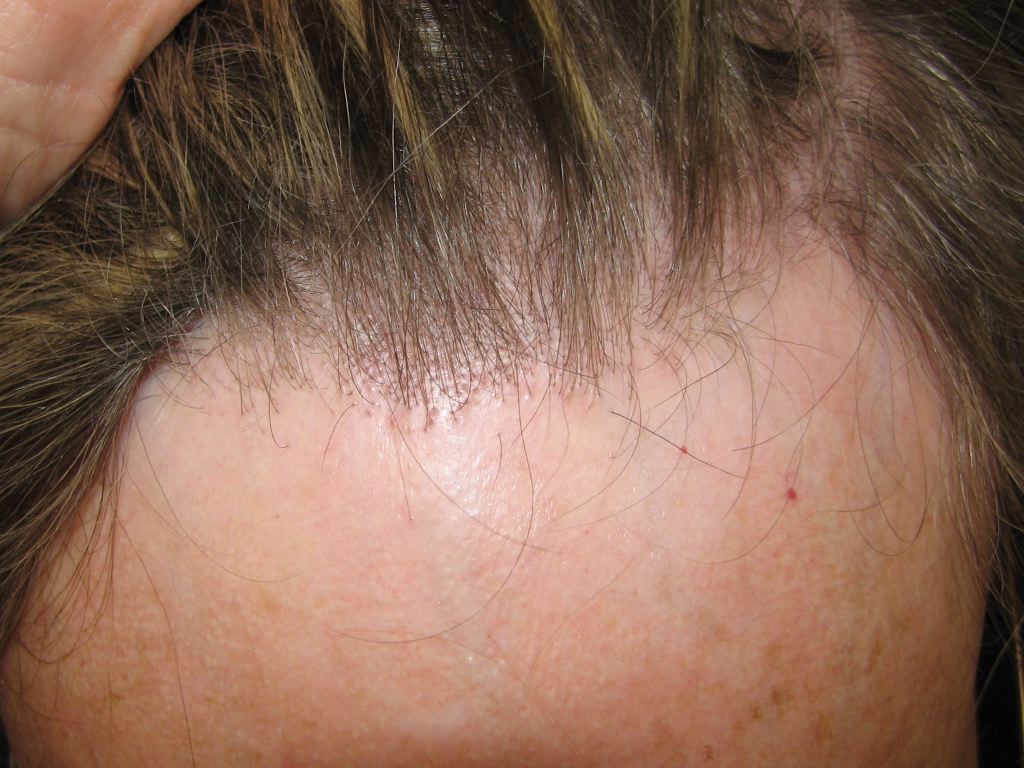
Frontal Fibrosing Alopecia. This cicatricial alopecia involves the frontal line and can extend to the preauricular and retroauricular regions of the scalp.
References
Jouanique C, Reygagne P. [Frontal fibrosing alopecia]. Annales de dermatologie et de venereologie. 2014 Apr:141(4):272-8. doi: 10.1016/j.annder.2014.01.020. Epub 2014 Mar 6 [PubMed PMID: 24703641]
Samrao A, Chew AL, Price V. Frontal fibrosing alopecia: a clinical review of 36 patients. The British journal of dermatology. 2010 Dec:163(6):1296-300. doi: 10.1111/j.1365-2133.2010.09965.x. Epub [PubMed PMID: 20698851]
Level 2 (mid-level) evidenceMacDonald A, Clark C, Holmes S. Frontal fibrosing alopecia: a review of 60 cases. Journal of the American Academy of Dermatology. 2012 Nov:67(5):955-61. doi: 10.1016/j.jaad.2011.12.038. Epub 2012 Apr 13 [PubMed PMID: 22503342]
Level 2 (mid-level) evidenceTan KT, Messenger AG. Frontal fibrosing alopecia: clinical presentations and prognosis. The British journal of dermatology. 2009 Jan:160(1):75-9. doi: 10.1111/j.1365-2133.2008.08861.x. Epub 2008 Sep 22 [PubMed PMID: 18811690]
Level 3 (low-level) evidenceNakamura M, Tokura Y. Expression of Snail1 in the fibrotic dermis of postmenopausal frontal fibrosing alopecia: possible involvement of an epithelial-mesenchymal transition and a review of the Japanese patients. The British journal of dermatology. 2010 May:162(5):1152-4. doi: 10.1111/j.1365-2133.2010.09682.x. Epub 2010 Feb 3 [PubMed PMID: 20132204]
Level 3 (low-level) evidenceClayton R, Chaudhry S, Ali I, Cooper S, Hodgson T, Wojnarowska F. Mucosal (oral and vulval) lichen planus in women: are angiotensin-converting enzyme inhibitors protective, and beta-blockers and non-steroidal anti-inflammatory drugs associated with the condition? Clinical and experimental dermatology. 2010 Jun:35(4):384-7. doi: 10.1111/j.1365-2230.2009.03581.x. Epub 2009 Oct 23 [PubMed PMID: 19874335]
Level 2 (mid-level) evidenceChiang YZ, Tosti A, Chaudhry IH, Lyne L, Farjo B, Farjo N, Cadore de Farias D, Griffiths CE, Paus R, Harries MJ. Lichen planopilaris following hair transplantation and face-lift surgery. The British journal of dermatology. 2012 Mar:166(3):666-370. doi: 10.1111/j.1365-2133.2011.10692.x. Epub 2012 Jan 9 [PubMed PMID: 21985326]
Level 3 (low-level) evidenceTosti A, Piraccini BM, Iorizzo M, Misciali C. Frontal fibrosing alopecia in postmenopausal women. Journal of the American Academy of Dermatology. 2005 Jan:52(1):55-60 [PubMed PMID: 15627081]
Level 3 (low-level) evidenceKatoulis A, Georgala, Bozi E, Papadavid E, Kalogeromitros D, Stavrianeas N. Frontal fibrosing alopecia: treatment with oral dutasteride and topical pimecrolimus. Journal of the European Academy of Dermatology and Venereology : JEADV. 2009 May:23(5):580-2. doi: 10.1111/j.1468-3083.2008.02963.x. Epub [PubMed PMID: 19415810]
Level 3 (low-level) evidenceMiao YJ, Jing J, Du XF, Mao MQ, Yang XS, Lv ZF. Frontal fibrosing alopecia: A review of disease pathogenesis. Frontiers in medicine. 2022:9():911944. doi: 10.3389/fmed.2022.911944. Epub 2022 Jul 25 [PubMed PMID: 35957858]
Vaisse V, Matard B, Assouly P, Jouannique C, Reygagne P. [Postmenopausal frontal fibrosing alopecia: 20 cases]. Annales de dermatologie et de venereologie. 2003 Jun-Jul:130(6-7):607-10 [PubMed PMID: 13679696]
Level 2 (mid-level) evidenceda Silva Libório R, Trüeb RM. Case Report of Connubial Frontal Fibrosing Alopecia. International journal of trichology. 2018 Mar-Apr:10(2):76-79. doi: 10.4103/ijt.ijt_105_17. Epub [PubMed PMID: 29769781]
Level 3 (low-level) evidenceChew AL, Bashir SJ, Wain EM, Fenton DA, Stefanato CM. Expanding the spectrum of frontal fibrosing alopecia: a unifying concept. Journal of the American Academy of Dermatology. 2010 Oct:63(4):653-60. doi: 10.1016/j.jaad.2009.09.020. Epub [PubMed PMID: 20846567]
Level 2 (mid-level) evidencePoblet E, Jiménez F, Pascual A, Piqué E. Frontal fibrosing alopecia versus lichen planopilaris: a clinicopathological study. International journal of dermatology. 2006 Apr:45(4):375-80 [PubMed PMID: 16650161]
Kossard S. Postmenopausal frontal fibrosing alopecia. Scarring alopecia in a pattern distribution. Archives of dermatology. 1994 Jun:130(6):770-4 [PubMed PMID: 8002649]
Miteva M, Camacho I, Romanelli P, Tosti A. Acute hair loss on the limbs in frontal fibrosing alopecia: a clinicopathological study of two cases. The British journal of dermatology. 2010 Aug:163(2):426-8. doi: 10.1111/j.1365-2133.2010.09807.x. Epub 2010 Apr 15 [PubMed PMID: 20394630]
Level 3 (low-level) evidenceArmenores P, Shirato K, Reid C, Sidhu S. Frontal fibrosing alopecia associated with generalized hair loss. The Australasian journal of dermatology. 2010 Aug:51(3):183-5. doi: 10.1111/j.1440-0960.2010.00653.x. Epub [PubMed PMID: 20695856]
Level 3 (low-level) evidenceRudnicka L, Olszewska M, Rakowska A, Slowinska M. Trichoscopy update 2011. Journal of dermatological case reports. 2011 Dec 12:5(4):82-8. doi: 10.3315/jdcr.2011.1083. Epub [PubMed PMID: 22408709]
Level 3 (low-level) evidenceChiang C, Sah D, Cho BK, Ochoa BE, Price VH. Hydroxychloroquine and lichen planopilaris: efficacy and introduction of Lichen Planopilaris Activity Index scoring system. Journal of the American Academy of Dermatology. 2010 Mar:62(3):387-92. doi: 10.1016/j.jaad.2009.08.054. Epub 2010 Jan 12 [PubMed PMID: 20061052]
Level 2 (mid-level) evidenceDonati A, Assouly P, Matard B, Jouanique C, Reygagne P. Clinical and photographic assessment of lichen planopilaris treatment efficacy. Journal of the American Academy of Dermatology. 2011 Mar:64(3):597-8; author reply 598-9. doi: 10.1016/j.jaad.2010.04.045. Epub [PubMed PMID: 21315955]
Level 3 (low-level) evidenceWolff H, Fischer TW, Blume-Peytavi U. The Diagnosis and Treatment of Hair and Scalp Diseases. Deutsches Arzteblatt international. 2016 May 27:113(21):377-86. doi: 10.3238/arztebl.2016.0377. Epub [PubMed PMID: 27504707]
Donovan JC, Samrao A, Ruben BS, Price VH. Eyebrow regrowth in patients with frontal fibrosing alopecia treated with intralesional triamcinolone acetonide. The British journal of dermatology. 2010 Nov:163(5):1142-4. doi: 10.1111/j.1365-2133.2010.09994.x. Epub [PubMed PMID: 20716217]
Level 3 (low-level) evidenceMulinari-Brenner FA, Guilherme MR, Peretti MC, Werner B. Frontal fibrosing alopecia and lichen planus pigmentosus: diagnosis and therapeutic challenge. Anais brasileiros de dermatologia. 2017:92(5 Suppl 1):79-81. doi: 10.1590/abd1806-4841.20175833. Epub [PubMed PMID: 29267454]
Mirmirani P, Willey A, Price VH. Short course of oral cyclosporine in lichen planopilaris. Journal of the American Academy of Dermatology. 2003 Oct:49(4):667-71 [PubMed PMID: 14512914]
Level 3 (low-level) evidenceCho BK, Sah D, Chwalek J, Roseborough I, Ochoa B, Chiang C, Price VH. Efficacy and safety of mycophenolate mofetil for lichen planopilaris. Journal of the American Academy of Dermatology. 2010 Mar:62(3):393-7. doi: 10.1016/j.jaad.2009.05.018. Epub 2010 Jan 12 [PubMed PMID: 20061053]
Jiménez F, Poblet E. Is hair transplantation indicated in frontal fibrosing alopecia? The results of test grafting in three patients. Dermatologic surgery : official publication for American Society for Dermatologic Surgery [et al.]. 2013 Jul:39(7):1115-8. doi: 10.1111/dsu.12232. Epub 2013 Apr 30 [PubMed PMID: 23631596]
Level 3 (low-level) evidenceBlume-Peytavi U, Hillmann K, Constantinou A, Vogt A. [Frontal fibrosing alopecia-update]. Der Hautarzt; Zeitschrift fur Dermatologie, Venerologie, und verwandte Gebiete. 2022 May:73(5):344-352. doi: 10.1007/s00105-022-04983-w. Epub 2022 Apr 8 [PubMed PMID: 35394176]
Lacarrubba F, Micali G, Tosti A. Absence of vellus hair in the hairline: a videodermatoscopic feature of frontal fibrosing alopecia. The British journal of dermatology. 2013 Aug:169(2):473-4. doi: 10.1111/bjd.12316. Epub [PubMed PMID: 23496000]
Level 3 (low-level) evidenceConde Fernandes I, Selores M, Machado S. Frontal fibrosing alopecia: a review of eleven patients. European journal of dermatology : EJD. 2011 Sep-Oct:21(5):750-2. doi: 10.1684/ejd.2011.1419. Epub [PubMed PMID: 21697058]
Dlova NC, Jordaan HF, Skenjane A, Khoza N, Tosti A. Frontal fibrosing alopecia: a clinical review of 20 black patients from South Africa. The British journal of dermatology. 2013 Oct:169(4):939-41. doi: 10.1111/bjd.12424. Epub [PubMed PMID: 23647261]
Level 3 (low-level) evidenceBernárdez C, Saceda-Corralo D, Gil-Redondo R, Moreno-Arrones OM, Rodrigues-Barata AR, Hermosa-Gelbard A, Vañó-Galvan S. Beard loss in men with frontal fibrosing alopecia. Journal of the American Academy of Dermatology. 2022 Jan:86(1):181-183. doi: 10.1016/j.jaad.2021.01.032. Epub 2021 Jan 20 [PubMed PMID: 33484769]
Heymann WR. Confronting frontal fibrosing alopecia. Journal of the American Academy of Dermatology. 2021 Aug:85(2):319-320. doi: 10.1016/j.jaad.2021.05.015. Epub 2021 May 23 [PubMed PMID: 34033818]
Lis-Święty A, Brzezińska-Wcisło L. Frontal fibrosing alopecia: a disease that remains enigmatic. Postepy dermatologii i alergologii. 2020 Aug:37(4):482-489. doi: 10.5114/ada.2020.98241. Epub 2020 Sep 2 [PubMed PMID: 32994767]