
 Anatomy, Bony Pelvis and Lower Limb, Foot Veins
Introduction
The venous drainage of the foot involves several vessels, including the plantar and dorsal venous arches, the great saphenous vein, and the small saphenous vein. Similar to what occurs in the hand, the foot's plantar and dorsal venous arches have digital veins extending between each toe and draining into the arches. The foot also contains large medial and lateral plantar veins that eventually join posteriorly to form the deep tibial veins and anteriorly form the anterior tibial veins. These veins contribute to the plantar pump, a physiological mechanism to propel blood back toward the heart.[1] The foot veins are susceptible to several pathologies, including corona phelbectatica, varicose veins, and venous ulcers.[1] The great saphenous vein is a common graft source for specialized conduit/bypass surgeries to manage various cardiac, thoracic, neurovascular, and urological pathologies.[2]
Structure and Function
Register For Free And Read The Full Article
 Search engine and full access to all medical articles
- 10 free questions in your specialty
- Free CME/CE Activities
- Free daily question in your email
- Save favorite articles to your dashboard
- Emails offering discounts
Learn more about a Subscription to StatPearls Point-of-Care
Structure and Function
The venous drainage of the lower limb starts mainly on the dorsum of the foot, with proximal venous blood return occurring in both a superficial and deep venous pattern. The small saphenous vein drains the majority of the foot. The small saphenous vein begins at the lateral end of the dorsal venous arch. It then courses posterior to the lateral malleolus, ascending on the posterior side of the leg, running with the sural nerve before emptying into the popliteal vein. The great saphenous vein begins at the medial end of the dorsal venous arch of the foot. It then passes anterior to the medial malleolus and runs on the medial side of the lower limb before emptying into the femoral vein. The femoral vein then drains into the external iliac vein, which combines with the internal iliac vein to form the common iliac vein, draining into the inferior vena cava and right atrium of the heart.
Embryology
The limb buds of the embryo begin to form about 5 weeks after fertilization as the lateral plate mesoderm migrates into the limb bud region and condenses along the central axis to eventually form the vasculature and skeletal components of the lower limb (see Image. Vasculature of the Lower Limbs).[3][4][5] Several factors influence the formation of the limb bud vasculature and musculature, including retinoic acid, sonic hedgehog (SHH), HOX genes, apical ectodermal ridge (AER), and the zone of polarizing activity (ZPA).
Retinoic acid is a global organizing gradient that initiates the production of transcription factors that specify regional differentiation and limb polarization. The apical ectodermal ridge (AER) produces a fibroblast growth factor (Fgf), promoting the outgrowth of the limb buds by stimulating mitosis.[3][4][5] The fibroblast growth factor involved in hindlimb development is Fgf10, which Tbx4 stimulates.[6] The zone of polarizing activity (ZPA) produces sonic hedgehog (Shh), which promotes the organization of the limb bud along the anterior-posterior axis. Shh activates specific Hox genes (Hoxd-9, Hoxd-10, Hoxd-11, Hoxd-12, and Hoxd-13) that are important in limb polarization and regional specification.[3][4][5] These genes control patterning and, consequently, the morphology of the developing limb in the human embryo.[6] Errors in Hox gene expression can lead to malformations in the limbs.[6]
Blood Supply and Lymphatics
The distribution of lymphatic vessels in the lower limb is similar to the distribution of the veins of the lower limb. It can be divided into 2 major groups: superficial and deep. The superficial vessels are the great saphenous vein (medial) and the lesser saphenous vein (lateral). The great saphenous vein drains into the inguinal lymph nodes, while the lesser saphenous vein drains into the popliteal nodes. The deep lymphatic vessels contain the anterior, posterior, and peroneal vessels, which follow each respective artery and drain into the popliteal nodes.
Nerves
The innervation of the foot involves 2 terminal branches of the tibial nerve, including the medial and lateral plantar nerve. The medial plantar nerve innervates the abductor hallucis, flexor digitorum brevis, flexor hallucis brevis, and the first lumbrical. The lateral plantar nerve supplies the other foot muscles with a superficial and deep branch. The superficial branch provides muscular and cutaneous innervation, while the deep branch only provides muscular innervation.[7]
Muscles
The foot can be divided into a dorsal and plantar aspect, each containing unique musculature, blood supply, and innervation. The foot's dorsal aspect contains the extensor hallucis brevis and extensor digitorum brevis muscles and receives innervation from the deep fibular nerve. The plantar aspect of the foot can be divided into 4 layers, each containing different muscles and tendons. The most superficial layer (1st layer) contains the abductor hallucis, flexor digitorum brevis, and abductor digiti minimi muscles. The second layer includes the flexor hallucis longus tendon, flexor digitorum longus tendon, quadratus plantae muscle, and 4 lumbrical muscles. The 3rd layer contains the flexor hallucis brevis, adductor hallucis (oblique and transverse heads), and the flexor digiti minimi brevis muscles. The deepest layer (4th layer) contains the fibularis longus tendon, tibialis anterior tendon, tibialis posterior tendon, and the interosseous muscles (3 plantar interosseous muscles, 4 dorsal interosseous muscles).[7]
Physiologic Variants
The lesser saphenous and greater saphenous veins typically terminate in the popliteal and femoral veins, respectively. However, variable connections between these veins are common, so the flow patterns should never be considered absolute. In a study of 200 lower extremities using Doppler sonography, 107 (53.5%) subjects had a lateral accessory branch of the great saphenous vein, and 33 (16.5%) had a medial accessory branch of the great saphenous vein.[8] In the same study, the authors found variations in the termination point of the small saphenous vein. In 108 subjects (54%), the small saphenous vein terminated in the popliteal vein; however, in 32 subjects (16%), it terminated above the popliteal vein.[8] Each of these variations in branching and termination points merits noting in clinical and surgical venous operations.
Surgical Considerations
Occlusive arterial disease is the leading cause of death worldwide.[9] One of the most commonly performed revascularization techniques to treat occlusive arterial disease is coronary artery bypass graft surgery. The great saphenous vein is a common conduit source for surgical revascularization.[9] The great saphenous vein can be harvested using an open or minimally invasive technique.[10] The vein then gets sewn to the aorta on one end and past the obstruction into the coronary artery on the other.[10]
Another surgical consideration of the veins of the foot involves varicose vein surgery. Varicose vein surgery is one of the most common forms of surgery.[11] There are several ways to remove or close off varicose veins surgically, but the 2 most common ways to remove varicose veins are via vein stripping and phlebectomy.[11] Vein stripping involves completely removing the varicose vein by making 2 incisions proximal and distal to the varicose vein and pulling the vein out through the top incision.[11] A phlebectomy involves making small incisions of a few millimeters along the affected vein and using a small hook to pull the vein out through the incisions to cut and remove pieces of it.[11] Several other ways to surgically treat varicose veins include endovenous thermoablation, radiofrequency ablation, and foam sclerotherapy.[12]
Clinical Significance
The foot veins are susceptible to several pathologies, including corona phelbectatica, varicose veins, deep vein thrombosis, and venous ulcers.[1] Corona phelbectatica is the presence of abnormally visible cutaneous blood vessels at the ankle with 4 distinct components: "venous cups," blue and red telangiectasias, and capillary "stasis spots."[1][13] The veins in corona phelbectatica are frequently referred to as "ankle spider veins." They are a clear sign of serious chronic venous insufficiency that requires medical attention because of their potential to result in more serious conditions, including blood clots or leg ulcers.[1][13]
Varicose veins are a common pathology seen in the foot and ankle veins. They are ectatic, tortuous vessels of the superficial venous system that are at least 3 mm in size and arise from the failure of venous valves to close properly to allow the backward flow of blood.[14] The 2 main superficial leg veins (the great saphenous and small saphenous veins) and their branches are the most common sites of varicose veins.[14] Patients with varicose veins may present with no symptoms other than the enlarged vein, but some may experience pain, burning, itching, or leg swelling. These varicose veins should be taken seriously because they present a cosmetic problem and increase the risk of developing superficial vein thrombosis and venous thromboembolic disease.[14] The treatment of varicose veins includes conservative and surgical treatments. Conservative treatment of varicose veins includes elastic compression stockings to control swelling and weight loss and topical steroid cream to control inflammation. Surgical treatment includes varicose vein stripping, phlebectomy, endovenous thermoablation, radiofrequency ablation, and foam sclerotherapy.[12]
Deep vein thrombosis (DVT) is a frequent complication seen in orthopedic surgery and can cause significant morbidity and mortality.[15] The incidence of DVT is low in foot and ankle surgery. However, a DVT leading to a pulmonary embolism (PE) can still cause mortality following foot and ankle surgery.[15] There are several risk factors for venous thromboembolism, including family history, older age, cancer, lengthy surgical procedures, tobacco use, and immobilization.[15] Each of these factors merits consideration following foot and ankle surgery to help prevent DVT and PE.
Lastly, the veins in the foot can be involved in abscesses and ulcers due to diabetic complications as well as intravenous drug use. In each case, it is crucial to treat the underlying medical issue, such as diabetes or addiction, while also providing topical wound care therapies, antibiotics, or anti-platelet or anticlotting medications to prevent a blood clot.
Media
(Click Image to Enlarge)
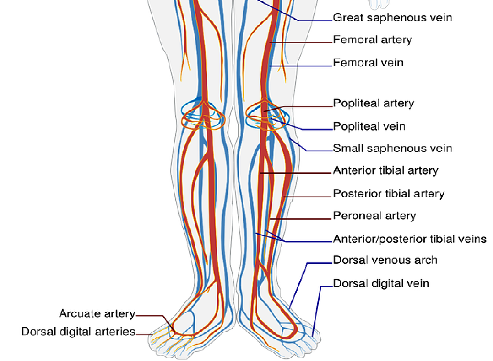
Vasculature of the Lower Limbs
Public Domain, via Clker Free Vector Images
References
Ricci S, Moro L, Antonelli Incalzi R. The foot venous system: anatomy, physiology and relevance to clinical practice. Dermatologic surgery : official publication for American Society for Dermatologic Surgery [et al.]. 2014 Mar:40(3):225-33. doi: 10.1111/dsu.12381. Epub 2013 Dec 23 [PubMed PMID: 24372905]
Yuan SM, Jing H. A reappraisal of saphenous vein grafting. Annals of Saudi medicine. 2011 Jan-Feb:31(1):62-71. doi: 10.4103/0256-4947.75781. Epub [PubMed PMID: 21245602]
Lezak B, Wehrle CJ, Summers S. Anatomy, Bony Pelvis and Lower Limb: Posterior Tibial Artery. StatPearls. 2024 Jan:(): [PubMed PMID: 30725666]
Mróz I, Kielczewski S, Pawlicki D, Kurzydło W, Bachul P, Konarska M, Bereza T, Walocha K, Kaythampillai LN, Depukat P, Pasternak A, Bonczar T, Chmielewski P, Mizia E, Skrzat J, Mazur M, Warchoł Ł, Tomaszewski K. Blood vessels of the shin - anterior tibial artery - anatomy and embryology - own studies and review of the literature. Folia medica Cracoviensia. 2016:56(1):33-47 [PubMed PMID: 27513837]
Gros J, Tabin CJ. Vertebrate limb bud formation is initiated by localized epithelial-to-mesenchymal transition. Science (New York, N.Y.). 2014 Mar 14:343(6176):1253-6. doi: 10.1126/science.1248228. Epub [PubMed PMID: 24626928]
Level 3 (low-level) evidenceBarham G, Clarke NM. Genetic regulation of embryological limb development with relation to congenital limb deformity in humans. Journal of children's orthopaedics. 2008 Feb:2(1):1-9. doi: 10.1007/s11832-008-0076-2. Epub 2008 Feb 7 [PubMed PMID: 19308596]
Bourne M, Talkad A, Varacallo M. Anatomy, Bony Pelvis and Lower Limb, Foot Fascia. StatPearls. 2024 Jan:(): [PubMed PMID: 30252299]
Kurkcuoglu A, Peker T, Gulekon N. Anatomical, radiological and histological investigation of the great and small saphenous veins. Saudi medical journal. 2008 May:29(5):672-7 [PubMed PMID: 18454212]
de Vries MR, Simons KH, Jukema JW, Braun J, Quax PH. Vein graft failure: from pathophysiology to clinical outcomes. Nature reviews. Cardiology. 2016 Aug:13(8):451-70. doi: 10.1038/nrcardio.2016.76. Epub 2016 May 19 [PubMed PMID: 27194091]
Level 2 (mid-level) evidenceFerrari E, von Segesser L, Berdajs D. Improving coronary artery bypass graft durability: use of the external saphenous vein graft support. Multimedia manual of cardiothoracic surgery : MMCTS. 2015:2015():. pii: mmv005. doi: 10.1093/mmcts/mmv005. Epub 2015 May 13 [PubMed PMID: 25976117]
Nesbitt C, Bedenis R, Bhattacharya V, Stansby G. Endovenous ablation (radiofrequency and laser) and foam sclerotherapy versus open surgery for great saphenous vein varices. The Cochrane database of systematic reviews. 2014 Jul 30:(7):CD005624. doi: 10.1002/14651858.CD005624.pub3. Epub 2014 Jul 30 [PubMed PMID: 25075589]
Level 1 (high-level) evidenceLin F, Zhang S, Sun Y, Ren S, Liu P. The management of varicose veins. International surgery. 2015 Jan:100(1):185-9. doi: 10.9738/INTSURG-D-14-00084.1. Epub [PubMed PMID: 25594661]
Uhl JF, Cornu-Thenard A, Satger B, Carpentier PH. Clinical analysis of the corona phlebectatica. Journal of vascular surgery. 2012 Jan:55(1):150-3. doi: 10.1016/j.jvs.2011.04.070. Epub 2011 Oct 5 [PubMed PMID: 21975063]
Jacobs BN, Andraska EA, Obi AT, Wakefield TW. Pathophysiology of varicose veins. Journal of vascular surgery. Venous and lymphatic disorders. 2017 May:5(3):460-467. doi: 10.1016/j.jvsv.2016.12.014. Epub [PubMed PMID: 28411716]
Chao J. Deep Vein Thrombosis in Foot and Ankle Surgery. The Orthopedic clinics of North America. 2016 Apr:47(2):471-5. doi: 10.1016/j.ocl.2015.10.001. Epub [PubMed PMID: 26772954]