Back To Search Results

Anthrax Infection

Editor: Josephin K. Mathai Updated: 7/7/2025 9:28:40 AM

Introduction

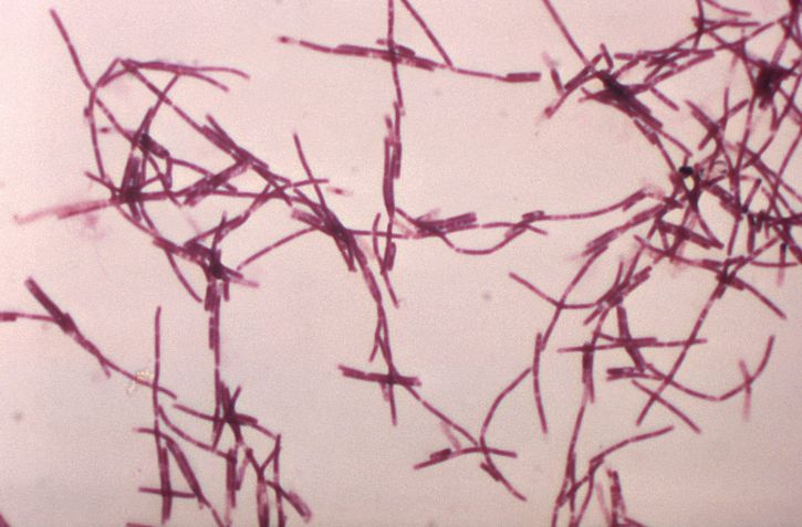
Anthrax is an acute zoonotic infection caused by the spore-forming, gram-positive Bacillus anthracis (see Image. Gram-Positive Rods).[1] The disease primarily affects herbivorous mammals, such as cattle, sheep, and goats. Humans can acquire the infection through direct or indirect contact with infected animals, contaminated animal products, ingesting undercooked meat, inhaling aerosolized spores, or, more rarely, by injecting contaminated drugs.[2] The United States (US) Centers for Disease Control and Prevention (CDC) notes that anthrax remains endemic in agricultural regions of the Americas, sub-Saharan Africa, Asia, and parts of Europe. However, sporadic outbreaks still occur in the US, particularly in areas with unvaccinated livestock.[3]

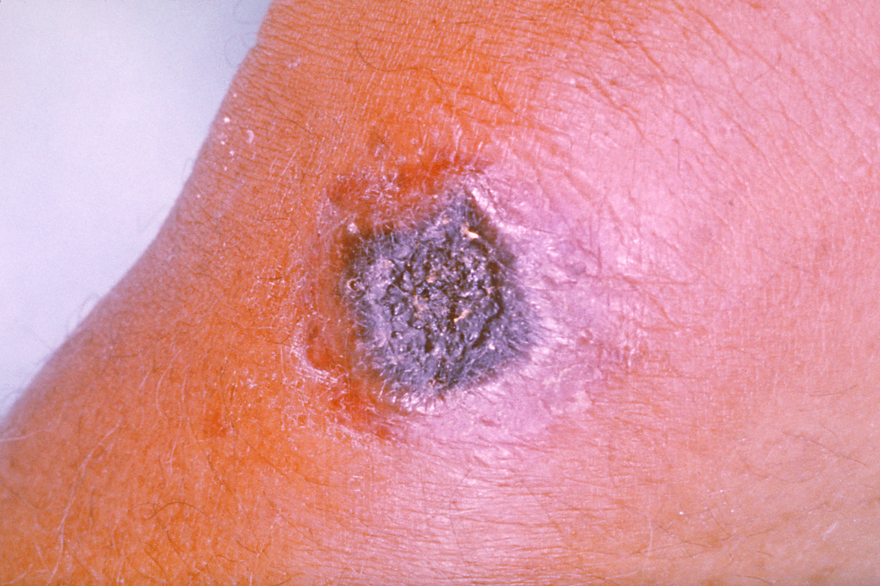
Anthrax manifests in 4 principal clinical forms, defined by the route of spore entry: cutaneous, inhalational, gastrointestinal (ingestion), and injectional. Cutaneous anthrax is the most common, accounting for over 95% of human cases, and typically presents as a painless ulcer with a characteristic black eschar (see Image. Eschar).[3][4] Inhalational anthrax, the most lethal form, is associated with rapid progression to severe respiratory distress and high mortality if untreated. Gastrointestinal anthrax results from ingestion of contaminated meat and can present as oropharyngeal or intestinal disease, while injected anthrax, recognized in recent outbreaks among heroin users, is characterized by severe soft tissue infection and sepsis.[4][5][6] 

The pathogenesis of anthrax is mediated by potent exotoxins and a poly-D-glutamic acid capsule, which together facilitate immune evasion, systemic dissemination, and rapid clinical deterioration. B anthracis is classified as a Tier 1 select agent due to its potential for use as a bioterrorism weapon, given the ease of spore production and environmental persistence.[5] Advances in antimicrobial therapy and critical care have improved outcomes, but anthrax remains a disease with significant morbidity and mortality, particularly in systemic and meningitic forms.[4][6]

Etiology

Register For Free And Read The Full Article
Get the answers you need instantly with the StatPearls Clinical Decision Support tool. StatPearls spent the last decade developing the largest and most updated Point-of Care resource ever developed. Earn CME/CE by searching and reading articles.
  • Dropdown arrow Search engine and full access to all medical articles
  • Dropdown arrow 10 free questions in your specialty
  • Dropdown arrow Free CME/CE Activities
  • Dropdown arrow Free daily question in your email
  • Dropdown arrow Save favorite articles to your dashboard
  • Dropdown arrow Emails offering discounts

Learn more about a Subscription to StatPearls Point-of-Care

Etiology

The bacterium B anthracis causes anthrax infection. The primary sources of human infection are contact with infected animals (especially herbivores such as cattle, sheep, and goats), their carcasses, or animal products (eg, hides, wool, meat) contaminated with B anthracis spores.[3] The spores are highly resistant and can persist in soil for decades, serving as a reservoir for infection in animals and humans.[7][8]

Transmission to humans occurs via 4 main routes:

  • Cutaneous anthrax (the most common form) results from direct inoculation of spores into the skin, typically through abrasions or cuts when handling contaminated animals or animal products.
  • Inhalation anthrax occurs after inhaling aerosolized spores, such as when processing contaminated wool or hides, and, more recently, with the intentional release (bioterrorism).[3][8][9]
  • Gastrointestinal (ingestion) anthrax follows the consumption of undercooked or raw meat from infected animals.[3]
  • Injection anthrax is rare and seen in individuals who inject drugs contaminated with B anthracis spores, particularly heroin.[6][10]

Person-to-person transmission is rare and not considered a significant route of spread. Anthrax is a zoonotic disease, with human cases now rare in countries with effective livestock and vaccination programs.[3]

Epidemiology

Anthrax remains a rare diagnosis in the US, with only 9 confirmed or probable cases reported to the CDC since 2006. Widespread livestock vaccination has contributed significantly to the near elimination of endemic transmission in the US; however, sporadic cases continue to occur, particularly in regions such as southwest Texas, Colorado, North and South Dakota, and Montana. Globally, individuals with direct occupational exposure to livestock or animal products, such as farmers, veterinarians, and those involved in handling or processing animal hides and wool, are at the highest risk for anthrax infection. In these populations, human outbreaks frequently coincide with epizootics in livestock, resulting in case clustering around such events.[8][11] There is no clear predilection for sex or age; rather, risk is determined by exposure patterns, which can vary from region to region due to customs and occupational roles. Pediatric cases are reported, particularly in endemic areas, but reflect exposure rather than age-specific vulnerability.[4]

The World Health Organization estimates there are between 2000 and 20,000 human cases of anthrax infection each year worldwide, and that the true number may be higher due to underreporting.[11][12][13] Globally, cutaneous anthrax accounts for 95% to 99% of reported human cases, with gastrointestinal and inhalational forms being much less common. Injectional anthrax has been reported almost exclusively among heroin users in Europe, with no documented cases in individuals who do not inject drugs. In the US, sporadic cases associated with contact with infected livestock or animal products have been reported. There have also been rare cases in laboratory or industrial settings, as well as in the context of bioterrorism.[10][14]

B anthracis spores persist in alkaline, calcium-rich soils, and outbreaks often follow environmental disturbances, such as heavy rains or droughts, that bring spores to the surface, increasing the exposure risk for grazing animals and, in turn, humans.[14] An example of this occurred in 2016 in Siberia (Yamal Peninsula), in which dozens of people became ill after melting permafrost released B anthracis spores from long-buried reindeer carcasses. The outbreak resulted in the infection of over 70 people and the death of thousands of reindeer, with genetic analysis confirming that the strains isolated from the outbreak were conserved in permafrost and released during the thawing event.[15][16]

The global distribution of anthrax risk is concentrated in arid and temperate regions, with an estimated 1.83 billion people living in at-risk areas; however, only a small fraction of these have significant occupational exposure.[11] Anthrax epidemiology is shaped by environmental, occupational, and socioeconomic factors rather than intrinsic host characteristics such as age or sex. Surveillance gaps in endemic regions likely lead to underreporting, and the potential for intentional release remains a public health concern.

Pathophysiology

B anthracis spores are highly resilient and can remain viable in soil or animal products for decades, facilitating zoonotic transmission in endemic regions and through contaminated materials such as hides, wools, or drums. Upon entry via skin abrasions, inhalation, ingestion, or injection, spores are phagocytosed by local macrophages and transported to regional lymph nodes, where they germinate into vegetative bacilli.[1] Pathogenesis is driven by the secretion of a tripartite exotoxin composed of protective antigen (PA), lethal factor (LF), and edema factor (EF). PA facilitates the cellular entry of LF and EF. Lethal toxin (LT), consisting of PA and LF—a zinc-dependent metalloprotease—cleaves mitogen-activated protein kinase kinases (MAPKKs), disrupting MAPK signaling. This impairs macrophage survival and function, induces apoptosis, and contributes to profound immunosuppression. LT also disrupts endothelial integrity by inhibiting MAPK and Tie-2 receptor signaling, promoting vascular leakage, tissue necrosis, shock, and multiorgan dysfunction.[17][18][19]

Edema toxin (ET), composed of PA and EF, a calmodulin-dependent adenylate cyclase, markedly elevates intracellular cyclic adenosine monophosphate levels. This disrupts neutrophil and macrophage function, suppresses innate immunity, and alters water homeostasis, resulting in significant local and systemic edema. LT and ET further impair endothelial cell junctions by inhibiting endocytic recycling and reducing cadherin expression, thereby destabilizing adherens junctions and exacerbating vascular permeability. Together, these effects underlie the fulminant shock, edema, and tissue damage observed in advanced anthrax.[20][21]

Clinical manifestations of anthrax vary by route of exposure. Cutaneous anthrax presents as a painless, pruritic papule that progresses to a vesicle and then a characteristic black eschar, often accompanied by significant edema and regional lymphadenopathy. Systemic toxicity is uncommon unless untreated. Inhalation anthrax follows a biphasic course, with an initial nonspecific prodrome (fever, malaise, cough) rapidly progressing to dyspnea, hypoxemia, and shock. Hemorrhagic mediastinitis, characterized by mediastinal widening and pleural effusions, is typically evident on imaging. Gastrointestinal anthrax may present as oropharyngeal (sore throat, neck swelling, dysphagia) or intestinal (abdominal pain, vomiting, bloody diarrhea, ascites) disease, both with high mortality. Injection anthrax, reported in heroin users, causes severe soft tissue infection and systemic toxicity without eschar formation.[4]

Histopathology

Histopathologic findings in anthrax vary by clinical form and reflect both toxin-mediated tissue injury and the organism’s immunosuppressive effects. In cutaneous anthrax, biopsies of the characteristic black eschar typically demonstrate epidermal ulceration, dermal coagulative necrosis, gelatinous edema, hemorrhage, and fibrinoid vascular damage dissecting collagen bundles, with a muted inflammatory infiltrate composed of neutrophils, lymphocytes, and histiocytes. This blunted response is attributed to the tripartite exotoxin.[22]

Hematoxylin and eosin, as well as Gram stains, reveal large, square-ended, gram-positive rods in chains. Giemsa, silver stains (eg, Warthin-Starry), and M’Fadyean staining highlight the poly-D-glutamic acid capsule as a pink halo, aiding differentiation from Bacillus cereus. Key features include the eschar, necrosis, edema, and presence of nonhemolytic, nonmotile bacilli, findings consistent with the Infectious Disease Society of America guidelines and established descriptions in the literature.[22][23]

Inhalational anthrax typically shows necrotizing hemorrhagic mediastinitis, hemorrhagic lymphadenitis, and large serosanguinous pleural effusions. Vascular necrosis and arteritis are common, leading to tissue-displacing hemorrhages and, in severe cases, shock. Lymphocyte apoptosis is prominent, and there is frequent evidence of vasculitis and arterial rupture. In cutaneous anthrax, there is ulceration, coagulative necrosis, marked edema, perivascular inflammation, and gram-positive bacilli in the lesion.

Injection anthrax, primarily associated with heroin use, histologically resembles necrotizing fasciitis. Biopsies reveal deep dermal and subcutaneous necrosis, hemorrhagic edema, and minimal inflammatory response. Gram and immunohistochemistry stains often show bacilli infiltrating fascial planes. Notably, purulence is limited despite abundant bacteria, reflecting toxin-mediated immune suppression. A hallmark of all forms of anthrax is extensive tissue necrosis with minimal purulence, a consequence of the immunosuppressive effects of anthrax exotoxins (LT and ET), which induce apoptosis in immune cells. This distinguishes anthrax from typical pyogenic infections, where robust neutrophilic infiltration and pus formation are expected.[17][24]

Toxicokinetics

B anthracis toxins (LT, ET) are produced locally after spore germination and are absorbed into the circulation, where they distribute systemically and exert their effects at distant sites. PA binds to cell surface receptors and facilitates endocytosis of LF and EF into host cells. Once internalized, LF and EF disrupt cellular signaling and immune responses: LF cleaves MAP kinase kinases, inducing apoptosis and immune suppression, while EF increases intracellular cyclic adenosine monophosphate, causing edema and further immune dysfunction.

These toxins are not metabolized in the classical sense but are degraded intracellularly after exerting their effects. Toxin distribution is systemic, accounting for the widespread tissue injury, shock, and multiple organ involvement observed in severe cases. While the clinical form—cutaneous, inhalational, gastrointestinal, or injectional—is determined by the portal of entry, systemic dissemination of toxins allows for distant and potentially fatal effects.

History and Physical

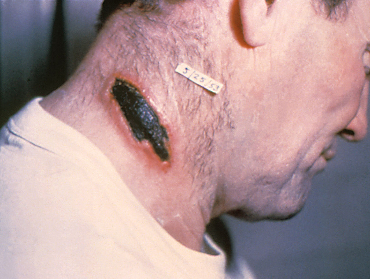
The clinical history and physical examination findings in anthrax vary by form, reflecting the route of exposure, toxin-mediated pathogenesis, and the characteristic muted inflammatory response. Each syndrome is defined by distinct diagnostic features based on symptomatology, exposure history, and laboratory evaluation. Cutaneous anthrax typically follows exposure to animal products or contaminated materials. Lesions begin as painless, pruritic papules that progress into vesicles over 1 to 2 days and then ulcerate, forming a characteristic black eschar to the lesion size (see Image. Black Eschar). Fever occurs in about one-third of cases. Regional lymphadenopathy may present, but pain and purulence are minimal. The infection remains localized with appropriate treatment, and mortality is less than 1%. Without treatment, systemic dissemination occurs in up to 20% of cases.[2]

Inhalation anthrax often follows exposure to aerosolized spores. Symptoms typically present after 1 to 7 days of incubation with a biphasic illness: an initial prodrome of fever, malaise, myalgia, and nonproductive cough, followed by abrupt progression to severe dyspnea, hypoxemia, diaphoresis, and shock. Physical findings may include tachycardia, hypotension, and, in advanced cases, evidence of mediastinal widening or pleural effusion on imaging, as well as meningeal signs if anthrax meningitis develops.

Gastrointestinal anthrax is associated with the ingestion of contaminated meat. The history includes severe, colicky abdominal pain, nausea, vomiting, anorexia, and bloody diarrhea. On examination, findings may include fever, abdominal tenderness, ascites, and signs of peritonitis. Oropharyngeal disease may present with severe sore throat, dysphagia, neck swelling, and cervical lymphadenopathy.

Injectional anthrax, seen in heroin users, presents with a history of recent injections. Physical findings include severe soft tissue infection with deep dermal and subcutaneous necrosis, extensive nonpitting edema, deep tissue necrosis, and minimal purulence despite extensive bacterial presence. Systemic toxicity is common, and the muted inflammatory response is notable despite the presence of bacteria.[25]

Evaluation

The diagnostic evaluation of anthrax involves a multimodal approach, incorporating microbiological culture, polymerase chain reaction (PCR), serologic testing, immunohistochemistry, and imaging, depending on the clinical presentation.[26] Blood cultures are essential in systemic disease, while lesion swabs or biopsies are useful in cutaneous cases. Imaging, such as chest radiography or computed tomography, is crucial in identifying inhalational anthrax, particularly in cases involving mediastinal widening and pleural effusions. Timely specimen collection before antimicrobial therapy improves diagnostic yield.

In high-risk individuals, a painless eschar with surrounding edema should raise suspicion for cutaneous anthrax. Oropharyngeal lesions may cause airway compromise, requiring prompt evaluation. Gastrointestinal anthrax should be considered in cases of unexplained ascites, peritonitis, or gastrointestinal hemorrhage. Inhalational anthrax may initially resemble influenza but can rapidly progress to sepsis, shock, and meningitis. A high index of suspicion is essential, with a focus on potential exposure risks in the history.

Laboratory Testing

Microbiological culture remains the diagnostic cornerstone. In cutaneous anthrax, specimens should be obtained from vesicular fluid beneath the eschar or via punch biopsy for Gram stain and culture. Blood cultures are essential in all cases with systemic symptoms, particularly inhalational, gastrointestinal, and injectional forms. Additional cultures from pleural fluid, ascitic fluid, cerebrospinal fluid (if meningitis is suspected), and tissue biopsies may aid in diagnosis. B anthracis typically grows as nonhemolytic, "medusa head" colonies on agar and appears as large, gram-positive rods with a characteristic "boxcar" morphology (see Image. Gram-Positive Rods).[1]

PCR assays targeting B anthracis-specific genes (eg, pagA, lef, capB) offer high sensitivity and specificity and can be performed on blood, tissue, or lesion swabs. PCR is especially useful when antibiotic therapy may compromise culture yield or when rapid diagnosis is critical.[27][28] Serologic testing, such as the enzyme-linked immunosorbent assay for anti-protective antigen antibodies, is primarily used for retrospective diagnosis and epidemiologic investigations, which require paired acute and convalescent sera. Detection of circulating toxin in acute-phase serum is possible but less commonly available.[26]

Immunohistochemistry can detect B anthracis in tissue specimens, particularly when cultures are negative or antibiotic therapy has already been initiated.[26] This lab technique is beneficial in formalin-fixed tissues, aiding in postmortem diagnosis or retrospective case confirmation. Other laboratory findings in severe anthrax may include hemoconcentration, thrombocytopenia, coagulopathy, and elevated transaminases. These abnormalities reflect systemic toxin effects and may aid in assessing disease severity and guiding supportive management. 

Imaging Studies

Imaging studies are essential in the evaluation of inhalational anthrax. Chest radiography or computed tomography typically demonstrates mediastinal widening, pleural effusions, and hemorrhagic mediastinal lymphadenopathy. Imaging also aids in detecting soft tissue involvement in injectional anthrax and in identifying complications associated with gastrointestinal disease.[10][29]

Treatment / Management

The CDC recommends a general approach to anthrax treatment, which involves the prompt initiation of effective antibiotics, with the regimen and duration tailored to the clinical form and severity of the disease and adjunctive measures as indicated.[3] Early treatment is critical, as delays are associated with significantly increased morbidity and mortality, particularly in systemic and inhalational forms. Antibiotic regimens and treatment duration should be tailored based on clinical form and severity. Localized cutaneous infections are typically treated for 7 to 14 days. However, systemic infections and postexposure prophylaxis require a minimum of 60 days of therapy due to the prolonged persistence of spores. A 60-day course is recommended for all clinical forms in cases of suspected bioterrorism or aerosol exposure. 

Treatment by Clinical Form

Cutaneous anthrax (uncomplicated, localized):

  • First-line options:
    • Ciprofloxacin 500 mg intravenously (IV) q 12h
    • Doxycycline 100 mg IV q 12 h
  • Alternative options:
    • Levofloxacin 750 mg daily
    • Moxifloxacin 400 mg daily
    • Penicillin (if susceptible)

Cutaneous anthrax (with systemic signs or head and neck involvement)

  • IV multidrug therapy followed by oral antibiotics for 14 days in total
  • Add antitoxin if toxemia is suspected:
    • Anthrasil (human immunoglobulin) 10-20 vials IV (weight-based) 
    • Raxibacumab 40 mg/kg IV (single dose)
    • Oblitoxacimab (dosing per label and clinical factors)  

Inhalational anthrax

  • IV combination therapy for ≥14 days, followed by oral therapy to complete 60 days
  • Regimen: Include 1 bactericidal agent and 1 protein synthesis inhibitor
    • Ciprofloxacin 400 mg every (q) 8 h IV + Clindamycin 600 mg q 8 h IV or Linezolid 600 mg q 12h
  • Alternatives:
    • Bactericidal: Levofloxacin, moxifloxacin, morpenem
    • Protein synthesis inhibitor: Doxycycline
  • Add antitoxin as above if toxemia is suspected.

Gastrointestinal anthrax (oropharyngeal or intestinal)

  • IV combination for ≥14 days, followed by oral therapy to complete 60 days
  • Same regimen and antitoxin indications as inhalational anthrax
  • Surgical intervention for perforation or hemorrhage 

Injection anthrax

  • IV combination therapy for ≥14 days, followed by oral therapy to complete 60 days
  • Same regimen and antitoxin indications as inhalational anthrax
  • Surgical debridement often required for necrotic tissue

Anthrax meningitis (complication of any form)

  • IV triple therapy for 2 to 3 weeks or until clinically stable
    • Regimen: 2 bactericidal agents plus 1 protein synthesis inhibitor
    • Ciprofloxacin 400 mg IV q 8h + meropenem 2 g q 8h + linezolid 600 mg q 12h
  • Adjunctive:
    • Anthrasil 10-20 vials IV (based on severity and weight)
    • Corticosteroids (Dexamethasone) for cerebral edema
    • CSF drainage if indicated

Postexposure Prophylaxis

Recommended for all suspected aerosol or bioterrorism exposures. The duration of therapy is 60 days. 

  • Antibiotics:
    • Doxycycline 100 mg orally twice daily (BID) (pediatric: 2.2 mg/kg BID)
    • Ciprofloxacin 500 mg BID (pediatric: 15 mg/kg BID)
  • Plus BioThrax vaccine
    • Administered at 0, 2, and 4 weeks

If multidrug-resistant B anthracis is suspected, treatment should be guided by susceptibility testing, with the addition of agents such as vancomycin or rifampin. In children and pregnant patients, dosing adjustments are required (eg, ciprofloxacin 10–15 mg/kg q 12 h IV in children). Despite potential risks, fluoroquinolones remain the preferred agents due to their efficacy.[30][31](B3)

Differential Diagnosis

The differential diagnosis of anthrax varies by clinical form and often overlaps with more common infectious and noninfectious conditions, making early recognition challenging. Accurate diagnosis requires careful consideration of exposure history, clinical presentation, and distinguishing features to differentiate anthrax from other causes of cutaneous, respiratory, or gastrointestinal symptoms.

  • Cutaneous anthrax:
    • Ecthyma
    • Spider bite
    • Tularemia
    • Necrotizing fasciitis
    • Bubonic plague
    • Cat scratch disease
    • Primary syphilis
    • Skin abscess
    • Erythematous granuloma annulare
    • Cellulitis 
  • Inhalational anthrax:
    • Influenza
    • Pneumonia
    • Pneumonic plague
    • Hemopneumothorax
    • Pneumothorax
    • Pulmonary embolism
    • Bronchitis
    • Viral syndrome
    • Tularemia
    • Q fever
  • Gastrointestinal anthrax:
    • Peptic ulcer disease with perforation
    • Typhoid fever
    • Shigellosis
    • Ischemic colitis
    • Peritonitis
    • Amebic dysentery 

Toxicity and Adverse Effect Management

Anthrax treatment includes antibiotics, antitoxins, and supportive therapies, all of which may pose risks of toxicity and adverse events that vary by drug class, dosage, duration, and patient-specific factors such as age, pregnancy, comorbidities, and drug intolerances.

  • Ciprofloxacin (fluoroquinolone): Gastrointestinal symptoms (nausea, diarrhea), tendinopathy—including Achilles tendon rupture (0.1%–1%), QT prolongation, and neurotoxicity (seizures, confusion), particularly in older adults or those with renal impairment [32]
  • Doxycycline (tetracycline): Associated with photosensitivity, esophageal irritation, and tooth discoloration in children younger than 8 years; however, its benefits outweigh risks in the context of anthrax
  • Clindamycin (protein synthesis inhibitor): The primary concern is Clostridioides difficile-associated diarrhea, with an incidence of 1% to 10% [33]
  • Linezolid: Risk of myelosuppression (anemia, thrombocytopenia) and peripheral neuropathy, especially with prolonged use beyond 2 weeks
  • Meropenem (carbapenem): May induce seizures (0.7%) and hypersensitivity reactions [34]
  • Anthrasil (anthrax immune globulin): Infusion-related reactions; fever, chills, or anaphylaxis (<1%) [35][36]
  • Raxibacumab (monoclonal antibodies): Rash, fatigue, and hypersensitivity (3%–8%); cardiac events (rare)
  • Obiltoxaximab (monoclonal antibodies): Rash, fatigue, and hypersensitivity (3%–8%)
  • Corticosteroids: Hyperglycemia and immunosuppression (particularly concerning with prolonged use)
  • BioThrax vaccine: 
    • Local reactions (pain, swelling, and erythema) occur in 20% to 60% of recipients; systemic effects, such as fatigue, headache, and myalgia, occur in 5% to 20%.
    • Hypersensitivity and rare neurologic events are reported in less than 1% of cases. According to CDC data, pregnant individuals exhibit similar reaction profiles, with no definitive evidence of fetal harm.[6][37][38]

Prognosis

Anthrax is a potentially fatal disease, but outcomes can vary widely depending on the form of infection and the timeliness of appropriate therapy. The prognosis of anthrax differs significantly based on the clinical form, timing of intervention, and extent of systemic involvement:

  • Uncomplicated cutaneous anthrax: Mortality is less than 1% with timely antibiotic therapy. Without treatment, progression to systemic disease occurs in up to 20% of cases, with mortality ranging from 10% to 20%. 
  • Inhalational anthrax: Untreated cases have a mortality rate of 85% to 90%. Prompt initiation of multidrug antibiotics and antitoxin reduces mortality by 40% to 50%. Delays beyond 48 hours significantly worsen outcomes.[8][39]
  • Gastrointestinal anthrax: Mortality ranges from 25% to 60% without treatment and decreases to 10% to 20% with aggressive IV antibiotics and antitoxin therapy.[39]
  • Injection anthrax: Despite treatment, mortality is 20% to 35%, though outcomes improve with surgical debridement. Untreated cases are nearly universally fatal.
  • Systemic complications like meningitis from any form of anthrax also have an elevated mortality rate of 50% to 90% even with triple antibiotic therapy, corticosteroids, and antitoxin administration.[5][10]

Complications

Anthrax can lead to severe complications, particularly in systemic or untreated cases. These complications primarily result from the organism’s potent exotoxins, which cause widespread vascular injury, immune suppression, and multiple organ dysfunction. Life-threatening manifestations may include:

  • Hemorrhagic mediastinitis
  • Fulminant gastrointestinal bleeding, intestinal perforation, ascites
  • Meningitis
  • Septic shock
  • Death

Deterrence and Patient Education

Deterrence and patient education are essential for reducing the risk of anthrax infections. The focus should be on prevention and promoting the early recognition of signs and symptoms of infection. Efforts should primarily target high-risk groups, such as military personnel, laboratory workers, and livestock handlers, who should consider vaccination.

Additionally, routine vaccinations for livestock and the proper disposal of animal carcasses through incineration in endemic areas can help minimize the spread of spores in the environment. Establishing appropriate occupational safeguards is crucial. To prevent infection, personal protective equipment, such as gloves and masks, should be provided to farmers, veterinarians, and wool sorters.

Moreover, pasteurization and the correct handling and cooking of meat can eliminate the risk of the gastrointestinal form of this illness. Patient education should emphasize the early recognition of all forms of anthrax infection symptoms, enabling individuals—especially those at high risk—to seek prompt medical treatment if exposed or symptomatic. They should also be informed about post-exposure prophylaxis and available vaccination options. Public campaigns in endemic regions should advise people to avoid contact with sick animals and to report livestock deaths, which can help control the disease and identify outbreaks early. Effective deterrence and education can significantly reduce risk and expedite lifesaving treatment.

Pearls and Other Issues

Key facts to keep in mind about anthrax infection include the following:

  • The infection is caused by B anthracis, a gram-positive, spore-forming rod.
  • Spores are stable in the environment and can enter the body through the skin, lungs, or gastrointestinal tract.
  • Cutaneous anthrax is the most common form of the disease; it typically presents as a painless papule that progresses to a vesicle and then a characteristic black eschar with surrounding edema. If left untreated, it may progress to systemic infection.
  • Inhalational anthrax presents as a biphasic illness, beginning with nonspecific flu-like symptoms followed by rapid clinical deterioration. Imaging typically reveals mediastinal widening and pleural effusions, findings associated with hemorrhagic mediastinitis.
  • Gastrointestinal anthrax results from ingestion of contaminated meat and presents with abdominal pain, bloody diarrhea, and ascites.
  • Injectional anthrax, typically seen in heroin users, causes severe soft tissue infection without eschar formation and carries a high risk of systemic involvement and sepsis.
  • Anthrax meningitis is a severe complication characterized by hemorrhagic cerebrospinal fluid and carries a high mortality rate, even with aggressive treatment.
  • The gold standard for diagnosis is a microbiological culture of B anthracis from the blood, lesion swabs, or sterile fluids, showing nonhemolytic “medusa head” colonies and gram-positive boxcar-shaped rods.
  • Imaging may show mediastinal widening in inhalational anthrax.

Enhancing Healthcare Team Outcomes

Effective management of anthrax requires a collaborative interprofessional approach that integrates skills, strategies, ethics, responsibilities, and communication to optimize patient-centered care, safety, and team performance. Clinicians use their diagnostic expertise to interpret histopathology and imaging, which allows them to initiate timely diagnoses and therapies. Meanwhile, pharmacists ensure accurate dosing of antibiotics (eg, ciprofloxacin) and antitoxins (eg, raxibacumab), minimizing the risk of toxicity and dosing errors for patients.

Nurses are vital in patient care by monitoring responses to treatment, watching for adverse events, managing IV lines and medications, and addressing patients' bedside needs. Their vigilant reporting of adverse effects enhances patient safety. Teams prioritize rapid responses and coordinate with public health officials to trace sources of zoonotic or bioterrorism threats, with treatment guided by CDC protocols for multidrug regimens and post-exposure prophylaxis.

Ethically, clinicians must balance informed consent—such as discussing the risks of vaccination in pregnancy—with the need for urgent intervention, ensuring transparency even under time constraints. Responsibilities are clearly defined, with physicians, advanced practice clinicians, and nurses directly managing patient care at the bedside. Additionally, lab technicians confirm the presence of Bacillus anthracis through PCR or culture while public health officials work to identify and contain potential threats to public health. Effective interprofessional communication, which includes shared electronic records and bedside discussions, aligns efforts between treating clinicians and support staff. This collaboration fosters trust and efficiency, empowers patients through education, reduces errors, and strengthens outcomes, especially in these critical cases.

Media


(Click Image to Enlarge)
<p>Eschar

Eschar. Cutaneous anthrax lesion with classic black eschar and surrounding erythema and edema, demonstrating the painless necrotic ulcer typical of Bacillus anthracis infection.

Contributed by The Centers for Disease Control and Prevention (CDC), James H. Steele (Public Domain)


(Click Image to Enlarge)
<p>Black Eschar

Black Eschar. Characteristic black eschar of cutaneous anthrax with surrounding edema and minimal inflammation—hallmark of localized infection caused by Bacillus anthracis exotoxin-mediated tissue necrosis.

Contributed by Wikimedia Commons, PD-USGov-HHS-CDC


(Click Image to Enlarge)
<p>Gram-Positive Rods

Gram-Positive Rods. Gram stain of Bacillus anthracis showing large, boxcar-shaped, Gram-positive rods arranged in short chains, characteristic of this spore-forming pathogen responsible for anthrax.

Contributed by Public Domain Images, Dr. William A. Clark, USCDCP

References


[1]

Sangwan N, Gangwal A, Jain P, Langtso C, Srivastava S, Dhawan U, Baweja R, Singh Y. Anthrax: Transmission, Pathogenesis, Prevention and Treatment. Toxins. 2025 Jan 24:17(2):. doi: 10.3390/toxins17020056. Epub 2025 Jan 24     [PubMed PMID: 39998073]


[2]

Harioputro DR, Kusumawardani A, Novita I, Nurhayatun E, Tunjungputri RN, Nelwan EJ. Cutaneous Anthrax: What is the Hallmark? Acta medica Indonesiana. 2024 Oct:56(4):555-556     [PubMed PMID: 39865052]


[3]

Bower WA, Yu Y, Person MK, Parker CM, Kennedy JL, Sue D, Hesse EM, Cook R, Bradley J, Bulitta JB, Karchmer AW, Ward RM, Cato SG, Stephens KC, Hendricks KA. CDC Guidelines for the Prevention and Treatment of Anthrax, 2023. MMWR. Recommendations and reports : Morbidity and mortality weekly report. Recommendations and reports. 2023 Nov 17:72(6):1-47. doi: 10.15585/mmwr.rr7206a1. Epub 2023 Nov 17     [PubMed PMID: 37963097]


[4]

Hendricks K, Person MK, Bradley JS, Mongkolrattanothai T, Hupert N, Eichacker P, Friedlander AM, Bower WA. Clinical Features of Patients Hospitalized for All Routes of Anthrax, 1880-2018: A Systematic Review. Clinical infectious diseases : an official publication of the Infectious Diseases Society of America. 2022 Oct 17:75(Suppl 3):S341-S353. doi: 10.1093/cid/ciac534. Epub     [PubMed PMID: 36251560]

Level 1 (high-level) evidence

[5]

Person MK, Cook R, Bradley JS, Hupert N, Bower WA, Hendricks K. Systematic Review of Hospital Treatment Outcomes for Naturally Acquired and Bioterrorism-Related Anthrax, 1880-2018. Clinical infectious diseases : an official publication of the Infectious Diseases Society of America. 2022 Oct 17:75(Suppl 3):S392-S401. doi: 10.1093/cid/ciac536. Epub     [PubMed PMID: 36251553]

Level 1 (high-level) evidence

[6]

Bower WA, Schiffer J, Atmar RL, Keitel WA, Friedlander AM, Liu L, Yu Y, Stephens DS, Quinn CP, Hendricks K, ACIP Anthrax Vaccine Work Group. Use of Anthrax Vaccine in the United States: Recommendations of the Advisory Committee on Immunization Practices, 2019. MMWR. Recommendations and reports : Morbidity and mortality weekly report. Recommendations and reports. 2019 Dec 13:68(4):1-14. doi: 10.15585/mmwr.rr6804a1. Epub 2019 Dec 13     [PubMed PMID: 31834290]


[7]

Oncü S, Oncü S, Sakarya S. Anthrax--an overview. Medical science monitor : international medical journal of experimental and clinical research. 2003 Nov:9(11):RA276-83     [PubMed PMID: 14586293]

Level 3 (low-level) evidence

[8]

Inglesby TV, O'Toole T, Henderson DA, Bartlett JG, Ascher MS, Eitzen E, Friedlander AM, Gerberding J, Hauer J, Hughes J, McDade J, Osterholm MT, Parker G, Perl TM, Russell PK, Tonat K, Working Group on Civilian Biodefense. Anthrax as a biological weapon, 2002: updated recommendations for management. JAMA. 2002 May 1:287(17):2236-52     [PubMed PMID: 11980524]

Level 1 (high-level) evidence

[9]

Shafazand S, Doyle R, Ruoss S, Weinacker A, Raffin TA. Inhalational anthrax: epidemiology, diagnosis, and management. Chest. 1999 Nov:116(5):1369-76     [PubMed PMID: 10559102]


[10]

Adalja AA, Toner E, Inglesby TV. Clinical management of potential bioterrorism-related conditions. The New England journal of medicine. 2015 Mar 5:372(10):954-62. doi: 10.1056/NEJMra1409755. Epub     [PubMed PMID: 25738671]


[11]

Carlson CJ, Kracalik IT, Ross N, Alexander KA, Hugh-Jones ME, Fegan M, Elkin BT, Epp T, Shury TK, Zhang W, Bagirova M, Getz WM, Blackburn JK. The global distribution of Bacillus anthracis and associated anthrax risk to humans, livestock and wildlife. Nature microbiology. 2019 Aug:4(8):1337-1343. doi: 10.1038/s41564-019-0435-4. Epub 2019 May 13     [PubMed PMID: 31086311]


[12]

Li Y, Yin W, Hugh-Jones M, Wang L, Mu D, Ren X, Zeng L, Chen Q, Li W, Wei J, Lai S, Zhou H, Yu H. Epidemiology of Human Anthrax in China, 1955-2014. Emerging infectious diseases. 2017 Jan:23(1):14-21. doi: 10.3201/eid2301.150947. Epub     [PubMed PMID: 27983489]


[13]

Tan LM, Hung DN, My DT, Walker MA, Ha HTT, Thai PQ, Hung TTM, Blackburn JK. Spatial analysis of human and livestock anthrax in Dien Bien province, Vietnam (2010-2019) and the significance of anthrax vaccination in livestock. PLoS neglected tropical diseases. 2022 Dec:16(12):e0010942. doi: 10.1371/journal.pntd.0010942. Epub 2022 Dec 20     [PubMed PMID: 36538536]


[14]

Sidwa T, Salzer JS, Traxler R, Swaney E, Sims ML, Bradshaw P, O'Sullivan BJ, Parker K, Waldrup KA, Bower WA, Hendricks K. Control and Prevention of Anthrax, Texas, USA, 2019. Emerging infectious diseases. 2020 Dec:26(12):2815-2824. doi: 10.3201/eid2612.200470. Epub     [PubMed PMID: 33219643]


[15]

Stella E, Mari L, Gabrieli J, Barbante C, Bertuzzo E. Permafrost dynamics and the risk of anthrax transmission: a modelling study. Scientific reports. 2020 Oct 7:10(1):16460. doi: 10.1038/s41598-020-72440-6. Epub 2020 Oct 7     [PubMed PMID: 33028874]


[16]

Timofeev V, Bahtejeva I, Mironova R, Titareva G, Lev I, Christiany D, Borzilov A, Bogun A, Vergnaud G. Insights from Bacillus anthracis strains isolated from permafrost in the tundra zone of Russia. PloS one. 2019:14(5):e0209140. doi: 10.1371/journal.pone.0209140. Epub 2019 May 22     [PubMed PMID: 31116737]


[17]

Moayeri M, Leppla SH, Vrentas C, Pomerantsev AP, Liu S. Anthrax Pathogenesis. Annual review of microbiology. 2015:69():185-208. doi: 10.1146/annurev-micro-091014-104523. Epub 2015 Jul 16     [PubMed PMID: 26195305]


[18]

Turk BE. Manipulation of host signalling pathways by anthrax toxins. The Biochemical journal. 2007 Mar 15:402(3):405-17     [PubMed PMID: 17313374]


[19]

Ghosh CC, Mukherjee A, David S, Knaus UG, Stearns-Kurosawa DJ, Kurosawa S, Parikh SM. Impaired function of the Tie-2 receptor contributes to vascular leakage and lethality in anthrax. Proceedings of the National Academy of Sciences of the United States of America. 2012 Jun 19:109(25):10024-9. doi: 10.1073/pnas.1120755109. Epub 2012 Jun 4     [PubMed PMID: 22665799]


[20]

Guichard A, McGillivray SM, Cruz-Moreno B, van Sorge NM, Nizet V, Bier E. Anthrax toxins cooperatively inhibit endocytic recycling by the Rab11/Sec15 exocyst. Nature. 2010 Oct 14:467(7317):854-8. doi: 10.1038/nature09446. Epub     [PubMed PMID: 20944747]


[21]

Guichard A, Nizet V, Bier E. New insights into the biological effects of anthrax toxins: linking cellular to organismal responses. Microbes and infection. 2012 Feb:14(2):97-118. doi: 10.1016/j.micinf.2011.08.016. Epub 2011 Sep 8     [PubMed PMID: 21930233]


[22]

Shieh WJ, Guarner J, Paddock C, Greer P, Tatti K, Fischer M, Layton M, Philips M, Bresnitz E, Quinn CP, Popovic T, Perkins BA, Zaki SR, Anthrax Bioterrorism Investigation Team. The critical role of pathology in the investigation of bioterrorism-related cutaneous anthrax. The American journal of pathology. 2003 Nov:163(5):1901-10     [PubMed PMID: 14578189]


[23]

Stevens DL, Bisno AL, Chambers HF, Dellinger EP, Goldstein EJ, Gorbach SL, Hirschmann JV, Kaplan SL, Montoya JG, Wade JC. Practice guidelines for the diagnosis and management of skin and soft tissue infections: 2014 update by the infectious diseases society of America. Clinical infectious diseases : an official publication of the Infectious Diseases Society of America. 2014 Jul 15:59(2):147-59. doi: 10.1093/cid/ciu296. Epub 2014 Jun 18     [PubMed PMID: 24947530]

Level 1 (high-level) evidence

[24]

Hicks CW, Sweeney DA, Cui X, Li Y, Eichacker PQ. An overview of anthrax infection including the recently identified form of disease in injection drug users. Intensive care medicine. 2012 Jul:38(7):1092-104. doi: 10.1007/s00134-012-2541-0. Epub 2012 Apr 24     [PubMed PMID: 22527064]

Level 3 (low-level) evidence

[25]

Bower WA, Hendricks K, Pillai S, Guarnizo J, Meaney-Delman D, Centers for Disease Control and Prevention (CDC). Clinical Framework and Medical Countermeasure Use During an Anthrax Mass-Casualty Incident. MMWR. Recommendations and reports : Morbidity and mortality weekly report. Recommendations and reports. 2015 Dec 4:64(4):1-22. doi: 10.15585/mmwr.rr6404a1. Epub 2015 Dec 4     [PubMed PMID: 26632963]


[26]

Siegel JD, Rhinehart E, Jackson M, Chiarello L, Health Care Infection Control Practices Advisory Committee. 2007 Guideline for Isolation Precautions: Preventing Transmission of Infectious Agents in Health Care Settings. American journal of infection control. 2007 Dec:35(10 Suppl 2):S65-164     [PubMed PMID: 18068815]

Level 1 (high-level) evidence

[27]

Ochai SO, Hassim A, Dekker EH, Magome T, Lekota KE, Makgabo SM, de Klerk-Loris LM, van Schalkwyk LO, Kamath PL, Turner WC, van Heerden H. Comparing microbiological and molecular diagnostic tools for the surveillance of anthrax. PLoS neglected tropical diseases. 2024 Nov:18(11):e0012122. doi: 10.1371/journal.pntd.0012122. Epub 2024 Nov 21     [PubMed PMID: 39571005]


[28]

Dauphin LA, Marston CK, Bhullar V, Baker D, Rahman M, Hossain MJ, Chakraborty A, Khan SU, Hoffmaster AR. Swab protocol for rapid laboratory diagnosis of cutaneous anthrax. Journal of clinical microbiology. 2012 Dec:50(12):3960-7. doi: 10.1128/JCM.02076-12. Epub 2012 Oct 3     [PubMed PMID: 23035192]


[29]

Stern EJ, Uhde KB, Shadomy SV, Messonnier N. Conference report on public health and clinical guidelines for anthrax. Emerging infectious diseases. 2008 Apr:14(4):e1. doi: 10.3201/eid1404.070969. Epub     [PubMed PMID: 18394267]


[30]

Ashique S, Biswas A, Mohanto S, Srivastava S, Hussain MS, Ahmed MG, Subramaniyan V. Anthrax: A narrative review. New microbes and new infections. 2024 Dec:62():101501. doi: 10.1016/j.nmni.2024.101501. Epub 2024 Oct 10     [PubMed PMID: 39497912]

Level 3 (low-level) evidence

[31]

. Anthrax Vaccine. Drugs and Lactation Database (LactMed®). 2006:():     [PubMed PMID: 30000064]


[32]

Muanda FT, Sood MM, Weir MA, Sontrop JM, Ahmadi F, Yoo E, Kim RB, Silverman MS, Knoll GA, Garg AX. Association of Higher-Dose Fluoroquinolone Therapy With Serious Adverse Events in Older Adults With Advanced Chronic Kidney Disease. JAMA network open. 2022 Aug 1:5(8):e2224892. doi: 10.1001/jamanetworkopen.2022.24892. Epub 2022 Aug 1     [PubMed PMID: 35917124]


[33]

Teng C, Reveles KR, Obodozie-Ofoegbu OO, Frei CR. Clostridium difficile Infection Risk with Important Antibiotic Classes: An Analysis of the FDA Adverse Event Reporting System. International journal of medical sciences. 2019:16(5):630-635. doi: 10.7150/ijms.30739. Epub 2019 May 7     [PubMed PMID: 31217729]


[34]

Cannon JP, Lee TA, Clark NM, Setlak P, Grim SA. The risk of seizures among the carbapenems: a meta-analysis. The Journal of antimicrobial chemotherapy. 2014 Aug:69(8):2043-55. doi: 10.1093/jac/dku111. Epub 2014 Apr 16     [PubMed PMID: 24744302]

Level 1 (high-level) evidence

[35]

Mytle N, Hopkins RJ, Malkevich NV, Basu S, Meister GT, Sanford DC, Comer JE, Van Zandt KE, Al-Ibrahim M, Kramer WG, Howard C, Daczkowski N, Chakrabarti AC, Ionin B, Nabors GS, Skiadopoulos MH. Evaluation of intravenous anthrax immune globulin for treatment of inhalation anthrax. Antimicrobial agents and chemotherapy. 2013 Nov:57(11):5684-92. doi: 10.1128/AAC.00458-13. Epub 2013 Aug 26     [PubMed PMID: 23979731]


[36]

Golden DBK, Wang J, Waserman S, Akin C, Campbell RL, Ellis AK, Greenhawt M, Lang DM, Ledford DK, Lieberman J, Oppenheimer J, Shaker MS, Wallace DV, Abrams EM, Bernstein JA, Chu DK, Horner CC, Rank MA, Stukus DR, Collaborators, Burrows AG, Cruickshank H, Workgroup Contributors, Golden DBK, Wang J, Akin C, Campbell RL, Ellis AK, Greenhawt M, Lang DM, Ledford DK, Lieberman J, Oppenheimer J, Shaker MS, Wallace DV, Waserman S, Joint Task Force on Practice Parameters Reviewers, Abrams EM, Bernstein JA, Chu DK, Ellis AK, Golden DBK, Greenhawt M, Horner CC, Ledford DK, Lieberman J, Rank MA, Shaker MS, Stukus DR, Wang J. Anaphylaxis: A 2023 practice parameter update. Annals of allergy, asthma & immunology : official publication of the American College of Allergy, Asthma, & Immunology. 2024 Feb:132(2):124-176. doi: 10.1016/j.anai.2023.09.015. Epub 2023 Dec 18     [PubMed PMID: 38108678]


[37]

Conlin AMS, Sevick CJ, Gumbs GR, Khodr ZG, Bukowinski AT. Safety of inadvertent anthrax vaccination during pregnancy: An analysis of birth defects in the U.S. military population, 2003-2010. Vaccine. 2017 Aug 3:35(34):4414-4420. doi: 10.1016/j.vaccine.2017.06.032. Epub 2017 Jun 30     [PubMed PMID: 28673484]


[38]

Ryan MA, Smith TC, Sevick CJ, Honner WK, Loach RA, Moore CA, Erickson JD. Birth defects among infants born to women who received anthrax vaccine in pregnancy. American journal of epidemiology. 2008 Aug 15:168(4):434-42. doi: 10.1093/aje/kwn159. Epub 2008 Jul 2     [PubMed PMID: 18599489]


[39]

Sweeney DA, Hicks CW, Cui X, Li Y, Eichacker PQ. Anthrax infection. American journal of respiratory and critical care medicine. 2011 Dec 15:184(12):1333-41. doi: 10.1164/rccm.201102-0209CI. Epub 2011 Aug 18     [PubMed PMID: 21852539]