Back To Search Results

Carotid Artery Surgery

Editor: Pramod Theetha Kariyanna Updated: 5/2/2025 7:32:57 PM

Introduction

Symptoms of extracranial carotid disease are most often caused by embolization. Arterial emboli account for approximately one-quarter of strokes in Europe and North America, and 80% of these originate from atherosclerotic lesions in a surgically accessible artery in the neck. The most common lesion is at the bifurcation of the carotid artery. Lesions of atherosclerosis in the internal carotid artery occur along the wall of the carotid bulb opposite to the origin of the external carotid artery. Enlarging the bulb just distal to this major branch point creates a low wall shear stress area, resulting in flow separation and loss of unidirectional flow. This allows for greater interaction between atherogenic particles and the vessel walls at this site, accounting for the localized plaque at the carotid bifurcation. Transcranial Doppler (TCD) studies have shown that emboli are observed in approximately 20% of patients with moderate (>50% stenosis) lesions at the carotid bifurcation, and even higher rates are seen with more than 70% stenosis. The incidence and frequency of emboli are increased in patients who have recently become symptomatic.[1][2][3]

The neurologic dysfunction associated with microemboli may appear as sudden or transient neurologic symptoms, including unilateral motor and sensory loss, aphasia (difficulty finding words), or dysarthria (difficulty speaking due to motor dysfunction). These are referred to as transient ischemic attacks (TIA). Most TIAs are brief, lasting only a few minutes. By convention, 24 hours is the arbitrary limit of a TIA. If the symptoms persist, it is a stroke or cerebrovascular accident (CVA). An embolus to the ophthalmic artery, the first branch of the internal carotid artery, can produce a temporary monocular vision loss, known as amaurosis fugax, or permanent blindness. 

The prevalence of significant carotid artery stenosis (defined as≥50% narrowing) is approximately 1.2% to 1.8% globally.[4] The annual occurrence rate ranges from 2% to 6%.[5] The cumulative risk of stroke may be as high as 15% in the first year and 30% within 5 years.[6] Moreover, as high as one-fourth of patients harbor a risk of recurrent stroke within 5 years.[7] Stroke results from thromboembolism originating from vulnerable plaques and low-flow states, inducing hypoxic-inducible factor 3A.[4][8] A large lipid-rich necrotic core, intra-plaque hemorrhage, surface fissures, minimal calcification, and a thin fibrous cap are hallmarks of vulnerable plaques.[8][9] 

The study of lesion biology through contrast-enhanced ultrasound and vessel wall imaging is more critical than assessing luminal narrowing through angiography alone, as applied in the North American Symptomatic Carotid Endarterectomy Trial (NASCET) and European Carotid Surgery Trial (ECST) trials.[8][10][11][12] There is also an emerging role of proteomic analysis, artificial intelligence, and machine learning in determining the same.[10][13][14] The accessibility of this localized atheroma enables the effective removal of the plaque and a significant reduction in stroke risk. Without treatment, 26% of patients with TIAs and more than 70% with carotid artery stenosis will develop permanent neurological impairment from continued embolization at 2 years. The risk of CVA can be reduced to 9% with plaque removal, and is typically lower for patients presenting with amaurosis fugax.[15][16] 

Carotid revascularization procedures comprise:

  • Carotid endarterectomy (CEA)
  • Carotid artery stenting (CAS) 
  • Transcarotid artery revascularization (TCAR) [17]

CEA was first reported by Eascott et al in 1954 and described in 1975 by DeBakey.[4][18] Annually, 150,000 patients undergo CEA globally.[19] In the United States, the annual usage of the procedure decreased from 51.6 to 22.5 cases per 100,000 population from 2006 to 2020.[4] Carotid revascularization procedures have profound benefits if performed within 2 weeks of the index event.[4] Surgery is usually deferred in the initial 48 hours (except in crescendo TIAs or stroke in evolution), owing to increased risk of periprocedural thromboembolic events from plaque instability and vulnerability.[4] 

Revascularization is not recommended for cohorts with diminished consciousness, disabling strokes (modified Rankin Scale score ≥3), and infarctions involving >30% of the middle cerebral artery region.[4] ECST, NASCET, and Veteran Affairs Cooperative Study (VACS) proved the superiority of CEA plus medical therapy over medical therapy among symptomatic individuals with >70% carotid stenosis.[4] The 5-year risk reduction of 16% with 1-month stroke and mortality risk of 7.1% was observed. The risk reduction was a mere 4.6% for cohorts with 50% to 69% stenosis, with no benefits observed if the stenosis was less than 50%.[4] 

CEA is superior to CAS in symptomatic CAS.[18] CEA has also been shown to ameliorate carotid artery stenosis-induced cognitive dysfunction.[20] Only CEA improves visual acuity.[21] The endarterectomy versus stenting in patients with symptomatic severe carotid stenosis (EVA-3S) trial and International Carotid Stenting Study (ICSS) reported a higher risk (2- to 3-fold) of stroke or death associated with stenting.[4] The North American carotid revascularization endarterectomy versus stenting (CREST) trial revealed a high risk of stroke in CAS and myocardial infarction in CEA.[4] The European Stroke Organization (ESO) does not advocate stenting for patients older than 70.[4]

Anatomy and Physiology

Register For Free And Read The Full Article
Get the answers you need instantly with the StatPearls Clinical Decision Support tool. StatPearls spent the last decade developing the largest and most updated Point-of Care resource ever developed. Earn CME/CE by searching and reading articles.
  • Dropdown arrow Search engine and full access to all medical articles
  • Dropdown arrow 10 free questions in your specialty
  • Dropdown arrow Free CME/CE Activities
  • Dropdown arrow Free daily question in your email
  • Dropdown arrow Save favorite articles to your dashboard
  • Dropdown arrow Emails offering discounts

Learn more about a Subscription to StatPearls Point-of-Care

Anatomy and Physiology

Anatomy of Carotid Artery Surgery: Branches of the Aorta and Brain Blood Circulation

Carotid artery surgery, particularly CEA and CAS, requires a detailed understanding of vascular anatomy, as it involves the manipulation of key arteries that supply blood to the head and brain. The surgery aims to alleviate stenosis (narrowing) or blockages in the carotid arteries to prevent strokes.

  • Carotid artery and branches
    • The common carotid artery (CCA) originates from the aortic arch on the left side and the brachiocephalic trunk on the right side. The CCA then ascends in the neck and bifurcates into the internal carotid artery (ICA) and the external carotid artery (ECA) (see Image. Common Carotid Artery Bifurcation).
      • CCA
        • The CCA runs along the side of the neck and divides into the ECA and ICA at approximately the level of the upper border of the thyroid cartilage (at the C4 vertebral level). The bifurcation is typically located around the level of the cricoid cartilage.
      • ECA
        • The ECA supplies blood to the face, scalp, and neck, with several vital branches:
          • Superior thyroid artery
            • Supplies the thyroid gland and larynx
          • Lingual artery
            • Supplies the tongue and floor of the mouth
          • Facial artery
            • Supply the muscles of the face
          • Occipital artery
            • Supplies the posterior scalp
          • Maxillary and temporal arteries
            • Supply the deep face and scalp
          The ECA’s branches are essential in surgery because injury to these vessels can lead to significant bleeding, which may complicate the procedure.
      • ICA
        • The ICA does not branch in the neck until it enters the skull through the carotid canal. This artery supplies blood to the brain and parts of the eye. The ICA travels upward through the neck, posterior to the external carotid artery, and enters the skull through the foramen lacerum. Inside the cranial cavity, it gives rise to multiple critical branches, including:
          • Ophthalmic artery
            • Supplies the eye and parts of the orbit
          • Posterior communicating artery
            • Part of the circle of Willis
          • Middle cerebral artery (MCA)
            • Supplies a significant portion of the lateral brain
          • Anterior cerebral artery (ACA)
            • Supplies the medial brain, especially the frontal lobes
          The ICA is often the focus of surgery in cases of atherosclerotic stenosis because its narrowing leads to a high risk of stroke.
  • Aortic arch and branches
    • The aortic arch gives rise to several critical arteries that supply blood to the upper body (see Image. Branches of the Aorta). In the context of carotid artery surgery, 2 main branches are of importance:
      • Brachiocephalic trunk (innominate artery)
        • This artery arises only on the right side and bifurcates into the right subclavian and common carotid arteries. The right common carotid artery is vital for blood flow to the right side of the head.
      • Left CCA
        • This arises directly from the aortic arch, traveling upward to the left side of the neck.
    • These vessels are vital for supplying blood to the carotid arteries, and their anatomy must be carefully preserved during carotid surgeries to prevent adverse outcomes, such as ischemia.
  • Brain blood circulation and the circle of Willis
    • Blood flow to the brain is provided by 2 main arterial systems, the ICAs and the vertebral arteries. Both systems are interconnected via the circle of Willis, a crucial anastomotic ring of arteries located at the brain's base, ensuring a continuous blood supply (see Image. Diagram of the Brain Blood Circulation).
      • ICA
        • As mentioned, the ICA bifurcates into the ACA and MCA. These arteries supply the majority of the cerebral hemispheres, including motor and sensory areas and areas responsible for vision and higher cognitive functions. The posterior communicating artery, which connects the ICA with the vertebral arteries, is crucial in collateral circulation.
      • Vertebral arteries
        • The vertebral arteries arise from the subclavian arteries and ascend through the transverse foramina of the cervical vertebrae. They merge at the base of the brain to form the basilar artery, which supplies the brainstem, cerebellum, and posterior cerebral arteries involved in visual processing and the occipital lobe.
      • Circle of Willis
        • This circle is a critical structure that connects the ICA with the vertebrobasilar system. The circle of Willis allows for collateral circulation, ensuring that even if 1 artery is blocked or narrowed, blood flow to the brain can still be maintained via collateral pathways. The circle of Willis consists of:
          • Anterior communicating artery
            • Connects the left and right anterior cerebral arteries
          • Posterior communicating arteries
            • Connect the ICAs with the posterior cerebral arteries
           

Implications for Carotid Artery Surgery

During carotid surgery, it is essential to understand the anatomy of the carotid bifurcation, including the relationships between the ICA, ECA, and their branches. Damage to the ICA can lead to a catastrophic loss of brain perfusion, while injury to the ECA branches can result in significant bleeding. The surgeon must not inadvertently damage any external carotid artery branches, as this can compromise blood supply to the face and neck.

The circle of Willis plays an important role during surgery, especially if the internal carotid artery is significantly stenotic or if there is preexisting cerebrovascular disease. In cases of severe stenosis or occlusion of the ICA, the brain may rely on collateral circulation via the posterior communicating arteries and the vertebrobasilar system. Understanding the integrity of this collateral circulation can guide decision-making in surgical interventions, such as whether to proceed with surgery or use stenting.

Surgical Considerations

CEA involves opening the carotid artery, removing the atherosclerotic plaque, and carefully closing the artery. Monitoring of cerebral perfusion during the procedure is critical, and in some cases, shunts may be used to maintain blood flow to the brain during the surgery. CAS is an alternative, particularly in high-risk surgical patients. This procedure involves the insertion of a stent to open the narrowed carotid artery, usually through a percutaneous approach.

In both procedures, careful management of the vascular anatomy, including attention to the branches of the carotid arteries, is essential to minimizing complications such as stroke, hemorrhage, and nerve injury.

Indications

The following are indications for carotid artery surgery:

  • Symptomatic (CVA, TIA, or amaurosis fugax) >50% carotid artery stenosis.
    • The ECST and NASCET have provided evidence to treat symptomatic patients with ≥50% stenosis, with the maximum benefit seen in those with 70% to 99% stenosis.[4]
  • Asymptomatic or nonsevere stenosis but with biological features of plaque instability or vulnerable plaque
  • Contraindication to CAS, such as renal dysfunction or allergies to contrast agents [7][8][12]

CAS is justified for patients with:

  • High plaque
  • Radical neck dissection
  • Postradiation therapy
  • Restenosis [12][18]

Contraindications

Absolute Contraindications

  • Severe comorbid conditions with limited life expectancy
    • Advanced malignancy or end-stage organ failure (eg, heart, lung, liver) where life expectancy is <1 year, and the benefit of stroke prevention is negligible
  • Complete occlusion of the ipsilateral ICA
    • No flow to revascularize; surgical intervention poses more risk than benefit
  • Uncorrectable coagulopathy
    • International normalized ratio (INR) >1.5, platelets <50,000, or other bleeding diatheses not correctable preoperatively
  • Major disabling stroke with minimal or no salvageable brain function
    • In patients who already have massive cerebral infarction, intervention offers no benefit and may worsen edema or hemorrhagic conversion.

Relative Contraindications

  • High cervical carotid bifurcation or distal lesions
    • Difficult surgical access increases the procedural risk; CAS may be preferred.
  • Severe contralateral laryngeal nerve palsy
    • This increases the risk of bilateral vocal cord paralysis from nerve injury during surgery.
  • Prior neck irradiation or extensive neck surgery
    • Distorted anatomy and fibrosis make dissection hazardous, potentially favoring endovascular approaches.
  • Recent myocardial infarction or unstable coronary syndrome
    • Elective CEA should be deferred until cardiac status is optimized.
  • Uncontrolled hypertension or diabetes
    • Poorly managed systemic conditions increase perioperative risk.
  • Contralateral carotid occlusion with inadequate circle of Willis
    • There is a higher risk for perioperative stroke if cerebral collateral flow is insufficient.
  • Active infection near the surgical site
    • If prosthetic material is used, this increases the risk of wound complications and graft infection.
  • Severe dementia or functionally dependent status
    • Poor neurologic reserve and unclear benefit from intervention in nonindependent individuals are contraindications.

Clinical judgment and imaging play a critical role in evaluating candidacy for carotid artery surgery, as contraindications must be carefully balanced against several individualized risk factors. These include the degree of carotid stenosis, the presence and timing of neurological symptoms such as a TIA or stroke, the anatomic accessibility of the lesion, and the adequacy of cerebral perfusion reserve, which is often assessed using TCD, cerebral perfusion imaging, or intraoperative stump pressure measurements. In patients deemed high risk for surgical intervention due to unfavorable anatomy (eg, high carotid bifurcation), prior neck irradiation, or significant comorbidities, CAS may offer a safer and equally effective alternative.

Personnel

The following personnel are needed for carotid artery surgery: 

  • Vascular surgeon, general surgeon, or neurosurgeon trained in cerebrovascular arterial disease
  • Assistant (resident, fellow, or physician assistant)
  • Anesthesiologist
  • Nursing personnel (typically a scrub nurse technician and a circulator nurse)

Preparation

Medical Management and Risk Factor Control Optimal management of symptomatic carotid disease in patients awaiting surgery relies on intensive medical therapy aimed at reducing cardiovascular events and stabilizing atherosclerotic plaque progression. This includes antiplatelet therapy, lipid and blood pressure control, and lifestyle modifications. Key antiplatelet agents include aspirin (ASA), clopidogrel, ASA with dipyridamole, and ticagrelor. ASA remains the most widely used due to its accessibility and cost-effectiveness, although dual antiplatelet therapy (DAPT) is often initiated for patients following a TIA or minor stroke.

Trials such as Clopidogrel in High-Risk Patients with Acute Nondisabling Cerebrovascular Events (CHANCE) and Platelet-Oriented Inhibition in New TIA and Minor Ischemic Stroke (POINT) support the use of DAPT for 3 to 30 days post-event to reduce ischemic risk; however, the trade-off is a higher risk of hemorrhagic events. For patients with an intraluminal thrombus, DAPT is essential. Newer antiplatelet options, such as ticagrelor, have shown mixed results in stroke prevention compared to ASA, and the benefit of extended DAPT durations remains uncertain.[4]

Lipid and Hypertension Management Statins are the cornerstone for managing hyperlipidemia, which significantly reduces cardiovascular events in carotid artery disease. Ezetimibe and PCSK9 inhibitors (eg, evolocumab) are used in cases where statins alone are insufficient. Blood pressure management is equally critical, maintaining a blood pressure reading under 140 mm Hg to reduce the risk of stroke. Antihypertensive medications, including angiotensin-converting enzyme (ACE) inhibitors, diuretics, and calcium channel blockers, are commonly used; however, tailored blood pressure targets may be more appropriate in patients with severe stenosis or low-flow episodes.[4]

Technique or Treatment

Anesthetic Management

Anesthetic management during CEA is critical for optimizing cerebral perfusion, minimizing cardiac stress, and ensuring rapid neurologic assessment. General and regional anesthesia, typically via cervical plexus block, are accepted techniques, and the choice depends on patient factors, surgeon preference, and institutional experience. Local anesthesia facilitates continuous real-time neurologic monitoring, allowing immediate detection of cerebral ischemia; however, it may be unpleasant and claustrophobic for the patient, leading to anxiety or the need for sedation.[22] Under general anesthesia, endotracheal intubation is preferred over a laryngeal mask airway to secure the airway and allow better ventilation control and hemodynamics.[22]

Regardless of anesthetic technique, meticulous blood pressure control is essential. Mean arterial pressure should be maintained at least 20% above the patient’s preoperative baseline, particularly during carotid cross-clamping, to promote collateral cerebral perfusion across the circle of Willis.[22] Normocapnia is targeted because hypocapnia can cause ipsilateral vasoconstriction, reducing cerebral blood flow. At the same time, hypercapnia can trigger a "steal phenomenon," where vasodilation of the contralateral hemisphere diverts blood away from the ischemic side.[22]

Intraoperative neuromonitoring (IONM), including electroencephalography (EEG), somatosensory evoked potentials (SSEP), TCD, or near-infrared spectroscopy (NIRS), may be used to detect cerebral ischemia early. Anesthetic goals include maintaining normoxia, avoiding hypotension and bradycardia (especially during carotid sinus manipulation), and ensuring rapid emergence for immediate postoperative neurologic evaluation. After clamp removal, careful blood pressure management is critical to prevent hyperperfusion syndrome, which can result in intracerebral hemorrhage. The anesthetic strategy must be highly individualized to maintain cerebral perfusion, ensure patient safety, and optimize surgical outcomes.

Patient Positioning and Exposure

CEA is typically performed with the patient in a supine, semi-seated position, with a small roll placed across the shoulder blades to allow gentle neck extension. To optimize surgical exposure, the head is externally rotated at least 45 degrees to the contralateral side. At the same time, care is taken to avoid overrotation or hyperextension, which could kink the vertebral or contralateral carotid artery. The ipsilateral arm is tucked securely with padding at the elbow and wrist to prevent nerve injury. The prepared surgical field should include key landmarks such as the earlobe, angle of the mandible, mastoid process, sternal notch, and clavicle.

A longitudinal or oblique incision along the anterior border of the sternocleidomastoid (SCM) muscle is the most commonly preferred approach. A transverse incision may offer better cosmetic results and is technically feasible except in patients with high carotid bifurcations. Electrocautery is used to divide the platysma and dissect along the medial border of the SCM, extending from its superior tendon to the omohyoid muscle inferiorly. The internal jugular vein (IJV) is exposed via sharp dissection. The common facial and middle thyroid veins are ligated inferiorly, and hypoglossal veins superiorly, facilitating lateral retraction of the SCM and IJV.

Meticulous identification and preservation of cranial nerves are paramount. The vagus nerve, usually posterolateral to the carotid artery, may lie anteriorly and is vulnerable during early dissection. The ansa cervicalis is divided near its origin and followed cephalad to the hypoglossal nerve. The superior thyroid artery is dissected only to its origin to avoid injury to the external branch of the superior laryngeal nerve. Distal ICA exposure may require dividing the digastric muscle and ligating overlying veins for high lesions or long plaques. Cephalad mobilization of the hypoglossal nerve may involve division of the occipital or sternocleidomastoid arteries. Adjunctive techniques, such as mandibular osteotomy, cutting the occipital artery, or sectioning the ansa cervicalis, may improve exposure.

Intraoperative Monitoring and Cerebral Protection

Although no single monitoring modality has demonstrated clear superiority, dual IONM using EEG and SSEP is more sensitive in detecting perioperative strokes than TCD, identifying more ischemic events (17 vs 4).[19][23] NIRS enables the assessment of ACA ischemia, and, in conjunction with TCD, facilitates monitoring cerebral hyperperfusion syndrome (CHS). IONM may also help identify intraoperative cranial nerve injuries.[24] Despite these monitoring advancements, surgical techniques remain more critical than anesthetic or neuromonitoring strategies in preventing adverse outcomes.[19][23]

Selective shunting is indicated in patients with contralateral carotid stenosis, impaired interhemispheric collateralization via the circle of Willis, or significantly reduced intraoperative ICA stump pressure (<40 mm Hg), all of which indicate a higher risk of cerebral ischemia.[25] Shunting is also considered if intraoperative TCD reveals a marked reduction in MCA velocity during cross-clamping.[22] Vessels are clamped in the sequence of ICA, ECA, then CCA, and unclamped in reverse order. Local carotid sinus block is reserved for treating intraoperative hypotension or bradycardia.

Dissection and Endarterectomy

Systemic heparinization is performed after vessel control with loops and clips (eg, 2-0 silk on the superior thyroid artery, and Schwartz clip for distal ICA). A microscope or exoscope is used for precision. A longitudinal arteriotomy is made, and a shunt is placed if indicated. The atheroma is elevated in a medial plane, with the proximal plaque sharply divided from the healthy CCA and extending cephalad to a feathered endpoint in the ICA. In patients with redundant ICA, the artery may be shortened and reimplanted more proximally.

Eversion endarterectomy is used when appropriate and involves transecting the ICA and inverting it to allow blunt, circumferential medial dissection:

  • Type I transection
    • For plaque localized to the ICA
  • Type II transection
    • For plaque involving the distal CCA and proximal ICA, typically 3 mm below the flow divider

Lesions extending more than 2 to 3 cm into the ICA are better managed with patch angioplasty than eversion. The arteriotomy may be extended caudally on the CCA and cephalad on the ICA to accommodate a wide patch and allow a tension-free anastomosis. After the eversion or standard endarterectomy, any remaining distal plaque may be sutured to the vessel wall with 7-0 interrupted sutures.

Patch Angioplasty and Closure

Patch angioplasty using Dacron or bovine pericardium is preferred over primary closure to reduce the risk of stroke and restenosis. The shunt, if used, is removed before completing the patch. The ICA is allowed to backbleed and irrigated with heparinized saline. Flow is restored first to the ECA and then to the ICA. The controversy surrounding using protamine to reverse heparin remains unresolved, with no clear evidence of its benefit; routine drain placement has not demonstrated clinical utility.

Postoperative Management and Follow-up

Blood pressure control is critical to prevent CHS, and postoperative hypertension must be strictly avoided.[18][26] Duplex ultrasound remains the imaging modality of choice for post-carotid revascularization surveillance.[4]

Complications

CEA, while effective, carries risks that must be weighed carefully against its benefits. The 2 most feared perioperative complications are stroke and myocardial infarction, with stroke rates reported up to 3.4% and myocardial infarction rates around 2.2% in meta-analyses comparing regional versus general anesthesia, and a pooled 1-month mortality rate of 1.1% based on combined data from NASCET, ESCT, and the VACS trial.[9][22] This high-risk profile is partly attributable to the fact that 50% to 75% of patients with significant carotid stenosis also have concomitant atherosclerotic coronary artery disease.[9][22] Perioperative blood pressure management is crucial; systolic pressures exceeding 180 mm Hg preoperatively or 220 mm Hg postoperatively are strongly associated with increased stroke and mortality risk.[22]

CHS, often due to poorly controlled postoperative hypertension, is another potentially devastating complication and is associated with seizures in approximately 1% of cases.[27][28] Cranial nerve injuries—most commonly to the vagus and hypoglossal nerves—are typically caused by excessive retraction and remain a notable source of postoperative morbidity.[18] Local complications include wound hematoma, laryngeal edema, and infection.[22] Specific anesthetic complications, particularly from deep cervical plexus blocks, include arterial injury, inadvertent intraarterial or intrathecal injection (leading to brainstem anesthesia), and phrenic nerve paralysis.[22]

Although protective in cases of poor collateral flow or low intraoperative stump pressure, the use of shunts carries its risks, including embolization (air or plaque), intimal tearing, and arterial dissection.[22] Carotid restenosis is another long-term complication, for which TCAR appears to offer more favorable outcomes than redo CEA (rCEA).[5][29] IONM, although valuable, has several limitations. EEG is less sensitive to deep cortical ischemia and can be challenging to interpret. SSEPs, though useful, are less specific and sensitive than EEG. NIRS lacks precision in assessing MCA flow and can be confounded by extracranial circulation and ambient light. TCD, while helpful for detecting emboli, is operator-dependent and limited by poor acoustic windows in some patients; fortunately, 90% of observed microemboli are benign.[22]

When comparing revascularization strategies, transfemoral carotid artery stenting (TFCAS) is associated with a significantly higher risk of perioperative stroke, approximately 70% greater, and nearly 3 times the risk of in-hospital death compared to CEA.[28] Both hospital and 1-year stroke/death outcomes are inferior with TFCAS relative to CEA and TCAR, with odds and hazard ratios of 1.31 and 1.4, respectively.[30] TCAR, while slightly increasing stroke risk compared to CEA, offers advantages in patients with high bifurcations or reoperative necks.[28] Urgent CEA performed within 6 hours of a crescendo TIA can achieve comparable neurologic outcomes to elective CEA, but carries a higher risk of perioperative myocardial infarction and overall mortality.[31] These considerations underscore the importance of patient selection, surgical expertise, meticulous technique, and perioperative hemodynamic control in optimizing outcomes.

Clinical Significance

Carotid artery surgery, particularly CEA, remains a cornerstone in the management of extracranial cerebrovascular disease due to its proven efficacy in stroke prevention. The primary purpose of CEA is to prevent stroke, and its benefits depend heavily on achieving low morbidity and mortality rates. This procedure is indicated primarily for symptomatic high-grade stenosis, symptomatic moderate stenosis, and selected cases of asymptomatic high-grade stenosis of the internal carotid artery.[32][33][34] Landmark trials such as the NASCET demonstrated that patients with hemispheric or retinal TIAs or nondisabling strokes and 70% to 99% stenosis had a significantly reduced 2-year ipsilateral stroke risk with surgery—9% in surgical patients versus 26% in medical patients (P <.001), with a major or fatal stroke risk of 2.5% versus 13.1%, respectively.

Subsequent findings also confirmed benefit even in patients with moderate (50%–69%) stenosis. The VACS similarly found a significant reduction in stroke and TIAs at 1 year (7.7% with CEA vs 19.4% with medical therapy). The ECST demonstrated that, in patients with 70% to 99% stenosis, surgery reduced the cumulative 3-year risk of stroke compared to medical therapy (12.3% vs 21.9%, P <.01), with a fatal or disabling ipsilateral stroke risk of 6.0% versus 11.0% (P <.05).

The clinical significance of CEA extends to asymptomatic carotid artery disease as well. The Asymptomatic Carotid Atherosclerosis Study (ACAS) demonstrated a 5.9% absolute risk reduction at 2.7 years, leading to early trial termination, with a perioperative stroke and death rate of 2.3%.[22] The Asymptomatic Carotid Surgery Trial (ACST) further supported a 5.4% reduction in risk with surgery.[22] However, these trials used different endpoints—ACAS focused on ipsilateral strokes, while ACST included contralateral and vertebrobasilar strokes.[22] Despite the more modest benefit in asymptomatic patients, CEA remains indicated in selected cases with high-grade disease and an acceptable operative risk.

Achieving favorable outcomes with CEA requires technical expertise and high procedural volumes. Centers performing 30 or more CEAs annually have significantly lower stroke/death rates (1.3%) compared to those with lower volumes (4.2%).[18][22] The current combined stroke and death risk is approximately 2% to 4% for symptomatic individuals, and less than 3% for those who are asymptomatic in many high-performing institutions.[35][36][37] Maintaining these low complication rates is critical for ensuring that the benefits of surgery outweigh the risks.

CEA also has a role in urgent situations. Performing CEA urgently after crescendo TIAs or minor strokes may prevent early recurrent strokes, but it is associated with an increased risk of perioperative myocardial infarction. Thus, timing, patient selection, and interdisciplinary coordination are crucial. Additionally, CEA directly removes unstable plaques, often the source of microemboli, providing immediate protection against ischemic events in patients with high-risk plaque morphology.

Alternative approaches such as CAS are reserved for patients with contraindications to surgery, including prior neck radiation, high carotid bifurcation, or significant comorbidities. However, TFCAS has been shown to have a 70% higher risk of stroke and nearly triple the in-hospital death rate compared to CEA. TCAR, employing flow reversal technology, offers stroke prevention rates approaching those of CEA and is particularly beneficial in redo cases and restenosis, where reoperative CEA outcomes are less favorable. Ultimately, carotid artery surgery is clinically significant not only for its impact on stroke prevention but also as part of a comprehensive strategy for reducing cerebrovascular risk. These procedures require careful patient selection, expert surgical technique, vigilant perioperative management, and robust interdisciplinary collaboration to maximize their benefits and minimize risks.

Enhancing Healthcare Team Outcomes

Successful carotid artery surgery demands technical surgical skill and seamless interprofessional collaboration to optimize patient outcomes, enhance patient safety, and ensure efficient team performance. Physicians and advanced clinicians must work closely to evaluate candidacy for surgery, taking a detailed history, performing neurological assessments, and ordering appropriate imaging studies to ensure accurate diagnosis and treatment. Surgeons require meticulous operative technique, anesthesiologists must manage blood pressure tightly before, during, and after the procedure to prevent hypoperfusion or hyperperfusion injuries, and nurses play a crucial role in preoperative education, intraoperative assistance, and vigilant postoperative monitoring for neurologic changes or wound complications. Pharmacists contribute by managing antiplatelet and anticoagulation therapy, ensuring optimal medication timing to balance the risks of thrombosis and bleeding. Physical therapists and rehabilitation specialists may be involved early in the postoperative period for stroke prevention and functional recovery if neurologic deficits occur.

Effective communication and clearly defined roles among the interdisciplinary team are vital. Structured handoffs, standardized protocols, and regular preoperative briefings ensure that all team members anticipate potential complications, understand the surgical plan, and are prepared to intervene rapidly if issues arise. Regular team-based simulation training, focusing on carotid procedures, stroke management, and emergency response, fosters trust, reinforces individual responsibilities, and enhances situational awareness. Patient-centered care is further supported by involving patients and families in shared decision-making, providing clear education about risks and benefits, and coordinating follow-up care through a multidisciplinary approach. Such comprehensive, team-based strategies ultimately improve patient satisfaction, reduce perioperative complications, and enhance long-term outcomes following carotid artery surgery.

Nursing, Allied Health, and Interprofessional Team Monitoring

An improved stroke risk stratification (not merely degree of stenosis) is of paramount importance for patients with carotid artery stenosis.[4][8][38] Careful patient and procedure selection are the cornerstones of improving carotid revascularization outcomes.[30] Utilizing the CEA pathway can help reduce the financial burden without compromising the clinical outcomes of CEA.[39]

Media


(Click Image to Enlarge)
<p>Branches of the Aorta

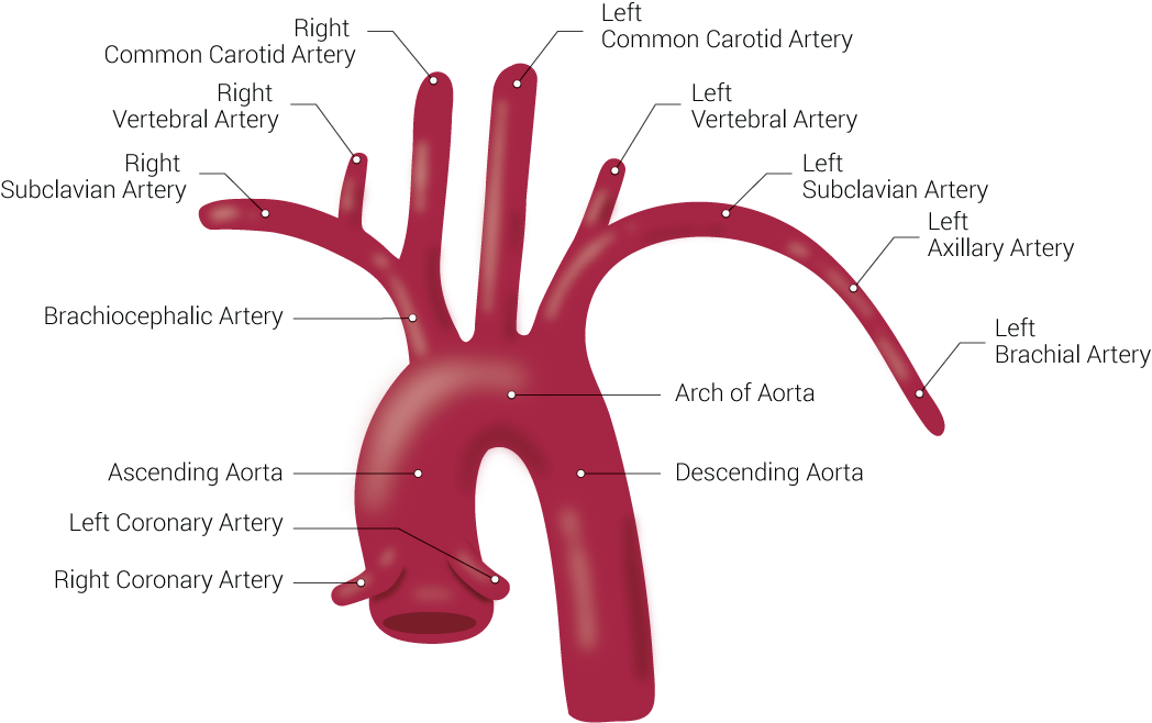
Branches of the Aorta. This illustration includes the right common carotid artery, right vertebral artery, right subclavian artery, brachiocephalic artery, ascending aorta, left coronary artery, right coronary artery, left common carotid artery, left vertebral artery, left subclavian artery, left axillary artery, left brachial artery, arch of aorta, and descending aorta.

Contributed by Beckie Palmer


(Click Image to Enlarge)
<p>Common Carotid Artery Bifurcation

Common Carotid Artery Bifurcation. This image shows the bifurcation of the common carotid artery into the external and internal carotid arteries.

Contributed by Erion Qaja, Wyckoff Heights Medical Center


(Click Image to Enlarge)
<p>Diagram of the Brain Blood Circulation

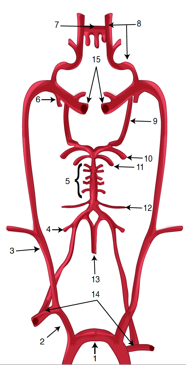
Diagram of the Brain Blood Circulation. Each number corresponds to the following neuroanatomy: 1) aortic arch; 2) brachiocephalic artery; 3) common carotid artery; 4) posterior inferior cerebellar artery; 5) pontine arteries; 6) anterior choroidal artery; 7) anterior communicating artery; 8) anterior cerebral artery; 9) posterior communicating artery; 10) posterior cerebral artery; 11) superior cerebellar artery; 12) anterior inferior cerebellar artery; 13) anterior spinal artery; 14) arches of vertebral arteries; and 15) internal carotid arteries.

Contributed by O Kuybu, MD 

References


[1]

Vercelli G, Sorenson TJ, Giordan E, Lanzino G, Rangel-Castilla L. Nuances of carotid artery stenting under flow arrest with dual-balloon guide catheter. Neurosurgical focus. 2019 Jan 1:46(Suppl_1):V4. doi: 10.3171/2019.1.FocusVid.18417. Epub     [PubMed PMID: 30611184]


[2]

Liao CH, Chen WH, Lee CH, Shen SC, Tsuei YS. Treating cerebrovascular diseases in hybrid operating room equipped with a robotic angiographic fluoroscopy system: level of necessity and 5-year experiences. Acta neurochirurgica. 2019 Mar:161(3):611-619. doi: 10.1007/s00701-018-3769-4. Epub 2019 Jan 4     [PubMed PMID: 30610374]


[3]

Lai Z, Guo Z, Shao J, Chen Y, Liu X, Liu B, Qiu C. A systematic review and meta-analysis of results of simultaneous bilateral carotid artery stenting. Journal of vascular surgery. 2019 May:69(5):1633-1642.e5. doi: 10.1016/j.jvs.2018.09.033. Epub 2018 Dec 19     [PubMed PMID: 30578074]

Level 2 (mid-level) evidence

[4]

Sebastian IA, Barakhanov K, Ganesh A. Symptomatic Cervical Carotid Artery Stenosis: Evolving Paradigms in Risk Stratification and Intervention. Annals of Indian Academy of Neurology. 2025 Jan 1:28(1):1-9. doi: 10.4103/aian.aian_838_24. Epub 2025 Jan 24     [PubMed PMID: 39865110]


[5]

Morishima Y, Kawabori M, Ito YM, Gekka M, Furukawa K, Niiya Y, Fujimura M. Validity of E-PASS Score for Evaluating Perioperative Minor Complications Associated with Carotid Endarterectomy. Neurologia medico-chirurgica. 2025 Jan 15:65(1):9-14. doi: 10.2176/jns-nmc.2024-0035. Epub 2024 Nov 25     [PubMed PMID: 39581620]


[6]

Guo L, Zhang J, Lv K, Li X, Guo M, Li C. Multimodal monitoring of cerebral perfusion in carotid endarterectomy patients: a computational fluid dynamics study. Frontiers in neurology. 2024:15():1455401. doi: 10.3389/fneur.2024.1455401. Epub 2024 Dec 5     [PubMed PMID: 39703353]


[7]

Akhtar KH, Metzger DC, Latif F. Carotid Disease and Management. Interventional cardiology clinics. 2025 Apr:14(2):191-204. doi: 10.1016/j.iccl.2024.11.006. Epub 2025 Jan 24     [PubMed PMID: 40049847]


[8]

Cyréus P, Wadén K, Hellberg S, Bergman O, Lengquist M, Karlöf E, Buckler A, Matic L, Roy J, Marlevi D, Chemaly M, Hedin U. Atherosclerotic plaque instability in symptomatic non-significant carotid stenoses. JVS-vascular science. 2025:6():100280. doi: 10.1016/j.jvssci.2025.100280. Epub 2025 Jan 17     [PubMed PMID: 40034249]


[9]

Henning RJ, Hoh BL. The diagnosis and treatment of asymptomatic and symptomatic patients with carotid artery stenosis. Current problems in cardiology. 2025 Jan 18:50(6):102992. doi: 10.1016/j.cpcardiol.2025.102992. Epub 2025 Jan 18     [PubMed PMID: 39832540]


[10]

Madden BJ, Polania-Sandoval C, Pujari GP, Mangalaparthi KK, Charlesworth MC, Prudencio M, Gendron T, Sandhu SJS, Nassar A, Petrucelli L, Meschia JF, Pandey A, Erben Y. Proteomic analysis of carotid artery plaques with and without vulnerable features on magnetic resonance angiography with vessel wall imaging: a pilot study. JVS-vascular science. 2025:6():100281. doi: 10.1016/j.jvssci.2025.100281. Epub 2025 Jan 21     [PubMed PMID: 40034248]

Level 3 (low-level) evidence

[11]

Cao J, Zeng Y, Zhou Y, Yao Z, Tan Z, Huo G, Zhang L, Zhou D. The value of contrast-enhanced ultrasound in assessing carotid plaque vulnerability and predicting stroke risk. Scientific reports. 2025 Feb 18:15(1):5850. doi: 10.1038/s41598-025-90319-2. Epub 2025 Feb 18     [PubMed PMID: 39966491]


[12]

Kumagai K, Takahashi T, Nishida S, Hayashi S, Ishihara H, Wada K, Kato H. Novel Fusion Imaging of Muscle, Plaque, Arteries, and Bone Using MR Imaging for Carotid Endarterectomy Patients. World neurosurgery. 2025 Mar:195():123735. doi: 10.1016/j.wneu.2025.123735. Epub 2025 Feb 19     [PubMed PMID: 39884352]


[13]

Madison M, Luo X, Silvey J, Brenner R, Gannamaneni K, Sawchuk AP. Clinical Decision Support for Patient Cases with Asymptomatic Carotid Artery Stenosis Using AI Models and Electronic Medical Records. Journal of cardiovascular development and disease. 2025 Feb 6:12(2):. doi: 10.3390/jcdd12020061. Epub 2025 Feb 6     [PubMed PMID: 39997495]

Level 3 (low-level) evidence

[14]

Pias AD, Pereira-Macedo J, Marreiros A, António N, Rocha-Neves J. Advancing Vascular Surgery: The Role Of Artificial Intelligence And Machine Learning In Managing Carotid Stenosis. Portuguese journal of cardiac thoracic and vascular surgery. 2024 Oct 12:31(3):55-64. doi: 10.48729/pjctvs.411. Epub 2024 Oct 12     [PubMed PMID: 39820882]


[15]

Arya S, Girotra S. Long-Term Mortality in Carotid Revascularization Patients. Circulation. Cardiovascular quality and outcomes. 2018 Nov:11(11):e004875. doi: 10.1161/CIRCOUTCOMES.118.004875. Epub     [PubMed PMID: 30571342]

Level 2 (mid-level) evidence

[16]

Patelis N, Diakomi M, Maskanakis A, Maltezos K, Schizas D, Papaioannou M. General versus local anesthesia for carotid endarterectomy: Special considerations. Saudi journal of anaesthesia. 2018 Oct-Dec:12(4):612-617. doi: 10.4103/sja.SJA_10_18. Epub     [PubMed PMID: 30429745]


[17]

Garcia F, Jácome F, Sousa J, Mansilha A. Transcarotid artery revascularization in symptomatic carotid stenosis: a systematic review. International angiology : a journal of the International Union of Angiology. 2025 Mar 6:():. doi: 10.23736/S0392-9590.24.05275-1. Epub 2025 Mar 6     [PubMed PMID: 40047790]

Level 1 (high-level) evidence

[18]

Uno M, Takai H, Yagi K, Matsubara S. Surgical Technique for Carotid Endarterectomy: Current Methods and Problems. Neurologia medico-chirurgica. 2020 Sep 15:60(9):419-428. doi: 10.2176/nmc.ra.2020-0111. Epub 2020 Aug 15     [PubMed PMID: 32801277]


[19]

de Carvalho CC, Guedes IHL, Holanda ALS, Costa YSC. Safety of anaesthesia techniques in patients undergoing carotid endarterectomy: a systematic review with meta-analysis of randomised clinical trials. Anaesthesia. 2025 Jan:80(1):109-111. doi: 10.1111/anae.16456. Epub 2024 Oct 22     [PubMed PMID: 39436018]

Level 1 (high-level) evidence

[20]

Cheng Y, Chen B, Zhang M, Chen Z, Liu M, Zhang Z, Tang H, Wang D, Lv W, Li B, Dai Y, Shang H. Carotid Endarterectomy Ameliorates Cognitive Impairment in Clinical and Experimental Unilateral Carotid Artery Stenosis. Journal of the American Heart Association. 2025 Jan 21:14(2):e038388. doi: 10.1161/JAHA.124.038388. Epub 2025 Jan 16     [PubMed PMID: 39817552]


[21]

Oya S, Yoshida S, Saito A, Iihoshi S, Obata H, Yamasaki A, Koizumi T, Shojima M, Suzuki K, Ooigawa H, Kohyama S, Kikkawa Y, Kurita H. Comparative Effects of Carotid Endarterectomy and Stenting on Visual Recovery in Patients With Carotid Artery Stenosis. Neurosurgery. 2025 Feb 17:():. doi: 10.1227/neu.0000000000003379. Epub 2025 Feb 17     [PubMed PMID: 39960289]

Level 2 (mid-level) evidence

[22]

Howell SJ. Carotid endarterectomy. British journal of anaesthesia. 2007 Jul:99(1):119-31     [PubMed PMID: 17556351]


[23]

Stasiowski MJ, Zmarzły N, Grabarek BO. Evaluating Anesthesia Guidance for Rescue Analgesia in Awake Patients Undergoing Carotid Endarterectomy with Cervical Plexus Blocks: Preliminary Findings from a Randomized Controlled Trial. Journal of clinical medicine. 2024 Dec 28:14(1):. doi: 10.3390/jcm14010120. Epub 2024 Dec 28     [PubMed PMID: 39797203]

Level 1 (high-level) evidence

[24]

León Jorba A, Velescu A, Álvarez López-Herrero N, Mases Fernández A, Romero Montana L, Clarà Velasco A. Early detection of cranial nerve dysfunction during carotid endarterectomy through intraoperative neurophysiological monitoring. Clinical neurophysiology : official journal of the International Federation of Clinical Neurophysiology. 2025 Apr:172():10-16. doi: 10.1016/j.clinph.2025.01.013. Epub 2025 Feb 1     [PubMed PMID: 39952002]


[25]

Czinege Z, Sándor ÁD, Gyürki D, Varga A, Csípő T, Székely A, Ungvári Z, Banga P, Sótonyi P, Horváth T. Understanding perioperative risk determinants in carotid endarterectomy: the impact of compromised circle of Willis morphology on inter-hemispheric blood flow indices based on intraoperative internal carotid artery stump pulse pressure and backflow patterns. GeroScience. 2025 Apr:47(2):2159-2177. doi: 10.1007/s11357-024-01390-y. Epub 2024 Oct 26     [PubMed PMID: 39460849]

Level 3 (low-level) evidence

[26]

Joo SP, Cho YH, Lee YJ, Kim YS, Kim TS. Modified Suturing Techniques in Carotid Endarterectomy for Reducing the Cerebral Ischemic Time. Journal of Korean Neurosurgical Society. 2020 Nov:63(6):834-840. doi: 10.3340/jkns.2020.0058. Epub 2020 Sep 10     [PubMed PMID: 32906227]


[27]

Rajesh K, Shen H, Bhaskar SMM. Seizures Following Carotid Endarterectomy: A Comprehensive Meta-Analysis of 69,479 Patients and Evidence-Based Recommendations for Perioperative Care. Diagnostics (Basel, Switzerland). 2024 Dec 24:15(1):. doi: 10.3390/diagnostics15010006. Epub 2024 Dec 24     [PubMed PMID: 39795534]

Level 1 (high-level) evidence

[28]

Abdelkarim A, Hamouda M, Real M, Zarrintan S, Magee GA, Malas MB. Cerebral Hyperperfusion Syndrome after Carotid Revascularization; Predictors and Complications. Annals of vascular surgery. 2025 Jun:115():13-22. doi: 10.1016/j.avsg.2025.02.001. Epub 2025 Mar 4     [PubMed PMID: 40044075]


[29]

Caron E, Yadavalli SD, Manchella M, Jabbour G, Mandigers TJ, Gomez-Mayorga JL, Bloch RA, Malas MB, Motaganahalli RL, Schermerhorn ML. Outcomes of redo vs primary carotid endarterectomy in the transcarotid artery revascularization era. Journal of vascular surgery. 2025 Feb 19:():. pii: S0741-5214(25)00339-8. doi: 10.1016/j.jvs.2025.02.014. Epub 2025 Feb 19     [PubMed PMID: 39984141]


[30]

Aridi HD, Frank G, Gutwein AR, Madison M, Schermerhorn ML, Kashyap VS, Wang G, Eldrup-Jorgensen J, Malas M, Motaganahalli R. Physicians' preference for carotid revascularization impacts postoperative stroke and death outcomes. Journal of vascular surgery. 2025 May:81(5):1092-1103.e2. doi: 10.1016/j.jvs.2024.12.125. Epub 2024 Dec 27     [PubMed PMID: 39733781]


[31]

Gajin P, Petrovic J, Pesic S, Atanasijevic I, Babic A, Neskovic M, Dabic P, Babic S, Matic P, Tanaskovic S, Ilijevski N. Urgent carotid endarterectomy for crescendo transient ischemic attack. VASA. Zeitschrift fur Gefasskrankheiten. 2025 Mar 6:():. doi: 10.1024/0301-1526/a001188. Epub 2025 Mar 6     [PubMed PMID: 40045545]


[32]

Murphy SJX, Naylor AR, Ricco JB, Sillesen H, Kakkos S, Halliday A, de Borst GJ, Vega de Ceniga M, Hamilton G, McCabe DJH. Optimal Antiplatelet Therapy in Moderate to Severe Asymptomatic and Symptomatic Carotid Stenosis: A Comprehensive Review of the Literature. European journal of vascular and endovascular surgery : the official journal of the European Society for Vascular Surgery. 2019 Feb:57(2):199-211. doi: 10.1016/j.ejvs.2018.09.018. Epub 2018 Nov 7     [PubMed PMID: 30414802]


[33]

McArdle MJ, Abbott AL, Krajcer Z. Carotid Artery Stenosis in Women. Texas Heart Institute journal. 2018 Aug:45(4):243-245. doi: 10.14503/THIJ-18-6711. Epub 2018 Aug 1     [PubMed PMID: 30374237]


[34]

Aber A, Howard A, Woods HB, Jones G, Michaels J. Impact of Carotid Artery Stenosis on Quality of Life: A Systematic Review. The patient. 2019 Apr:12(2):213-222. doi: 10.1007/s40271-018-0337-1. Epub     [PubMed PMID: 30328068]

Level 2 (mid-level) evidence

[35]

Wang SK, Fajardo A, Sawchuk AP, Lemmon GW, Dalsing MC, Gupta AK, Murphy MP, Motaganahalli RL. Outcomes associated with a transcarotid artery revascularization-centered protocol in high-risk carotid revascularizations using the ENROUTE neuroprotection system. Journal of vascular surgery. 2019 Mar:69(3):807-813. doi: 10.1016/j.jvs.2018.06.222. Epub 2018 Oct 6     [PubMed PMID: 30301690]


[36]

Gaba K, Ringleb PA, Halliday A. Asymptomatic Carotid Stenosis: Intervention or Best Medical Therapy? Current neurology and neuroscience reports. 2018 Sep 24:18(11):80. doi: 10.1007/s11910-018-0888-5. Epub 2018 Sep 24     [PubMed PMID: 30251204]


[37]

Thomas MA, Pearce WH, Rodriguez HE, Helenowski IB, Eskandari MK. Durability of Stroke Prevention with Carotid Endarterectomy and Carotid Stenting. Surgery. 2018 Dec:164(6):1271-1278. doi: 10.1016/j.surg.2018.06.041. Epub 2018 Sep 18     [PubMed PMID: 30236609]


[38]

Hameed H, Khan S, Nasir S. Advancing Risk Stratification for Ischaemic Patients Undergoing Carotid Endarterectomy: A Personalised Approach for Safer Outcomes. European journal of vascular and endovascular surgery : the official journal of the European Society for Vascular Surgery. 2025 Feb 26:():. pii: S1078-5884(25)00171-6. doi: 10.1016/j.ejvs.2025.02.037. Epub 2025 Feb 26     [PubMed PMID: 40021115]


[39]

Grunebach H, Madeira T, Bose S, Holscher C, Aru RG, Abularrage CJ, Black JH 3rd, Lum YW, Perler BA, Hicks CW. A standardized carotid endarterectomy care pathway is associated with lower ICU admission rates and a significant reduction in hospital charges. American journal of surgery. 2025 Jan:239():116056. doi: 10.1016/j.amjsurg.2024.116056. Epub 2024 Nov 10     [PubMed PMID: 39572277]