
Introduction
There are three primary anterior knee fat pads, such as the quadriceps suprapatellar, pre-femoral suprapatellar, and infrapatellar retro-patellar tendon (Hoffa's fat pad), all of which may experience symptomatic impingement.[1] The Hoffa pad, also known as the infrapatellar fat pad (IFP), is an extra-synovial, intracapsular structure that occupies the majority of the anterior knee compartment.[2]
The IFP can become one of many causes of anterior knee pain when it becomes inflamed, leading to impingement at the tibiofemoral joint or the lateral aspect of the patellofemoral joint, known as Hoffa pad impingement syndrome.[3] This structure is richly vascularized and innervated. Its arterial supply is from an anastomotic network of branches of the medial and lateral superior geniculate and medial and lateral inferior geniculate arteries.[4] It is innervated by the posterior articular nerve, a branch of the posterior tibial nerve that courses through the external borders of the menisci, synovium, cruciate ligaments, and the IFP.[5]
A normal IFP attaches to the proximal patellar tendon, the inferior pole of the patella, anterior horns of the medial and lateral menisci, transverse meniscal ligament, and periosteum of the anterior tibia.[3] The posterior aspect of this structure is lined with synovium and extends posteriorly, connecting to the intercondylar notch and, in some structures, is continuous with the anterior cruciate ligament.[6][7]
Etiology
Register For Free And Read The Full Article
 Search engine and full access to all medical articles
- 10 free questions in your specialty
- Free CME/CE Activities
- Free daily question in your email
- Save favorite articles to your dashboard
- Emails offering discounts
Learn more about a Subscription to StatPearls Point-of-Care
Etiology
Injury to the IFP is usually the result of a single traumatic event or repetitive microtrauma due to blunt impact, shear injury with anterior cruciate ligament (ACL) tear, dislocation of the patella, or a twisting injury at the knee.[8] Hoffa pad impingement syndrome has been well-documented as a source of anterior knee pain, commonly found in runners, cyclists, and soldiers.[9]
The IFP is a potent source of pain due to its robust innervation by nociceptive Type IVa-free nerve endings and fibers targeted by substance P.[10] Substance P causes vasodilation, thereby promoting the recruitment of immune cells and potentially contributing to edema development within the tissue. This inflammatory process can cause thickening and fibrosis leading to the loss of elastic properties of the IFP.[3]
The scar tissue gradually accumulates and impinges at the intercondylar notch and trochlea, leading to a mechanical and/or painful block to full extension.[11] The IFP produces fibroblast growth factor, vascular endothelial growth factor, tumor necrosis factor, and interleukin-6, all of which interact to contribute to the development of inflammation, fibrosis, and pain within the IFP.[12]
Epidemiology
The epidemiology of Hoffa pad impingement syndrome is unknown. The low documented incidence could be due to the difficulty in diagnosing the condition and its tendency to be adequately treated with conservative management.[13] It is believed that Hoffa pad impingement syndrome is underdiagnosed by imaging because it is only present in 1% of patients undergoing knee arthroscopy.[8][14]
Pathophysiology
Hoffa first described the most accepted etiology of Hoffa pad impingement syndrome in 1904, which entailed IFP injury followed by associated hemorrhage, inflammation, hypertrophy, and eventual fibrosis.[15] Acute injury, repetitive microtrauma, and iatrogenic injury can lead to the initial cascade of events ending in fibrosis. Iatrogenic causes of IFP pathology include fibrosis due to arthroscopy portals, bone-patella tendon-bone autograft harvest during ACL reconstruction, or fat pad resection.[16]
The clinical manifestations of other lesions inside IFP are comparable and often include swelling, knee discomfort, restricted range of motion, and an occasional palpable lump. Synovial lipomas, pigmented villonodular synovitis, glomus tumors, synovial chondromatosis, synovial haemangiomas, and ganglion cysts are among the possible causes.[17][18] It results from repeated pinching of the exterior part of the fat pad between the cartilage of the lateral facet of the trochlea and the lateral patellofemoral ligament. Any pathology that reduces the space between these two structures might encourage local impingement. HFP impingement is also linked to a higher patella position (patella alta).[19]
Histopathology
On MRI, IFP appears similar to subcutaneous adipose tissue and has fibrous structures dispersed throughout the adipocytes.[20] Histologically, non-diseased tissue resembles visceral adipose tissue.[21] Inflamed IFP demonstrates fibro-fatty tissue and degenerative change, consistent with chronic injury and inflammation.[3] The dense fibrous tissue gradually replaces lipocytes and occasionally transforms into cartilaginous tissue with ossifications.[22]
Obvious visible fibrotic changes and/or calcifications of the diseased IFP are rare, although when present, are specific for Hoffa pad impingement.[22] The synovial lining of the inferior patellar fat pad has type IVa free nerve terminals, which are the primary source of anterior knee discomfort.[23]
History and Physical
Patients with Hoffa pad impingement syndrome report a "burning" or "aching" sensation to the anterior knee localized deep to and on either side of the patellar tendon adjacent to the inferior pole of the patella. This most commonly occurs with the knee at full extension, dynamic extension, or prolonged flexion. The pain usually occurs over a prolonged period and may be associated with a previous knee injury or may be idiopathic.
Physical examination reveals discomfort and tenderness around the patellar tendon, typically located close to the inferior pole of the patella. All patients have some degree of range of motion restriction, and one study reported severe limiting at 20 degrees of flexion. Pain at the end of the extension is also common in patients with fat pad impingement.[3]
The patient's symptoms can be reproduced with the Hoffa test, which is performed by applying firm pressure with the thumb inferior to the patella just outside the margin of the patellar tendon with the knee in 30 degrees of flexion. With steady pressure, the knee is fully extended. Increased pain with this maneuver is a positive test. The test is then repeated on the medial or lateral side of the tendon.[15]
Care must be taken to differentiate this anterior knee pain from joint line pain secondary to anterior meniscal pathology.[24] Pain may also be reproduced with isometric quadriceps contraction with the knee in full extension. The diseased IFP may be enlarged, firm, or tender to palpation. IFP pathology is also associated with an extension block and decreased patellar mobility.[25]
The arthroscopic release has been devised for anterior interval release followed by rehabilitation, which focuses on preventing re-scarring with extension exercises and patellar mobilization.[15] An "infrapatellar plica" is the term used to describe the thickening and fibrosis of the ligamentum mucosum. Nonspecific symptoms such as continuous agonizing knee discomfort made worse by movement, sporadic popping, snapping, and effusion have been reported in patients with symptomatic infrapatellar plica.[26]
Evaluation
Sagittal MRI is the gold standard imaging modality for assessing IFP pathology. On T1-weighted MRI of the normal IFP, the appearance is similar to surrounding subcutaneous fat except for the foci of lower signal intensity representing the interposed fibrous septae.[25]
Hypointense vertical clefts are present in the proximal aspect of the fat pad, and horizontal clefts are present in its posterior aspect. The hypointense signal on T1 adjacent to the anterior horns of the medial and lateral menisci represents the transverse meniscal ligament.[27] T2 fat saturation MRI portrays the IFP as isointense to muscle.[28] The posterior continuation of the Hoffa fat pad synovium, also known as ligamentum mucosum, can occasionally be appreciated as a T2 hyper-intense band anterior to the ACL and inserted within the intercondylar notch. Blood vessels are also visualized on T2 MRI, taking a vertical course in the medial and lateral aspects of the IFP.[28] In Hoffa pad impingement syndrome, an increased signal on T1 or hypo-intense T2 signal within the IFP indicates fibrosis. Intra-fat pad ossification can be differentiated from fibrosis using an X-ray. Hyper-intense T2 signal within the IFP indicates inflammation and its associated edema and hemorrhage.[25]
A positive Hoffa's test and loss of extension and flexion are the usual symptoms of anterior interval scarring, which is scar tissue that binds the Infrapatellar fat pad to the anterior tibia. Other symptoms include discomfort during extension and anterior knee pain.
In a comparative study, Arthur et al. demonstrated that individuals with clinical fat pad impingement had edema on MRI in the superolateral area of the Hoffa fat pad. However, similar edema may also be present in people without signs of fat pad impingement. Therefore clinical assessment is very important for diagnosis rather than sole reliance on MRI findings.[29]
Treatment / Management
Nonoperative Management
Physical therapy is the mainstay of treatment for Hoffa pad impingement syndrome.[30] The goal of physical therapy is to restore the biomechanics of patellar tracking via vastus medialis obliquus strengthening, taping to offload the patella tendon and fat pad, stretching, and improving pelvic control with training focused on gluteal strengthening to optimize lower extremity mechanics.[31] (A1)
The goal of taping is to tilt the inferior portion of the patella anteriorly to decrease impingement and subsequent inflammation of the IFP. This is accomplished by applying tape across the proximal half of the patella with the knee fully extended. Two separate strips are applied in a "V" formation, with the apex at the tibial tubercle. The first part of the V tape starts at the tibial tubercle and is applied over the medial epicondyle while the patella and surrounding soft tissue are pulled inferiorly. The second strip is applied beginning at the tibial tubercle and towards the lateral epicondyle in the same fashion. The tape is to remain in place all day, every day until the patient is https://www.citethisforme.com/vancouverpain-free. Patients are also instructed to avoid hyperextension and rapid extension maneuvers to decrease impingement of the IFP.[31]
Muscle training focuses on closed-chain exercises targeting the quadriceps, namely the vastus medialis obliquus, to improve patellar tracking.[32] Resistance training of the posterior fibers of the gluteus medius to decrease internal rotation of the hip and the resultant valgus force at the knee can be implemented to better align the patella within the trochlea. The patient may stretch the anterior hip structures to increase available external rotation if the patient has marked femoral internal rotation. (B3)
House and Connell reported an improved visual analog score after injecting a mean of four injections of alcohol and bupivacaine under ultrasound guidance.[33] Infrapatellar fat pad pain usually responds to 1 to 3 injections of 6 ccs of 2% lidocaine combined with 40 mg of methylprednisolone acetate or 50 mg of hydrocortisone at intervals of 4 to 6 weeks. Corticosteroid injections have also been used to treat Hoffa pad impingement syndrome and have demonstrated satisfactory results.[16](B2)
Operative Management
Operative intervention is indicated once the patient has exhausted conservative management. Arthroscopic resection of the posterior aspect of the IFP is the primary surgical treatment of the inflamed, symptomatic fat pad.[34] It has been recommended to use a high anterolateral portal and a standard anteromedial working portal during arthroscopic resection. A high anteromedial portal is sometimes necessary to best visualize and access the lateral ala impingement. Symptomatic infrapatellar plica has been demonstrated to respond well to arthroscopic excision. The majority (86 to 91%) of patients with solitary infrapatellar plica release experienced few to no post-operative complaints. Regardless of the approach, hemostasis is precisely maintained.[35] (B2)
Patients with discomfort and swelling around the inferior pole of the patella may find relief with arthroscopic denervation of the nociceptive nerves innervating those areas. The inferior fat pad is often surgically removed from where it inserts on the inferior patellar pole using an electrothermal cautery method.[36] After arthroscopic excision, patients with significant impingement of the fat pad having no other concurrent disease may anticipate remission or long-term improvement in their symptoms and function.[34] (B2)
Kim et al. reported improved clinical results with either partial or subtotal arthroscopic excision of the impinged fat pad between the patella and femoral trochlea. Given that partial resection was just as successful as subtotal resection and kept more of the fat pad, it may thus be a suitable course of therapy.[37](B2)
Differential Diagnosis
Hoffa pad impingement syndrome is a diagnosis of exclusion, mainly due to the numerous conditions that cause anterior knee pain and extension block. Soft tissue pathology, such as bursitis, must be ruled out. The anterior knee has four bursae: the prepatellar, superficial, infrapatellar, deep infrapatellar, and pes anserine bursae, and can become inflamed with repetitive microtrauma or sports requiring repetitive knee use such as cycling.[38]
Patellar tendinosis must also be considered and is typically seen in high-impact jumping sports. It is characterized by activity-related pain localized to the inferior pole of the patella and occurs more prominently with eccentric quadriceps contraction. Intra-articular pathologies such as plica syndrome, meniscal tears, articular cartilage injuries, and osteochondral lesions must also be ruled out. Plica syndrome occurs when the incompletely-resorbed embryonic septa separating the knee joint into medial, lateral, and suprapatellar compartments become inflamed following trauma, overuse, long-standing patellar mal-tracking, diabetes, and inflammatory arthropathy.[39]
Meniscus tears usually occur following a traumatic injury to the knee, resulting in pain and an intra-articular clicking/catching sensation. Mechanical blocks to full extension can also occur from hypertrophic fibrosis following ACL reconstruction. Also known as a "cyclops lesion," this nodule or hypertrophied graft tissue has been reported to occur between 1% and 10% of all ACL reconstructions and is the second most common cause of restricted knee extension following graft impingement.[40][41]
Anterior interval scarring is another commonly known block to full knee extension, often causing pain, decreased patellar mobility, and quadriceps atrophy. The anterior interval is the space between the IFP and the anterior tibia and is also often implicated as the cause of extension block following arthroscopic ACL reconstruction.[42]
Prognosis
Hoffa pad impingement syndrome is most often successfully treated with conservative management such as physical therapy and/or steroid injections.[30] In those requiring operative intervention, the vast majority return to their pre-injury level of sporting activities following arthroscopic debridement.[34]
Complications
Missed or delayed diagnosis of Hoffa pad impingement syndrome can lead to diminished quality of life, missed playing time, and progression of inflammation into the development of fibrosis. An enlarged and fibrotic IFP can cause a knee flexion contracture which alters gait mechanics.
Deterrence and Patient Education
Patients, especially athletes, can help avoid the development of Hoffa pad impingement syndrome by focusing on closed-chain quadriceps exercises that will, in turn, promote appropriate patellar tracking and avoid the initial impingement event. Those with ligamentous laxity, a history of patellar dislocation, or prior ligamentous knee injury are at increased risk of developing this pathology. Increased awareness of this disease serves as the best preventative measure.
Enhancing Healthcare Team Outcomes
Hoffa pad impingement syndrome is one of the many causes of infrapatellar knee pain. Due to the prevalence of anterior knee pain, familiarity with this disease is paramount inappropriate diagnosis, treatment, and recovery for patients with this condition. The healthcare professional must be thoughtful and intentional during the history and physical examination to arrive at a diagnosis in a timely fashion. It has been reported that many cases of IFP syndrome have been incorrectly diagnosed and treated as meniscal pathology, which leads to inefficient use of resources and improper treatment. This pathology requires effective interprofessional communication between the clinician, athletic trainer, nurses, and physical therapist, as the majority of these cases can successfully be treated with physical therapy alone.[30] [Level I]
Improving the healthcare professional's understanding of how to promptly evaluate and treat this condition will lead to better patient outcomes.
Media
(Click Image to Enlarge)
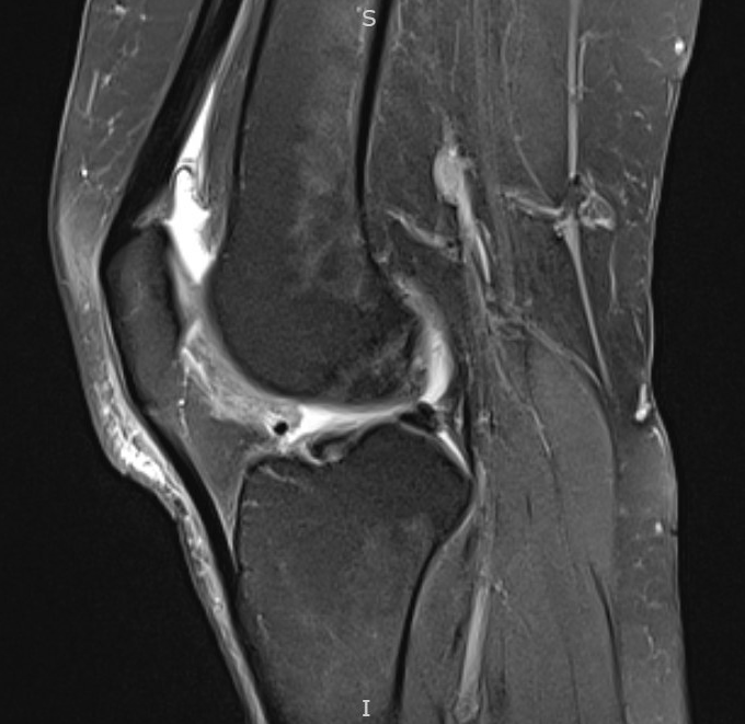
Sag PD FS MRI Hoffa pad impingement syndrome
Contributed by Matthew DuBose, MD
References
Borja MJ,Jose J,Vecchione D,Clifford PD,Lesniak BP, Prefemoral fat pad impingement syndrome: identification and diagnosis. American journal of orthopedics (Belle Mead, N.J.). 2013 Jan [PubMed PMID: 23431544]
Biedert RM,Sanchis-Alfonso V, Sources of anterior knee pain. Clinics in sports medicine. 2002 Jul; [PubMed PMID: 12365231]
Radu A,Discepola F,Volesky M,Munk PL,Le H, Posterior Hoffa's fat pad impingement secondary to a thickened infrapatellar plica: a case report and review of the literature. Journal of radiology case reports. 2015 Mar; [PubMed PMID: 25926930]
Level 3 (low-level) evidenceEymard F,Chevalier X, Inflammation of the infrapatellar fat pad. Joint bone spine. 2016 Jul; [PubMed PMID: 27068617]
Kennedy JC,Alexander IJ,Hayes KC, Nerve supply of the human knee and its functional importance. The American journal of sports medicine. 1982 Nov-Dec; [PubMed PMID: 6897495]
Brooker B,Morris H,Brukner P,Mazen F,Bunn J, The macroscopic arthroscopic anatomy of the infrapatellar fat pad. Arthroscopy : the journal of arthroscopic [PubMed PMID: 19664502]
Gallagher J,Tierney P,Murray P,O'Brien M, The infrapatellar fat pad: anatomy and clinical correlations. Knee surgery, sports traumatology, arthroscopy : official journal of the ESSKA. 2005 May; [PubMed PMID: 15678298]
Grando H,Chang EY,Chen KC,Chung CB, MR imaging of extrasynovial inflammation and impingement about the knee. Magnetic resonance imaging clinics of North America. 2014 Nov; [PubMed PMID: 25442030]
Pharis H,Kong A,Robbins M,Waranch C,Wissman R, Friction Syndromes of the Knee. The journal of knee surgery. 2022 Apr; [PubMed PMID: 35189665]
Bohnsack M,Meier F,Walter GF,Hurschler C,Schmolke S,Wirth CJ,Rühmann O, Distribution of substance-P nerves inside the infrapatellar fat pad and the adjacent synovial tissue: a neurohistological approach to anterior knee pain syndrome. Archives of orthopaedic and trauma surgery. 2005 Nov; [PubMed PMID: 15891922]
Kim SJ,Kim JY,Lee JW, Pathologic infrapatellar plica. Arthroscopy : the journal of arthroscopic [PubMed PMID: 12632039]
Level 3 (low-level) evidenceUshiyama T,Chano T,Inoue K,Matsusue Y, Cytokine production in the infrapatellar fat pad: another source of cytokines in knee synovial fluids. Annals of the rheumatic diseases. 2003 Feb; [PubMed PMID: 12525378]
Rooney A,Wahba AJ,Smith TO,Donell ST, The surgical treatment of anterior knee pain due to infrapatellar fat pad pathology: A systematic review. Orthopaedics [PubMed PMID: 25935799]
Level 1 (high-level) evidenceOgilvie-Harris DJ,Giddens J, Hoffa's disease: arthroscopic resection of the infrapatellar fat pad. Arthroscopy : the journal of arthroscopic [PubMed PMID: 8003146]
Steadman JR,Dragoo JL,Hines SL,Briggs KK, Arthroscopic release for symptomatic scarring of the anterior interval of the knee. The American journal of sports medicine. 2008 Sep; [PubMed PMID: 18753680]
Paulos LE,Wnorowski DC,Greenwald AE, Infrapatellar contracture syndrome. Diagnosis, treatment, and long-term followup. The American journal of sports medicine. 1994 Jul-Aug; [PubMed PMID: 7943507]
Level 2 (mid-level) evidenceKrebs VE,Parker RD, Arthroscopic resection of an extrasynovial ossifying chondroma of the infrapatellar fat pad: end-stage Hoffa's disease? Arthroscopy : the journal of arthroscopic & related surgery : official publication of the Arthroscopy Association of North America and the International Arthroscopy Association. 1994 Jun [PubMed PMID: 8086026]
Level 3 (low-level) evidenceHardy P,Muller GP,Got C,Lortat-Jacob A,Benoit J, Glomus tumor of the fat pad. Arthroscopy : the journal of arthroscopic & related surgery : official publication of the Arthroscopy Association of North America and the International Arthroscopy Association. 1998 Apr [PubMed PMID: 9586981]
Level 3 (low-level) evidenceDraghi F,Ferrozzi G,Urciuoli L,Bortolotto C,Bianchi S, Hoffa [PubMed PMID: 27000624]
Vahlensieck M,Linneborn G,Schild H,Schmidt HM, Hoffa's recess: incidence, morphology and differential diagnosis of the globular-shaped cleft in the infrapatellar fat pad of the knee on MRI and cadaver dissections. European radiology. 2002 Jan; [PubMed PMID: 11868081]
Level 2 (mid-level) evidenceIoan-Facsinay A,Kloppenburg M, An emerging player in knee osteoarthritis: the infrapatellar fat pad. Arthritis research [PubMed PMID: 24367915]
von Engelhardt LV,Tokmakidis E,Lahner M,Dàvid A,Haage P,Bouillon B,Lichtinger TK, Hoffa's fat pad impingement treated arthroscopically: related findings on preoperative MRI in a case series of 62 patients. Archives of orthopaedic and trauma surgery. 2010 Aug; [PubMed PMID: 20556618]
Level 2 (mid-level) evidenceBiedert RM,Stauffer E,Friederich NF, Occurrence of free nerve endings in the soft tissue of the knee joint. A histologic investigation. The American journal of sports medicine. 1992 Jul-Aug [PubMed PMID: 1415886]
Smith BW,Green GA, Acute knee injuries: Part I. History and physical examination. American family physician. 1995 Feb 15; [PubMed PMID: 7863957]
Jacobson JA,Lenchik L,Ruhoy MK,Schweitzer ME,Resnick D, MR imaging of the infrapatellar fat pad of Hoffa. Radiographics : a review publication of the Radiological Society of North America, Inc. 1997 May-Jun; [PubMed PMID: 9153705]
Boyd CR,Eakin C,Matheson GO, Infrapatellar plica as a cause of anterior knee pain. Clinical journal of sport medicine : official journal of the Canadian Academy of Sport Medicine. 2005 Mar; [PubMed PMID: 15782055]
Level 2 (mid-level) evidencePatel SJ,Kaplan PA,Dussault RG,Kahler DM, Anatomy and clinical significance of the horizontal cleft in the infrapatellar fat pad of the knee: MR imaging. AJR. American journal of roentgenology. 1998 Jun; [PubMed PMID: 9609172]
Saddik D,McNally EG,Richardson M, MRI of Hoffa's fat pad. Skeletal radiology. 2004 Aug; [PubMed PMID: 15221217]
De Smet AA,Davis KW,Dahab KS,Blankenbaker DG,del Rio AM,Bernhardt DT, Is there an association between superolateral Hoffa fat pad edema on MRI and clinical evidence of fat pad impingement? AJR. American journal of roentgenology. 2012 Nov [PubMed PMID: 23096185]
Level 2 (mid-level) evidenceCrossley K,Bennell K,Green S,Cowan S,McConnell J, Physical therapy for patellofemoral pain: a randomized, double-blinded, placebo-controlled trial. The American journal of sports medicine. 2002 Nov-Dec; [PubMed PMID: 12435653]
Level 1 (high-level) evidenceDragoo JL,Johnson C,McConnell J, Evaluation and treatment of disorders of the infrapatellar fat pad. Sports medicine (Auckland, N.Z.). 2012 Jan 1; [PubMed PMID: 22149697]
Doucette SA,Child DD, The effect of open and closed chain exercise and knee joint position on patellar tracking in lateral patellar compression syndrome. The Journal of orthopaedic and sports physical therapy. 1996 Feb; [PubMed PMID: 8808512]
Level 3 (low-level) evidenceHouse CV,Connell DA, Therapeutic ablation of the infrapatellar fat pad under ultrasound guidance: a pilot study. Clinical radiology. 2007 Dec [PubMed PMID: 17981168]
Level 3 (low-level) evidenceKumar D,Alvand A,Beacon JP, Impingement of infrapatellar fat pad (Hoffa's disease): results of high-portal arthroscopic resection. Arthroscopy : the journal of arthroscopic [PubMed PMID: 17986405]
Level 2 (mid-level) evidenceDemirag B,Ozturk C,Karakayali M, Symptomatic infrapatellar plica. Knee surgery, sports traumatology, arthroscopy : official journal of the ESSKA. 2006 Feb; [PubMed PMID: 16059707]
Level 2 (mid-level) evidenceOgon P,Maier D,Jaeger A,Suedkamp NP, Arthroscopic patellar release for the treatment of chronic patellar tendinopathy. Arthroscopy : the journal of arthroscopic & related surgery : official publication of the Arthroscopy Association of North America and the International Arthroscopy Association. 2006 Apr [PubMed PMID: 16581464]
Level 2 (mid-level) evidenceKim YM,Joo YB, Arthroscopic Treatment of Infrapatellar Fat Pad Impingement between the Patella and Femoral Trochlea: Comparison of the Clinical Outcomes of Partial and Subtotal Resection. Knee surgery [PubMed PMID: 30871293]
Level 2 (mid-level) evidenceKuwabara A,Fredericson M, Narrative: Review of Anterior Knee Pain Differential Diagnosis (Other than Patellofemoral Pain). Current reviews in musculoskeletal medicine. 2021 Jun; [PubMed PMID: 33818700]
Lee PYF,Nixion A,Chandratreya A,Murray JM, Synovial Plica Syndrome of the Knee: A Commonly Overlooked Cause of Anterior Knee Pain. Surgery journal (New York, N.Y.). 2017 Jan; [PubMed PMID: 28825013]
Recht MP,Piraino DW,Cohen MA,Parker RD,Bergfeld JA, Localized anterior arthrofibrosis (cyclops lesion) after reconstruction of the anterior cruciate ligament: MR imaging findings. AJR. American journal of roentgenology. 1995 Aug; [PubMed PMID: 7618562]
Level 2 (mid-level) evidenceKambhampati SBS,Gollamudi S,Shanmugasundaram S,Josyula VVS, Cyclops Lesions of the Knee: A Narrative Review of the Literature. Orthopaedic journal of sports medicine. 2020 Aug; [PubMed PMID: 32923503]
Level 3 (low-level) evidenceRose M,McNeilan R,Genuario J,Schlegel T, Surgical Technique for Release of Anterior Interval Scarring of the Knee After Anterior Cruciate Ligament Reconstruction. Arthroscopy techniques. 2018 Sep; [PubMed PMID: 30258768]