
Introduction
Exotropia is the outward deviation of eyes, i.e., away from the nose. Exodeviations can be congenital or acquired. These can be concomitant or incomitant. Concomitant deviation means that the divergent deviation of the visual axis remains the same in all positions of gaze. Incomitant deviation implies that the divergent deviation of the visual axis is variable in different gazes. These are often associated with the limitation of extraocular movements resulting either from paralytic or a restrictive etiology. The concomitant exotropia is further divided into congenital, primary, sensory, or consecutive exotropia.[1]
Incomitant exotropia is divided into paralytic (third nerve paralysis, isolated medial rectus paralysis, paralysis of convergence), A, V, or X pattern, or restrictive (Duane retraction syndrome, thyroid myopathy, restriction due to trauma or following surgery). The intermittent or constant deviation can be graded as exophoria, intermittent exotropia, or exotropia (constant exodeviation).[2] In this activity, our prime focus will be on the concomitant exodeviations.
Etiology
Register For Free And Read The Full Article
 Search engine and full access to all medical articles
- 10 free questions in your specialty
- Free CME/CE Activities
- Free daily question in your email
- Save favorite articles to your dashboard
- Emails offering discounts
Learn more about a Subscription to StatPearls Point-of-Care
Etiology
The etiology for the concomitant divergent exodeviations varies for each type.
Congenital (Infantile) Exotropia
Congenital (infantile) exotropia is a rare condition compared to congenital esotropia.[3] It is often associated with craniofacial anomalies, ocular albinism, or cerebral palsy.
Primary Exotropia
This can be associated with mechanical or innervational factors. The mechanical factors which can lead to exotropia include shape and axes of orbit, interpupillary distance, the mechanical property of the conjunctiva or tenon's capsule, extraocular muscle characteristics. The innervational factors include an imbalance between the convergence and divergence mechanisms. Thus an excessive divergence or insufficient convergence can lead to an exodeviation.
Sensory Exotropia
This often results from poor visual acuity in one eye. Sensory exotropia is more common when unilateral vision loss occurs in infancy or adulthood. It may appear secondary to anisometropia, unilateral medial opacity secondary to corneal opacity or lenticular changes, unilateral aphakia, optic atrophy, or macular pathology.[4]
Consecutive Exotropia
Consecutive exotropia may result from a surgical overcorrection of esotropia or a spontaneous change of esotropia to exotropia.[5]
Epidemiology
The incidence of exotropia was studied by Govindan et al. in their ten-year retrospective study from Minnesota. They found the annual age and gender-adjusted incidence of 64.1/100,000 patients younger than 19 years. Of these, 86% had an intermittent exotropia or convergence insufficiency.[6]
Hashemi et al., in their meta-analysis of 56 articles, found the pooled prevalence of exotropia as 1.23%. They also reported that the heterogeneity among exotropia patients varied directly with age.[7] Another study from China done among preschool children found the prevalence of intermittent exotropia as 3.24%. They additionally found no significant differences with age (p-value = 0.19), or sex (p-value = 0.89). Among these, the "basic type" intermittent exotropia was the most common present among 74.7%, followed by "divergence excess" among 19.9%, and the least common type was "convergence weakness" among 5.4% of the study population.[8]
Pathophysiology
The exact pathophysiology of congenital exotropia is not well known. But strabismus is very commonly seen to run in families, which points towards an autosomal dominant inheritance pattern. The exodeviations are related to the excessive divergence tone that may exceed the convergence tone, leading to drifting of eyes outwards.[9] The deviations often start as an exophoria. In this stage, the eyes are well aligned most of the time, and an exodeviation is noted only on breaking the fusion with a cover test. Patients have good bifoveal fixation in this stage.[10]
In due course of time, these patients often progress to intermittent exotropia. In the stage of intermittent exotropia, eyes are well aligned most of the time and dissociate spontaneously, especially when the patient is daydreaming or physically or mentally exhausted. The next stage is of constant exotropia, in which the patient is not able to fuse at a single object with both his eyes at one point.
Histopathology
Yao et al. studied the ultrastructure of medial rectus muscle harvested surgically from patients with intermittent exotropia.[11] The muscle samples were examined after Hematoxylin and eosin staining and studied under transmission electron microscopy. They found that muscle fibers had variable thickness, were atrophied, and were misaligned when visualized under an optical microscope. On electron microscopy, there was sarcomere destruction and disintegration of the myofilaments. Additionally, there were unclear dark and light bands and fibrosis of the muscle fibers.
Through histological and immunofluorescence studies, Kim et al. studied the differences in muscle bundles and satellite cells in medial rectus muscle among intermittent exotropia patients.[12] They reported that the mean muscle bundle diameter was significantly greater in the exotropia group (60.21 +/- 1.48) as compared to the control group (52.27 +/- 0.74) (p-value <0.001). The PAX7(+)/muscle bundle number ratio and the PCNA(+)/muscle bundle number ratio were significantly higher among the control group in comparison to the exotropia group (p-value <0.001 each).
History and Physical
The history in patients with exotropia is crucial as it gives a clue towards the amount stage of exodeviation and thus helps in better management. The essential points to include in history are the age of onset, duration of squinting, intermittent or constant, frequency of manifest deviation, change in the pattern of squinting, any individual predisposing factors like fatigue, health problem, bright light, day-dreaming, drowsiness, or visual inattention.[13] Also, a history of refractive errors, any significant peri-natal history, or family history of squinting is essential.
The characteristics of congenital exotropia include a large angle constant exodeviation of more than 35 prism diopters and poor fusional reserves. Amblyopia incidence is much higher in these patients as compared to intermittent exotropia. Like infantile esotropia, these patients also require surgical intervention in most cases, but a higher level of binocular function is achieved. Associations like oblique muscle overaction and dissociated vertical deviation are common, though these often manifest after one year.[14]
The patients with intermittent exotropia may have variable presentation depending on the stage and presence or lost bifoveal fixation.[15] Often, the patients might be asymptomatic, and the only complaint might be occasional deviation noticed by the patient, parents, friends, or neighbors. Patients may present with the following complaints as well.
- Transient diplopia - Occasional double vision can be the presenting complaint among the patients in the early stages of intermittent exotropia. Over time, suppression and abnormal retinal correspondence develop, thus preventing double vision.
- Diplophotobia – Bright light often affects the fusional convergence, thus disrupting the balance between convergence and divergence forces and leading to the exodeviation. The usual history is the closure of one eye in bright light. It might happen as a response to prevent transient double vision also.[16]
- Asthenopia – Patients often complain of eye strain, headache, or other asthenopic symptoms, especially after prolonged reading. These are more often experienced when the eyes deviate momentarily in the early phases. Asthenopic symptoms result when the patient attempts to return the eyes to fusion.[17]
- Micropsia – Though a less common complaint, this results from the accommodative convergence to keep the deviation of eyes under control.[18]
- Exodeviation – This is often the most common presenting complaint by the parents. The deviation is typically noticed for distance in the beginning. Over years as the disease progresses, the deviation becomes more frequent and of prolonged duration. Ultimately, the deviation starts appearing for near also before it becomes a constant deviation.[18]
Factors that Affect the Progression of Different Stages of Exotropia
- Decreasing tonic convergence with increasing age
- Gradual loss of accommodation power
- Development of sensory adaptations like suppression, abnormal retinal correspondence
- Divergence of orbits with advancing age
There have been many classification systems for intermittent exotropia. The first classification was given by Burian, which Kushner later modified.[19]
Kushner Describes the Following Types
- Basic – Near and distance deviations are equal
- Tenacious proximal fusion – Distance deviation exceeds the near deviation in initial measurements, but near deviation increases following the occlusion test.
- High AC/A ratio – Distance deviation exceeds the near deviation, and a high AC/A ratio is present.
- Proximal convergence - Distance deviation exceeds the near deviation, which remains the same even after the patch test. AC/A ratio is normal
- Low AC/A ratio - Near deviation exceeds the distance deviation. AC/A ratio is low
- Fusional convergence insufficiency - Near deviation exceeds the distance deviation, and patients exhibit low fusional convergence amplitudes
- Pseudo-convergence insufficiency - Near deviation exceeds the distance deviation, but distance deviation increases following the occlusion test.
Additionally, the New Castle scoring system assesses the control among intermittent exotropia patients. This is divided into scores for home control and clinic control for near and distance.[20]
Scores for Home Control
- Squinting/ monocular eye occlusion never noticed
- Squinting/ monocular eye occlusion occasionally noticed
- Squinting/ monocular eye occlusion is frequently noticed for distance (50% of the waking hours)
- Squinting/ monocular eye occlusion frequently noticed for distance and near
Scores for Clinic Control for Near
- Squinting is noticed only after the cover test and regains fusion without the need for blinking
- Squinting is noticed only after the cover test and regains fusion after blinking
- Squinting is noticed after fusion disruption but do not regain fusion
- Squinting manifests spontaneously
Scores for Clinic Control for Distance
- Squinting is noticed only after the cover test and regains fusion without the need for blinking
- Squinting is noticed only after the cover test and regains fusion after blinking
- Squinting is noticed after fusion disruption but do not regain fusion
- Squinting manifests spontaneously
Total score = Score for Home Control + Scores for Clinic Control for Near + Scores for Clinic Control for Distance
Evaluation
A patient with exotropia should undergo a detailed physical and ocular examination. The crucial clinical evaluation points in any patient with exotropia should include the following:
- Visual acuity – It is imperative to record the visual acuity or fixation patterns using age-appropriate methods. Any anisometropia or amblyopia should always be ruled out.
- Cycloplegic refraction - should ideally be repeated every six months in any patient presenting squinting eyes.
- Corneal reflex/Hirschberg test should assess the ocular alignment at 1/3 meter, 6 meters, and far distance.
- Cover test – should be done to assess eyes' alignment following breakage of fusion. This also needs to be done at 1/3 meter, 6 meters, and far distance.
- Prism cover test – prism bar cover test should be performed in all cardinal positions of gaze and for both near and distance. It is essential to look for lateral gaze incomitance or any associated A or V pattern. Lateral gaze incomitance is diagnosed when there is a 20% reduction in squint angle in the lateral gazes.
- Measurement of squint should also be done at a far distance beyond 6 meters. At times a larger angle of deviation may be detected when tested at a far distance.
- Binocular single vision (BSV) – Should be assessed for distance and near separately with worth four dots test. BSV should be repeated at each visit and documented, as it might worsen over time.
- Stereopsis – Should be tested during the phoric phase. A progressive decline significantly in the distance stereopsis is an early indication for surgical intervention.
- An occlusion test is essential when the patient presents with an intermittent exotropia that is present only for distance or a distance deviation that exceeds near deviation by 15 prism diopters or more. In this test, one eye is occluded for a minimum of 45 minutes duration, and the squint measurements are repeated using the prism bar cover test. 18929306 If the post occlusion near the angle of deviation increases markedly, this is labeled as simulated divergence excess exotropia. If the near deviation remains unchanged, this is labeled as true divergence excess exotropia. This helps in differentiating between true and simulated divergence excess.
- +3 diopter spherical lens test – In this test, the exodeviations for near are measured with and without a +3 diopter spherical lens placed in front of the exotropic eye while using an accommodative target, and the AC/A ratio can be estimated. The AC/A ratio predicts how a patient will respond to plus lenses should a surgical overcorrection result.[19]
- Fusional amplitudes – Convergence and divergence amplitudes should be measured. Most patients with intermittent exotropia have good convergence amplitudes for near and poor to good amplitudes for distance. Divergence amplitudes are normal in these patients.
Treatment / Management
The management of exotropia depends on the type of exodeviation and associated ocular findings, if any.
Congenital Exotropia
Amblyopia therapy should be started at the earliest possible. Surgical treatment should be planned after six months of age and usually better before 24 months. Bilateral lateral rectus recession or unilateral recession- resection procedure may be preferred in the amblyopic or the non-dominant eye.
Intermittent Exotropia
The management of intermittent exotropia can be divided into non-surgical and surgical heads.
Non-Surgical
- Refractive error correction – Cycloplegic refraction should be done to assess any underlying myopia, hyperopia, astigmatism, or anisometropia. If refractive errors are not rectified, they can impair fusion and thus lead to a manifest deviation. Minus lenses, in particular, helps in regaining the fusion control. These patients need to be followed up closely at a gap of 3 months to check compliance and improvement in control after spectacle usage.
- Over minus therapy – Minus lenses work by stimulating the accommodative convergence. This is particularly useful in patients with a high AC/A ratio.[21]
- Antisuppression exercises – In the form of part-time occlusion, are particularly beneficial for very young children who cannot perform active orthoptic exercises. This may help in improving the stage of intermittent exotropia to exophoria. Occlusion can be advised for either the dominant eye or an alternate eye occlusion in patients with alternating exotropia t prevent amblyopia. These passive antisupression exercises are also helpful during waiting periods before surgical correction or in young children until accurate measurements are recorded.
- Prismotherapy – Prisms have a limited role in patients with intermittent exotropia. These are used to promote fusional convergence. Base in prisms are used to promote bifoveal stimulation. Prims might also be useful in the immediate postoperative period as a temporary measure if patients complaint of transient disturbing diplopia.[22]
- Orthoptics – Active anti-suppression exercises and diplopia awareness exercises like bar reading, cheiroscope, or synaptophore help make the patient aware whenever the deviation becomes manifest.[23] This helps in promoting the fusional convergence amplitudes. The aim is to obtain a near-normal point of convergence. (A1)
Surgical
Surgical intervention might be needed when the exodeviation occurs for more than 50% of the waking hours, and there is a recorded gradual loss of fusional control, progressive increase of primary deviation, asthenopic symptoms not relieved by convergence, or fusional exercises, development of suppression or gradual deterioration of distance stereopsis.
Surgical Options
- Bilateral lateral rectus recession
- Unilateral medial rectus resection with lateral rectus recession
- Augmented bilateral lateral rectus recession
- Supermaximal unilateral recession resection procedure
The surgery of choice depends on the type and degree of exodeviation, the patient's preference, and the surgeon's experience and expertise. In general, the consensus is to perform unilateral lateral rectus recession with medial rectus resection procedure or bilateral lateral rectus recessions in patients with basic and simulated divergence excess types of intermittent exotropia, and a bilateral medial rectus resection in convergence insufficiency type of intermittent exotropia.[24] (A1)
The role of Botulinum toxin has also been evaluated in patients with intermittent exotropia.[25] Su et al. assessed the role of botulinum toxin type A versus strabismus surgery in children with intermittent exotropia. They found botulinum toxin type A to be equally effective, but the fusion recovery was lower than surgery.[26]
Sensory Exotropia
In these cases, the alignment is mainly for cosmetic reasons and to help them improve their quality of life. It is common for patients with sensory exodeviations to expect eyes' alignment in adolescence, especially when going for a high school education or before marriage. The usual choice of surgery in these patients will be a recession resection procedure in the amblyopic eye.[27] A super maximal recession of the range of 14mm of the lateral rectus and super maximal resection up to 11 mm of the medial rectus has been described in the literature for large-angle exotropia.(B3)
Consecutive Exotropia
Details of the surgical procedure performed should always be checked. Any underlying refractive errors should be corrected, and a trial of amblyopia therapy, if needed should be started.
The patient should be assessed for binocular sensory cooperation.
- Normal retinal correspondence and bifoveal fusion – Exercises to overcome suppression, improve fusional convergence, and later planned for surgical correction.
- No bifoveal fusion – Assess cosmetic appearance. If good – observation alone with regular follow-ups can be advised. If poor – plan for cosmetic surgical correction.
Ceylan et al., in their study, compared lateral rectus recession versus medial rectus advancement for the treatment of patients with consecutive exotropia. Although there were no statistically significant differences, they found better results with medial rectus advancement than lateral rectus recession in treating consecutive exotropia.[28]
Differential Diagnosis
The differential diagnosis for exotropia can be broadly divided into comitant and incomitant variants.
Comitant Exotropia Differentials
- Decompensated intermittent exotropia
- Sensory exotropia
- Consecutive exotropia
- Convergence insufficiency
- Paralysis.
Incomitant Exotropia Differentials
- Third nerve palsy
- Duane retraction syndrome
- Crouzon syndrome
- Restrictive pathologies (thyroid eye disease, medial wall blowout fracture, myositis, myasthenia gravis)
Prognosis
The alignment on a long-term basis following either non-surgical or surgical management depends on a few underlying factors. The essential prognostic factors determining long term outcomes include:
- Fusion control in the pre-operative period
- Duration of misalignment of eyes – longer the gap between the onset of exodeviation and treatment, the higher the chances of recurrence
- Tenacious proximal fusion – associated with better postoperative results
- Correction of refractive error and orthoptics exercises – better compliance means better alignment
Complications
The complications in patients with exotropia vary depending on the stage of presentation and control of exodeviation. Patients may suffer from frequent headaches, problems with prolonged reading, eye strain, blurred vision. Patients might progress from exophoria to intermittent and subsequently permanent exotropia if left untreated in the early stages. With each successive stage of disease progression, an associated worsening of fusional capacity occurs, thus predisposing to loss of binocular vision, stereopsis, and ultimately amblyopia.[29]
Apart from the complications related to the disease process, the patient is also at a higher risk of developing other conditions like attention deficit hyperactive disorder, learning disabilities, short attention span, social inhibitions, dyslexia, etc.[30]
Complications associated with surgery among exotropia are the same as for any other squint procedure. The anesthesia-related complications include malignant hyperthermia, oculocardiac reflex, or anaphylactic reactions. Surgical procedure-related complications include slipped or lost muscle, exposed tenons capsule, inadvertent advancement of plica semilunaris, scleral perforation. Immediate postoperative complications might include chemosis, lid edema, Dellen, pyogenic or suture-related granuloma, epithelial inclusion cyst, anterior segment ischemia, under or overcorrection, stretched scar syndrome, fat adherence syndrome, double vision, eyelid retraction or ptosis, very rarely endophthalmitis.[31]
Postoperative and Rehabilitation Care
The postoperative care in patients who undergo surgical intervention should focus on alignment, any unexpected gross misalignment, conjunctival opposition. Patients with exotropia might occasionally complain of double vision in the immediate postoperative period, which needs to be carefully observed with close follow-up. For most patients, diplopia should settle down within six weeks.
If persistent, temporary use of prism glasses or occlusion glasses should be advised. Most surgeons prefer using short-term topical steroids and antibiotics, with or without oral analgesics, on a case-to-case basis. Younger children can be advised infusion dosage of analgesics if needed. Follow-up visits should focus on assessing the alignment of eyes, ocular motility, and correction of any underlying refractive errors. Patients might need to be maintained on orthoptic exercises to prevent recurrence of exodeviations.
Consultations
Any patient presenting to the outpatient department with an outward deviation of the eyeball, whether congenital or acquired, should be thoroughly evaluated by an ophthalmologist. The ophthalmologists should be well worse with the basic squint evaluation like the cover test, uncover test, and the alternate cover test.
Ideally, all patients should be evaluated by a pediatric ophthalmologist to pinpoint the correct diagnosis and decide about the management. Prompt diagnosis, meticulous management, and timely intervention are essential in these cases to prevent amblyopia and cosmetic disfigurement.
Deterrence and Patient Education
Timely presentation and managing exotropia are vital to preserving binocular vision and stereopsis. Detailed evaluation can help identify the type of exotropia and plan management accordingly. Conservative management in the form of orthoptic exercises, fusion exercises, and minus therapy has shown to improve alignment and help delay the need for surgical intervention.
Parents need to be made aware of the condition to make an early diagnosis, and appropriate management is started. Among patients requiring surgical intervention, it is essential to educate them properly about the type and nature of surgery. They should be clearly explained about the cosmetic nature of the procedures and expected associations like pain, redness, lid edema in the immediate postoperative period.
Changes in lid fissure size residual squint in large angles should also be discussed with the patients. Those undergoing adjustable sutures should be explained pre-operatively about the need for adjustment and making the knots permanent to avoid confusion in the postoperative period. It is also essential to describe chances of immediate diplopia in the postoperative period the need for common optical correction and orthoptic exercises, with regular follow-ups. Patient with sensory exotropia needs to be explained clearly about chances of recurrence over the years due to poor visual acuity in the exotropic eye.
Enhancing Healthcare Team Outcomes
Patients with exotropia need careful and detailed assessment by an optometrist and an ophthalmologist. Evaluation of binocular vision, stereopsis, control of fusion are essential to decide the management plan for the patient. The patient might need the pediatrician and psychologist's assessment to help deal with the social stigma associated with strabismus.
Genetic consultations are essential, particularly when parents have further family plans, as exotropia like other squints runs in families. These patients might present first to primary health care, a nurse, or an optometrist. All these professionals need to be aware of the condition's usual presentations and should be able to give parents gross insights about the disease and further prognosis.
The orthoptist would be responsible for keeping track of the stereopsis binocular vision. Progressive deterioration in the distance stereopsis is often the first indication of surgical intervention. Further, appropriate referrals should be made to the ophthalmologists, who should then do a detailed examination and take charge of managing the patient. Interprofessional care is needed to follow up with the patients. The nurses and counselors would ensure correct usage of postoperative drops, compliance with glasses, and orthoptic exercises advised to the patient.
Media
(Click Image to Enlarge)
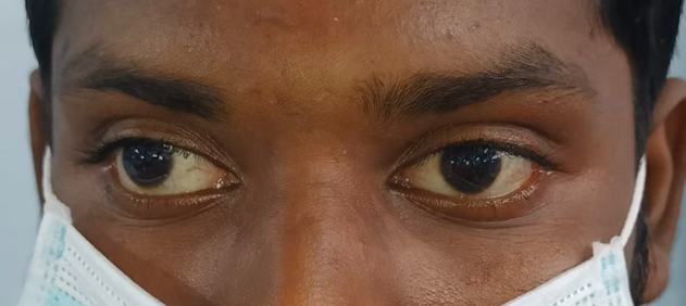
Digital image of the patient depicting an exotropia of approximately 40 degrees in the right eye while fixing with the left eye
Contributed by Dr. Kirandeep Kaur, MBBS, DNB, FPOS, FICO, MRCS Ed, MNAMS
(Click Image to Enlarge)
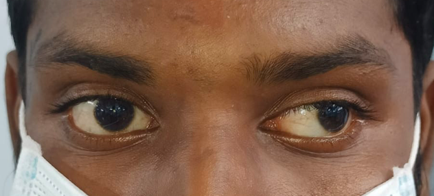
Digital image of the patient depicting an exotropia of approximately 40 degrees while the patient is fixing with his right eye
Contributed by Dr. Kirandeep Kaur, MBBS, DNB, FPOS, FICO, MRCS Ed, MNAMS
(Click Image to Enlarge)
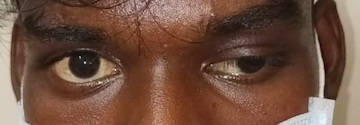
Digital image of the patient depicting left eye upper lid moderate ptosis with an exotropia and hypotropia suggestive of third nerve palsy
Contributed by Dr. Kirandeep Kaur, MBBS, DNB, FPOS, FICO, MRCS Ed, MNAMS
References
Chougule P,Kekunnaya R, Surgical management of intermittent exotropia: do we have an answer for all? BMJ open ophthalmology. 2019; [PubMed PMID: 30997406]
Hatt SR, Gnanaraj L. Interventions for intermittent exotropia. The Cochrane database of systematic reviews. 2013 May 31:2013(5):CD003737. doi: 10.1002/14651858.CD003737.pub3. Epub 2013 May 31 [PubMed PMID: 23728647]
Level 1 (high-level) evidenceHunter DG,Ellis FJ, Prevalence of systemic and ocular disease in infantile exotropia: comparison with infantile esotropia. Ophthalmology. 1999 Oct; [PubMed PMID: 10519591]
Level 1 (high-level) evidenceCiftci S,Simsek A,Dogan E,Ciftci L, Sensory exotropia due to keratoconus and review of the literature. Clinical ophthalmology (Auckland, N.Z.). 2013; [PubMed PMID: 24204108]
Level 3 (low-level) evidenceTaylan Sekeroglu H,Erkan Turan K,Karakaya J,Sener EC,Sanac AS, Clinical risk factors for the development of consecutive exotropia: a comparative clinical study. International journal of ophthalmology. 2016; [PubMed PMID: 27366693]
Level 2 (mid-level) evidenceGovindan M,Mohney BG,Diehl NN,Burke JP, Incidence and types of childhood exotropia: a population-based study. Ophthalmology. 2005 Jan; [PubMed PMID: 15629828]
Level 2 (mid-level) evidenceHashemi H,Pakzad R,Heydarian S,Yekta A,Aghamirsalim M,Shokrollahzadeh F,Khoshhal F,Pakbin M,Ramin S,Khabazkhoob M, Global and regional prevalence of strabismus: a comprehensive systematic review and meta-analysis. Strabismus. 2019 Jun; [PubMed PMID: 31012389]
Level 1 (high-level) evidencePan CW,Zhu H,Yu JJ,Ding H,Bai J,Chen J,Yu RB,Liu H, Epidemiology of Intermittent Exotropia in Preschool Children in China. Optometry and vision science : official publication of the American Academy of Optometry. 2016 Jan; [PubMed PMID: 26583796]
Economides JR,Adams DL,Horton JC, Capturing the Moment of Fusion Loss in Intermittent Exotropia. Ophthalmology. 2017 Apr; [PubMed PMID: 28081943]
Kelkar JA,Gopal S,Shah RB,Kelkar AS, Intermittent exotropia: Surgical treatment strategies. Indian journal of ophthalmology. 2015 Jul; [PubMed PMID: 26458472]
Yao J,Wang X,Ren H,Liu G,Lu P, Ultrastructure of medial rectus muscles in patients with intermittent exotropia. Eye (London, England). 2016 Jan; [PubMed PMID: 26514242]
Kim CZ,Lee SJ, Increased myofiber size and reduced satellite cell numbers in medial rectus muscle of patients with intermittent exotropia. Strabismus. 2020 Dec; [PubMed PMID: 33085552]
O'Dowd C, Evaluating squints in children. Australian family physician. 2013 Dec; [PubMed PMID: 24324989]
Biglan AW,Davis JS,Cheng KP,Pettapiece MC, Infantile exotropia. Journal of pediatric ophthalmology and strabismus. 1996 Mar-Apr; [PubMed PMID: 8965243]
Morrison D,McSwain W,Donahue S, Comparison of sensory outcomes in patients with monofixation versus bifoveal fusion after surgery for intermittent exotropia. Journal of AAPOS : the official publication of the American Association for Pediatric Ophthalmology and Strabismus. 2010 Feb; [PubMed PMID: 20227623]
Level 2 (mid-level) evidenceChoi WJ,Jang Y,Kim SJ,Jung JH, Investigation of transient eye closure evoked with bright light in the patients with intermittent exotropia. BMC ophthalmology. 2021 Jul 31; [PubMed PMID: 34332561]
Audren F, [Intermittent exotropia]. Journal francais d'ophtalmologie. 2019 Nov; [PubMed PMID: 31301849]
Oh BL,Suh SY,Choung HK,Kim SJ, Squinting and photophobia in intermittent exotropia. Optometry and vision science : official publication of the American Academy of Optometry. 2014 May; [PubMed PMID: 24727823]
Level 2 (mid-level) evidenceKushner BJ,Morton GV, Distance/near differences in intermittent exotropia. Archives of ophthalmology (Chicago, Ill. : 1960). 1998 Apr; [PubMed PMID: 9565045]
Haggerty H,Richardson S,Hrisos S,Strong NP,Clarke MP, The Newcastle Control Score: a new method of grading the severity of intermittent distance exotropia. The British journal of ophthalmology. 2004 Feb; [PubMed PMID: 14736781]
Level 2 (mid-level) evidenceChen AM,Erzurum SA,Chandler DL,Hercinovic A,Melia BM,Bhatt AR,Suh DW,Vricella M,Erickson JW,Miller AM,Marsh JD,Bodack MI,Martinson SR,Titelbaum JR,Gray ME,Holtorf HL,Kong L,Kraker RT,Rahmani B,Shah BK,Holmes JM,Cotter SA,Pediatric Eye Disease Investigator Group., Overminus Lens Therapy for Children 3 to 10 Years of Age With Intermittent Exotropia: A Randomized Clinical Trial. JAMA ophthalmology. 2021 Apr 1; [PubMed PMID: 33662112]
Level 1 (high-level) evidencePiano M,O'Connor AR, Conservative management of intermittent distance exotropia: a review. The American orthoptic journal. 2011; [PubMed PMID: 21856878]
Ma MM,Kang Y,Chen C,Su C,Tian Z,Le M, Vision therapy for intermittent exotropia: A case series. Journal of optometry. 2021 Jul-Sep; [PubMed PMID: 32800454]
Level 2 (mid-level) evidencePang Y,Gnanaraj L,Gayleard J,Han G,Hatt SR, Interventions for intermittent exotropia. The Cochrane database of systematic reviews. 2021 Sep 13; [PubMed PMID: 34516656]
Level 1 (high-level) evidenceEscuder AG,Hunter DG, The Role of Botulinum Toxin in the Treatment of Strabismus. Seminars in ophthalmology. 2019; [PubMed PMID: 31177893]
Su H,Fu J,Wu X,Sun A,Zhao B,Hong J, Comparison of Botulinum toxin type A with surgery for the treatment of intermittent exotropia in children. BMC ophthalmology. 2022 Feb 4; [PubMed PMID: 35114960]
Chang JH,Kim HD,Lee JB,Han SH, Supermaximal recession and resection in large-angle sensory exotropia. Korean journal of ophthalmology : KJO. 2011 Apr; [PubMed PMID: 21461229]
Level 3 (low-level) evidenceCeylan OM,Oğuz YG,Ayyıldız Ö,Köksal S,Yumuşak E,Mutlu FM, Surgical management of consecutive exotropia: Long-term outcomes. European journal of ophthalmology. 2021 May; [PubMed PMID: 33426920]
Lavrich JB, Intermittent exotropia: continued controversies and current management. Current opinion in ophthalmology. 2015 Jul; [PubMed PMID: 26204476]
Level 3 (low-level) evidenceAdams GG,McBain H,MacKenzie K,Hancox J,Ezra DG,Newman SP, Is strabismus the only problem? Psychological issues surrounding strabismus surgery. Journal of AAPOS : the official publication of the American Association for Pediatric Ophthalmology and Strabismus. 2016 Oct; [PubMed PMID: 27651232]
Olitsky SE,Coats DK, Complications of Strabismus Surgery. Middle East African journal of ophthalmology. 2015 Jul-Sep; [PubMed PMID: 26180463]