Continuing Education Activity
Total knee arthroplasty (TKA) is one of the most highly effective, cost-efficient, and consistently successful surgical procedures in orthopedics. This procedure provides reliable outcomes for patients suffering from end-stage degenerative knee osteoarthritis. This condition is characterized by severe joint pain, stiffness, and reduced mobility, which can significantly impact a patient's quality of life. Candidates for TKA typically include those who have not responded to conservative treatments such as medications, physical therapy, or injections. TKA aims to alleviate these symptoms and restore function by replacing the damaged knee joint with prosthetic components. This results in improved pain relief and enhanced overall well-being for patients.
This activity comprehensively reviews the indications, contraindications, and techniques pertinent to TKA, emphasizing the crucial role of the interprofessional healthcare team in collaborative patient care throughout this procedure. This activity facilitates a better understanding of the principles and considerations of TKA, thereby enabling healthcare professionals to optimize patient outcomes and enhance patient care and satisfaction. In addition, this activity provides participants with essential knowledge and skills for effectively treating patients with knee osteoarthritis, thereby fostering improved patient care and satisfaction.
Objectives:
Identify appropriate candidates for total knee arthroplasty based on a comprehensive assessment of clinical history, physical examination findings, and radiographic evidence of end-stage degenerative knee osteoarthritis.
Implement evidence-based preoperative optimization strategies, including patient education, medical management, and shared decision-making, to enhance perioperative care and patient satisfaction.
Apply advanced surgical techniques and technologies, such as computer-assisted navigation or patient-specific instrumentation, to enhance the precision and accuracy of total knee arthroplasty procedures.
Collaborate with interdisciplinary healthcare teams to optimize perioperative care pathways and promote optimal functional outcomes and patient satisfaction.
Introduction
Total knee arthroplasty (TKA) is one of the most effective, cost-efficient, and consistently successful surgeries in orthopedics. Patient-reported outcomes underscore its significant impact on pain relief, functional restoration, and improved quality of life. TKA provides reliable outcomes for patients with end-stage, tricompartmental, or degenerative osteoarthritis. Considering osteoarthritis affects millions of people in the United States, the knee bears the brunt of this progressive condition, which is characterized by a gradual degeneration and loss of articular cartilage.
Estimates indicate an annual incidence of symptomatic knee osteoarthritis at 240 per 100,000 patients, with approximately 400,000 primary TKA surgeries performed annually in the United States. Although primary osteoarthritis is the most common clinical diagnosis associated with TKA, other potential underlying diagnoses include inflammatory arthritis, fracture (posttraumatic osteoarthritis or deformity), dysplasia, and malignancy.[1][2][3]
Anatomy and Physiology
The knee comprises 2 distinct joints—the tibiofemoral and patellofemoral joints.[4][5][6] These joints work together to facilitate smooth movement and support weight-bearing activities of the knee.
Patellofemoral Joint
The patellofemoral joint increases the extensor mechanism's lever arm. The patella transmits the tensile forces generated by the quadriceps tendon to the patellar tendon. The maximum contact force between the patella and femoral trochlea occurs at a knee flexion of 45°, and joint reaction forces reach up to 7 times the body weight in the position of deep squatting.
The quadriceps muscles provide dynamic stability to the patellofemoral joint, and passive anatomic restraints include:
- Medial patellofemoral ligament: This is the primary passive restraint against lateral translation at 20° of flexion.
- Medial patellomeniscal ligament: This contributes 10% to 15% of the total restraining force.
- Lateral retinaculum: This provides 10% of the total restraining force.
Tibiofemoral Articulation
The tibiofemoral articulation transfers body weight from the femur to the tibia and generates joint reaction forces of 3 and 4 times body weight during walking and climbing, respectively. Movement primarily occurs in the sagittal plane, ranging from 10° of hyperextension to approximately 140° to 150° of hyperflexion. However, extreme flexion is often limited due to direct contact between the posterior thigh and calf. With increased flexion, the tibiofemoral contact point and femoral center of rotation shift posteriorly to optimize knee flexion before impingement. In normal gait, the required range of motion is up to 75°.
Knee stability in the coronal plane is provided by the lateral collateral ligament, which resists varus stresses, and the medial collateral ligament, which resists valgus stress forces. In addition, the anterior and posterior cruciate ligaments offer resistance to anteriorly and posteriorly directed forces at the knee, respectively. The posterolateral corner structures provide resistance to external rotatory forces.
Indications
Once considered suitable mainly for the older and low-demand patient groups, primary TKA is now offered more frequently and provides consistent positive outcomes, even in younger cohorts of patients. Generally, the most common underlying diagnosis associated with performing TKAs across all patient age groups is primary, end-stage, and tricompartmental osteoarthritis.[7][8][9]
TKA is an elective procedure that is, in most cases, reserved for patients experiencing chronic, debilitating symptoms that continue to persist despite exhaustion of all conservative and nonoperative treatment modalities. Patients usually opt for TKA when their symptoms significantly affect their quality of life and daily activities.
Contraindications
TKA is contraindicated in the following clinical scenarios:
- Local knee infection or sepsis.
- Remote (extra-articular), active, ongoing infection or bacteremia.
- Severe cases of vascular dysfunction, which may compromise healing and recovery.
Equipment
In the 19th century, the reconstruction of articular surfaces involved soft tissue interposition. The evolution of TKA prosthesis designs began in the 1950s with Walldius' creation of the first hinged-knee replacement. In 1958, MacIntosh and McKeever proposed the first acrylic tibial plateau prosthesis. Then, in the 1960s, Gunston introduced the first cemented surface arthroplasty of the knee. In the early 1970s, the total condylar prosthesis emerged as the first TKA prosthesis designed to resurface all 3 knee compartments, featuring a posterior-stabilized design. The 4 main categories of TKA prosthesis designs, sorted by increasing levels of constraint by design, are outlined below.[10][11][12]
Cruciate-Retaining
The cruciate-retaining TKA prosthesis relies on an intact posterior cruciate ligament to provide stability in flexion, making it unsuitable for patients with preexisting or intraoperatively recognized posterior cruciate ligament insufficiency. Caution is warranted with patients exhibiting at least moderate instability in any plane of motion, especially those with instability in posterolateral corner structures. Instability in posterolateral corner structures instability predisposes the native posterior cruciate ligament in a cruciate-retaining TKA to abnormally high stresses and forces, ultimately leading to early failure and needing revision. Therefore, cruciate-retaining TKA is contraindicated in patients with inflammatory arthritic conditions due to the increased risk of early posterior cruciate ligament attenuation, such as rheumatoid arthritis.
Proposed advantages of the cruciate-retaining TKA design include:
- Prevention of tibial post-cam impingement and dislocation.
- Resemblance of more normal knee kinematics and anatomy, theoretically.
- Preservation of bone stock (less distal femur resected compared to posterior-stabilized TKA prosthesis).
- Retention of native posterior cruciate ligament proprioception.
Proposed disadvantages of the cruciate-retaining TKA design include:
- A tight posterior cruciate ligament that leads to early or accelerated polyethylene wear.
- A loose or ruptured posterior cruciate ligament that results in flexion instability and possible subluxation or dislocation.
Multiple meta-analyses have demonstrated satisfactory survivorship and similar outcomes when comparing the cruciate-retaining and posterior-stabilized TKA prosthesis designs. Additionally, cruciate-retaining TKA may be preferred in patients with higher functional demands and those involved in activities requiring an increased range of motion.
Posterior-Stabilized
The posterior-stabilized TKA design is slightly more constrained and requires the surgeon to sacrifice the posterior cruciate ligament. The femoral component contains a cam designed to engage with the tibial polyethylene post during knee flexion.
Proposed advantages of the posterior-stabilized TKA design include:
- Overall knee balancing in the setting of an absent posterior cruciate ligament.
- Better knee flexion, theoretically.
- Lower ranges of axial rotation and condylar translation
Proposed disadvantages of the posterior-stabilized TKA design include:
- Potential cam jump due to a loose flexion gap or knee hyperextension.
- Risk of patellar clunk syndrome.
- Possibility of tibial post-wear or fracture.
Constrained Nonhinged Design
The constrained nonhinged prosthesis incorporates a more extensive tibial post and deeper femoral box, providing increased stability and constraint (within 2° to 3°) in both varus-valgus and internal-external rotatory planes. Indications include collateral ligament attenuation or deficiency, flexion gap laxity, and moderate bone loss in neuropathic arthropathy. However, the drawbacks of this design include an increased risk of earlier aseptic loosening due to increased intercomponent constraint and the need for additional femoral bone resection to accommodate the components.
Constrained Hinged Design
The constrained hinged design consists of linked femoral and tibial components. Rotating hinge options allow the tibial bearing to rotate around a yoke, theoretically mitigating the risk of aseptic loosening but at the expense of increasing levels of prosthetic constraint. Indications include global ligamentous deficiencies, resections in the setting of tumors, and massive bone loss in neuropathic joint conditions.
Other Component Considerations
Modularity and mobile bearing designs are other noteworthy additional prosthetic design considerations. Mobile bearing designs allow polyethylene rotation on the tibial baseplate. Although this design concept remains controversial in generating reproducibly superior patient-reported outcome measures, proponents advocate for its use and specific indications in younger patient demographics due to enhanced wear rates. However, a notable disadvantage includes the potential for bearing spin-out, particularly in a loose flexion gap.
All-polyethylene tibial base plates differ from the traditional metal tray with polyethylene inserts (ie, tibial component modularity), allowing surgeons more flexibility in intraoperative adjustments for fine-tuning TKA stability. Surgeons can adjust the polyethylene size (ie, upsize or downsize) after final tibial implant fixation between the metal implant and cement (or bone) interfaces. This allows for a final check and balance step, which many TKA surgeons appreciate. In contrast, proponents of all-polyethylene base plates highlight significant cost savings and reduced rates of osteolysis in TKA cohorts, particularly among older patients undergoing TKA.[13][14]
Preparation
Preparation for TKA is crucial for achieving optimal outcomes in patients undergoing this procedure. This involves a comprehensive evaluation of the patient's medical history, physical condition, and readiness for surgery. Preoperative assessments aim to identify and address potential risk factors or medical conditions that could affect the surgical outcome or recovery process. Additionally, preparation for TKA involves patient education, optimizing medical management, and planning for postoperative care.
Nonoperative Treatment Modalities
According to the 2011 American Academy of Orthopaedic Surgeons (AAOS), evidence-based clinical guidelines for the treatment of symptomatic hip or knee osteoarthritis, strong or moderately strong recommendations for nonoperative treatment modalities include weight loss, physical activity, physical therapy programs, and nonsteroidal anti-inflammatory drugs (NSAIDs) and tramadol. Although other modalities lack moderate or strong evidence support, they are often considered reasonable alternative treatment options. These include but are not limited to acupuncture, chondroitin supplementation, hyaluronic acid injections, corticosteroid injections, lateral wedge insoles, and offloading braces.[15]
Preoperative Evaluation
Clinical examination: A comprehensive history and physical examination are required before performing a TKA on any patient. Patients should be questioned about previous interventions and treatments, including joint replacements, arthroscopic procedures, or other knee surgeries. Surgical scars from previous procedures should be considered, as they may affect the planned surgical approach. Patients with prior injuries or procedures may also exhibit mechanical axis deformities, retained hardware, or knee instability in various planes. These factors can significantly influence the selection of the most appropriate TKA prosthesis for the patient.
Every patient undergoing elective TKA surgery should receive a comprehensive medical evaluation with any appropriate medical optimization tests performed before the TKA procedure. Surgeons must consider the risks and potential benefits of performing TKA on a case-by-case basis.
During the physical examination, assessing the overall mechanical axis of the limb is crucial. Notably, it is essential to rule out or at least consider hip pathology before conducting any knee surgery. In addition, the vascular status of the limb should also be assessed by observing the skin for any chronic venous stasis changes, cellulitis, or even wounds or ulcerations that may be present on the extremity. Distally, ensuring symmetric and palpable pulses is essential.
In the preoperative setting, any patient presenting with peripheral vascular disease should be considered for consulting a vascular surgeon. Surgeons should also be aware of the potential for peripheral vascular disease to present as knee pain out of proportion in the setting of relatively benign radiographs.
Before surgery, the preoperative range of motion should be assessed at the knee and adjacent joints (hips and ankles). Soft tissues should be examined for signs of gross atrophy, overall symmetry, and ligamentous stability in all planes of the knee joint. In addition, it is crucial to document the presence of any laxity in the varus or valgus plane and the ability to correct deformities. These parameters help the surgeon anticipate any necessary soft tissue releases to facilitate mechanical axis correction and plan for potential additional bone resection in cases of significant contractures.
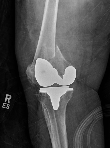
Radiographs: Preoperative radiographs, including a weight-bearing anteroposterior view, are evaluated for overall mechanical alignment, the presence of deformity, and bone loss (see Image 1. Anteroposterior View of a Periprosthetic Distal Femur Fracture). The tibiofemoral angle can help estimate the magnitude of coronal deformity. The femoral resection angle is calculated as the difference between the mechanical and anatomic axis of the femur. In addition, the lateral view of the knee is essential for appreciating the native posterior slope of the proximal tibia as well as the presence of posterior osteophytes on the femoral condyles (see Image 2. Lateral View of a Periprosthetic Distal Femur Fracture).
Although the patellofemoral radiographic view is unnecessary for TKA templating, it allows surgeons to evaluate the magnitude of patellofemoral arthritis and deformity. In cases of advanced patellofemoral deformity, osteophyte removal may be necessary before attempting to evert the patella during the procedure. In addition, a surgeon can plan for a possible lateral release to improve patellar tracking.
Technique or Treatment
TKA involves replacing damaged or diseased knee components with artificial parts to alleviate pain and enhance function. The technique has evolved significantly, with advancements in surgical approaches, implant designs, and perioperative care.
Surgical Approaches
The most common approaches for the standard primary TKA procedure include the medial parapatellar, midvastus, and subvastus approaches. The medial parapatellar approach is commonly utilized and entails proximal dissection through a medial cuff of the quadriceps tendon to facilitate superior tissue quality closure after the procedure. Distally, a meticulous, continuous medial subperiosteal dissection sleeve is performed while maintaining close proximity to the proximal tibial bone. The extent of dissection is typically determined by the anticipated degree of deformity correction needed. This medial release is generally more aggressive in cases of severe varus deformity and less extensive in cases of moderate to advanced valgus knee deformity. Additionally, the medial meniscus is resected along with this sleeve of soft tissue.[16][17][18]
Alternatives to the standard medial parapatellar arthrotomy include the midvastus and subvastus approaches. In the midvastus approach, the quadriceps tendon is spared, and dissection is directed towards the superomedial aspect of the proximal pole of the patella, preserving the vastus medialis obliquus muscle belly. The subvastus approach also spares the quadriceps tendon and lifts the muscle belly of the vastus medialis obliquus off the intermuscular septum. While it maintains the patella's vascularity, caution is warranted as it may restrict exposure in challenging cases or in patients with obesity.
Procedural Steps
The specific order of bone resections and soft tissue releases may vary based on surgeon preference. However, this technical summary aims to provide a general overview of the preferred method for the TKA procedure.
Once the arthrotomy is completed, the patella is everted, and the knee is flexed to enable additional soft tissue releases before achieving knee dislocation. If the surgeon opts to proceed with the femur first, an intramedullary drill is used to access the femoral canal for the insertion of a distal femoral intramedullary jig. The angle set on the guide is determined by the patient-specific preoperative evaluation (anteroposterior x-ray), typically resulting in 5° or 7° of valgus. Although system-specific, most surgeons prefer resecting 9 to 10 mm of the distal femur.
Next, the proximal tibia is cut utilizing either an intramedullary or extramedullary guide to cut the bone perpendicular to the tibial axis (or within 2° to 3° of varus for surgeons aiming for an "anatomic" TKA procedure). Preferably, an intramedullary guide and a perpendicular tibial cut are utilized. Rotation is set by referencing the medial one-third of the tibial tubercle (proximally) and a point slightly medial to the center of the ankle joint (distally). This alignment is also cross-referenced with the second ray of the foot and the tibial crest.
After performing the tibial cut, the extension gap is assessed. A spacer block is then inserted with the knee in full extension, and the overall balance of the knee is evaluated using an alignment rod to facilitate and confirm the achieved varus-valgus and tibial slope parameters.
Next, the flexion gap is attained by using an anteroposterior sizing guide positioned with respect to the bony landmarks on the femur (usually the Whiteside line or the native transepicondylar axis). Depending on the surgeon's preference for anterior or posterior referencing, the flexion gap is established and fine-tuned using system-specific incremental sizing adjustments available with the cutting guides. In addition, it is essential to visualize the flexion gap before making the bony cuts and to ensure proper soft tissue balancing.
A spacer block can facilitate this assessment. A surgeon must verify that a rectangular flexion gap will be achieved after the bone resections. Once satisfactory checks and balancing steps are confirmed, the anterior, posterior, and anterior and posterior chamfer cuts are executed. Care must be taken to protect the collateral soft tissue structures, such as the lateral collateral ligament and medial collateral ligament, using retractors.
Following that, the intercondylar notch cut is performed perpendicular to the transepicondylar axis. Attention is then directed back to the proximal tibia to complete preparation, sizing, and rotational alignment. Care must be taken to avoid internal rotation and component overhang, as these can result in suboptimal TKA outcomes. Subsequently, the femoral and tibial trial implants are impacted, and a provisional spacer trial is inserted. The knee is reduced and evaluated for stability from 0° of extension through mid-flexion stability.
When considering patellar resurfacing, it is advisable to perform the resection after carefully assessing the native anatomy and size of the patellofemoral joint. Inferior TKA outcomes can result from either over-resection, which can compromise implant bone stock and lead to patella fracture, or under-resection, resulting in chronic postoperative pain due to an overstuffed patellofemoral joint.
Finally, stability parameters are reassessed, and patellar tracking is evaluated, ensuring that intraoperative tracking tests are successfully completed. Surgeons commonly use either a natural range of motion tracking test or a towel clip technique to confirm that the TKA meets the criteria of the "no thumb" test.
Patellar maltracking, most commonly occurring laterally, can be addressed through a standard lateral release. However, in more severe cases or scenarios consistent with component malalignment, correcting component position(s) should be considered.
Hot Topics in TKA Techniques
Gap balancing versus measured resection: Achieving a well-balanced symmetric flexion and extension gap is paramount in TKA.[19] Precise bony cuts and accurate soft tissue balancing determine femoral component rotation.[20] Malrotation of the femoral component may result in anterior knee pain, patellofemoral joint instability, flexion gap instability, or arthrofibrosis.[21][22]
Gap balancing: Gap balancing involves ligament releases to correct any fixed deformities and align the limb to the most approximate correct alignment before making bony cuts. There are 2 approaches to gap balancing that are commonly used—flexion gap first or extension gap first—followed by balancing the flexion gap based on a balanced extension gap.[23]
Better knee flexion stability can be achieved with the gap balancing technique that creates a rectangular flexion gap. Additionally, this technique is considered more reliable than measured bone resection because it is independent of the transepicondylar or anteroposterior axes, which can vary and be inconsistent to determine.[24][25]
However, gap balancing requires precise accuracy in the proximal tibial cut. A varus tibial cut can cause internal rotation of the femoral component, while a valgus tibial cut can cause external rotation of the femoral component. Moreover, an imbalance between the flexion and extension gaps can occur due to over- or under-resection of the femoral or tibial bone. Moreover, precise ligament balancing is crucial. If the superficial medial collateral ligament is deficient, tensioning the medial flexion gap can lead to an excessive medial flexion gap, resulting in excessive internal rotation of the femoral component. Conversely, if the lateral collateral ligament is deficient, tensioning the lateral flexion gap can result in an excessive lateral flexion gap, leading to excessive external rotation of the femoral component.[23]
Measured resection technique: Measured resection technique involves creating bone cuts independently of soft tissue tension. In this technique, bony landmarks are used to set the femoral component rotation, such as the transepicondylar axis,[26] the anteroposterior axis,[27][28] and the posterior condylar axis. However, determining the transepicondylar axis intraoperatively has shown inconsistency in multiple studies.[26][29][30]
Anterior versus posterior referencing: The correct positioning and sizing of the femoral component ensure proper kinematic function in TKA. Anterior and posterior referencing are the 2 main strategies for setting the center of rotation and sagittal plane balancing in TKA, based on the reference used to determine the size and geometry of distal femur resection.[31] In anterior referencing, the anterior femoral cortex is the reference, whereas the resection of the posterior femoral condyle varies, posing challenges in flexion space balancing. Anterior referencing helps reduce the risk of AFC notching and patellofemoral joint overstuffing. However, it increases the risks of flexion gap instability with excessive posterior condylar resection or femoral offset reduction.[32]
In posterior referencing, the posterior femoral condyles are the reference, whereas the anterior femoral cut varies. This approach ensures precision in resecting the exact thickness from the posterior femoral condyles, maintaining posterior femoral offset and enabling deep knee flexion. However, this technique increases the risk of anterior femoral notching or overstuffing of the patellofemoral joint.[31]
Mobile bearing versus fixed bearing: The design of the polyethylene inserts in TKA has been a subject of debate in the literature.[33] The 2 types of inserts include:
- Fixed bearing inserts: These inserts are rigidly fixed with the tibial component and have been shown to offer satisfactory outcomes and long survivorship.[34][35][36][37] However, implant loosening is believed to be due to high contact stresses and polyethylene wear rates.[38][39]
- Mobile bearing inserts: These inserts simulate native knee kinematics by providing better conformity, lower contact stresses, and lower implant loosening rates.[40] However, these inserts carry the unique risk of bearing dislocation.[41]
The literature does not support a specific design over another.[42][34]
Wound Closure
The recent literature concerning the optimal knee position and suture material for closure following TKA remains controversial. Attention to detail is crucial, and a systematic closure is unanimously advocated. A preferred method includes closure with uni- or bi-directional barbed sutures for the arthrotomy, deep fascial, and deep dermal or subcutaneous layers. Staples or monofilament absorbable sutures can be used for the skin. A sterile dressing is applied and left in place without being changed for the first 7 days. In addition, a minimal cotton or ace soft wrap dressing is applied to the knee for at most 24 hours to facilitate the appropriate balance between wound healing and postoperative knee movement.
Other Considerations
Topical tranexamic acid is the preferred application while the cement is fully hardened before the tourniquet is dropped. Other controversial technical modalities in TKA include using a tourniquet and cementing the patella, femoral, and tibial components, as well as incorporating a betadine soak to the wound as part of the copious saline irrigation that is applied before the closure of the arthrotomy and surgical wound. However, preferred techniques involve using a tourniquet, cementing all components, and employing copious pulsatile saline irrigation before closing the arthrotomy.
Complications
Complications arising from TKA can lead to inferior outcomes and patient-reported satisfaction scores. Despite being a reliable and consistently successful procedure for individuals with severe degenerative arthritis, studies indicate that as many as 1 in 5 patients undergoing primary TKA may express dissatisfaction with the results.[43][44][45]
Periprosthetic Fracture
Periprosthetic fractures in TKA are further characterized by the location and residual stability of the implants. These fractures occur in the distal femur at a rate of 1% to 2%, with risk factors including compromised bone quality in patients and the use of more constrained TKA components. Although controversial, anterior femoral notching is a potential risk factor for postoperative fracture.
Tibial periprosthetic fractures occur at a rate of 0.5% to 1%, with risk factors including prior tibial tubercle osteotomy, component malposition or loosening, and the use of long-stemmed components. In unresurfaced TKA cases, patellar periprosthetic fractures occur less frequently, with incidence rates ranging from 0.2% to 15% or 20%. Risk factors for patellar fracture include osteonecrosis, technical errors in asymmetric or over-resection, and implant-related factors such as central, single-peg implants, uncemented fixation, and metal-backed components.
Aseptic Loosening
TKA aseptic loosening occurs secondary to a macrophage-induced inflammatory response, resulting in eventual bone loss and TKA component loosening. Patients typically experience increased pain during weight-bearing activities or recurrent effusions. Pain at rest or during range of motion may be minimal. Serial imaging and infectious labs are required to work up these conditions appropriately. If symptoms persist, revision surgery may be necessary, and the patient is evaluated as a potential candidate for surgery. Aseptic loosening involves particulate debris formation, macrophage-induced osteolysis, micromotion of components, and dissemination of particulate debris.
Wound Complications
The spectrum of TKA postoperative wound complications ranges from superficial surgical infections, such as cellulitis, superficial dehiscence, or delayed wound healing, to deep infections causing full-thickness necrosis. In severe cases, patients may require returns to the operating room for irrigation, debridement (incision and drainage), and rotational flap coverage.
Periprosthetic Joint Infection
The reported incidence of prosthetic total knee infection (TKA PJI) following primary TKA is approximately 1% to 2%. Risk factors include patient-specific lifestyle factors such as morbid obesity, smoking, intravenous drug use and abuse, alcohol abuse, and poor oral hygiene, as well as past medical history factors such as uncontrolled diabetes, chronic renal disease, liver disease, malnutrition, and HIV (CD4 counts less than 400). PJI represents the most common reason for revision surgery.
The most common bacterial organisms implicated in the acute setting include Staphylococcus aureus, S epidermidis, and coagulase-negative staphylococci in chronic TKA PJI cases. Treatment in the acute setting, defined as occurring within 3 weeks after the index surgery, may involve interventions such as incision and drainage, polyethylene exchange, and component retention. Intravenous antibiotics are typically administered for a duration of 4 to 6 weeks. Successful outcomes vary and are often influenced by intraoperative and patient-related factors, as well as the specific bacterial pathogens involved, with studies reporting a success rate of around 55%.
More aggressive treatments may be necessary in cases beyond the acute presentation (3-4 weeks after surgery). These include a 1- or 2-stage revision TKA procedure with interval antibiotic spacer placement. The surgeon must confirm and document evidence of infection eradication.
Other Complications and Considerations
Other potential complications after TKA extend beyond the scope of this review but include:
- TKA instability: This can occur in the coronal or sagittal plane(s). Additionally, when patients report persistent anterior knee pain in the postoperative setting, patellar maltracking or other patellofemoral joint issues (eg, joint overstuffing) are considered.
- Extensor mechanism disruption or rupture
- Patellar clunk syndrome: This condition often occurs 12 months after TKA and is characterized by popping and catching sensations during knee extension. Patellar clunk syndrome arises from nodule formation on the posterior quad tendon near its insertion on the patella and is often associated with posterior stabilized knee designs. Although the exact cause of scar tissue formation remains unclear, pain ensues from tissue entrapment in the intercondylar notch. Surgical intervention, either arthroscopic or open debridement/synovectomy, is the primary treatment, as conservative measures are usually ineffective. Physical therapy may aid in quad strengthening post-surgery but is not curative. Recurrence after surgical intervention is rare, and more aggressive measures, such as revision TKA, are often not warranted without component malposition.
- Vascular injury and bleeding
- Peroneal nerve palsy: This condition is one of the most common complications following TKA, especially when correcting valgus deformity. When balancing the soft tissues of a valgus knee, the iliotibial band primarily affects the extension space more than the flexion space and inserts on the Gerdy tubercle. Conversely, the popliteus predominantly affects the flexion space more than the extension space.
Clinical Significance
TKA is one of the most successful and cost-effective procedures in all orthopedics, primarily benefiting patients with debilitating, end-stage arthritic conditions of the knee. Once considered a procedure suitable only for older, low-demand patients, TKA is now gaining popularity among younger populations. Between 1991 and 2010, the annual primary TKA volume in the US Medicare population alone increased by 161.5%, from more than 93,000 to more than 226,000 cases.
Enhancing Healthcare Team Outcomes
After total knee replacement, an interprofessional healthcare team is necessary to prevent and manage the comorbidities. Due to the frailty or cognitive impairment of many patients, a dietary consultation is necessary. Additionally, a pain consultation is vital before rehabilitation. However, the chosen analgesic must balance pain control and prevent sedation. Furthermore, long-term pain control is not recommended as it may mask signs of fever, infection, or evidence of vascular compromise.
Constipation, a prevalent issue in this group of populations, needs to be properly addressed. Pharmacists should collaborate with clinicians in selecting and managing appropriate pain medications. The healthcare team, comprising physicians, advanced practitioners, and nurses, must ensure patients receive prophylaxis for deep vein thrombosis.
Finally, orthopedic nurses should educate patients to avoid crossing their legs, turning their legs inward, or bending over to reach objects. Additionally, nurses should aid the clinician in providing close follow-up and reporting any concerns about complications. Therapists should educate patients on using assistive devices if their gait is unbalanced. Follow-up with these patients is necessary until they can ambulate independently.[46][47][48]
Outcomes
Although recent reports indicate that up to 1 in 5 patients may remain dissatisfied following TKA, the risks of patient-reported dissatisfaction can be reduced by selectively operating on patients with other potentially relevant clinical pathologies, such as hip or back conditions ruled out as confounding pain generators. Success following TKA leads to significant improvements in patient-reported pain and functional outcome scores in both short- and long-term postoperative periods.
While various patient-related and prosthetic technical factors influence the overall longevity of the TKA prosthesis, typically, its lifespan is expected to be around 15 to 20 years. Clinicians are advised to ensure that surgical candidates have explored all nonoperative treatment modalities. With the rising rates of surgical procedures in both young and older populations, orthopedic surgeons can anticipate excellent outcomes in appropriately selected patient populations.[49][50]