Neurofibromatosis Type 2

Earn CME/CE in your profession:


Continuing Education Activity

Neurofibromatosis type 2 (NF2) is an autosomal dominant disorder characterized by the development of multiple tumors involving the central nervous system (CNS). Bilateral vestibular schwannomas are the hallmark feature of NF2 and are present in approximately 90 to 95 percent of patients. Meningiomas are seen in approximately 50 percent of patients with NF2. This activity reviews the presentation, evaluation, and management of NF2 and highlights the role of the interprofessional team in the care of these patients.

Objectives:

  • Identify the etiology of neurofibromatosis type 2.

  • Evaluate the typical presentation of a patient with neurofibromatosis type 2.

  • Assess the treatment and management options available for neurofibromatosis type 2.

  • Communicate interprofessional team strategies for improving care coordination and communication to advance the treatment of neurofibromatosis type 2 and improve patient outcomes.

Introduction

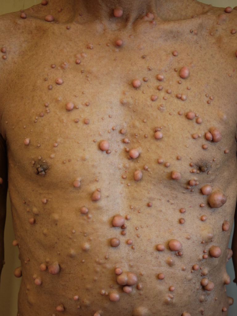
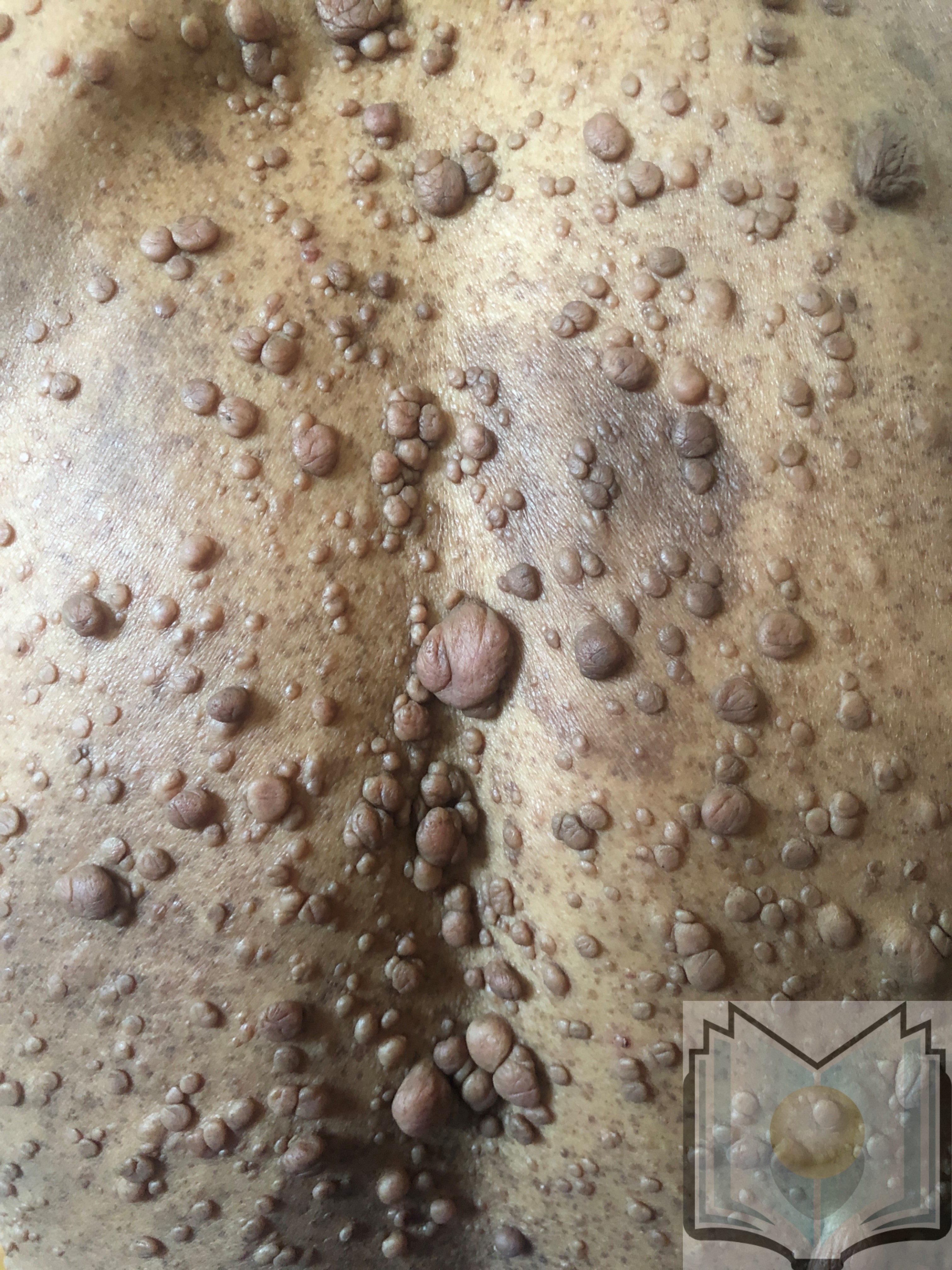
Neurofibromatosis type 2 (NF2) is an autosomal dominant disorder characterized by the development of multiple tumors involving the central nervous system (CNS).[1] See Image. Neurofibromatosis.

Etiology

NF2 is inherited as an autosomal dominant trait in some patients. The abnormal gene can be inherited from either of the parents, and the risk of passing the gene to offspring from a parent is 50%. There is no family history in some patients with NF2; the disease is caused by a de novo mutation in the NF2 gene. NF2 is caused by mutations in the NF2 gene located in the long arm of chromosome number 22 (22q12.2). The NF2 gene encodes for the protein known as merlin, which acts as a tumor suppressor gene.[2] Merlin is found in the Schwann cells in the nervous system.[3]

Epidemiology

The incidence of NF2 is about 1 in 25,000 to 40,000 individuals.

Pathophysiology

Bilateral vestibular schwannomas are the hallmark feature of NF2 and are present in approximately 90% to 95% of patients. Meningiomas are seen in approximately 50% of patients with NF2.

Histopathology

Vestibular schwannomas are benign tumors involving the vestibular portion of the vestibulocochlear nerve (CN VIII). The most common location is an inferior vestibular nerve. They are well-circumscribed encapsulated tumors arising from the perineural elements of the Schwann cells. They usually cause splaying and displacement of the nerve fibers rather than engagement. Microscopically, 2 types of cellular architecture can be seen in these tumors: Antoni A and B.[4] Antoni A-type regions have dense cellularity with closely packed elongated nuclei alternated with clear zones devoid of nuclei (Verocay bodies). Antoni B-type regions are less cellular and contain more loosely arranged cells. The volume of Antoni B regions in any given tumor is variable and may be absent.

History and Physical

NF2 patients typically present with tumor-related symptoms around 20 years of age. The most common intracranial tumor associated with NF2 is vestibular schwannoma, which is typically bilateral in these patients. Patients typically present with tinnitus, sensorineural hearing loss, and balance problems. The cutaneous features in NF2 are less common and more subtle than NF1. The most common skin finding is an elevated plaque-like lesion, which may be hyperpigmented compared to the surrounding skin. The other cutaneous abnormalities are subcutaneous nodules representing swelling of the nerves and cutaneous tumors, which generally represent schwannomas rather than neurofibromas. Patients with meningiomas develop symptoms based on location, including a headache, seizures, or focal neurological symptoms. NF2 patients tend to develop meningiomas earlier than those with sporadic meningiomas. Approximately 20% of children with meningiomas have NF2. Patients with intraspinal tumors present with pain, muscular weakness, and paresthesia.

The clinical diagnosis of NF2 is based on the presence of any 1 of the following criteria:

  • Bilateral vestibular schwannomas less than 70 years of age
  • Unilateral vestibular schwannoma before age 70 years and a first-degree relative with NF2
  • Any 2 of the following: meningioma, schwannoma (non-vestibular), neurofibroma, glioma, cerebral calcification, cataract AND first-degree relative to NF2 OR unilateral vestibular schwannoma and negative LZTR1 testing
  • Multiple meningiomas and unilateral vestibular schwannoma or any 2 of the following: schwannoma (non-vestibular), neurofibroma, glioma, cerebral calcification, cataract
  • Constitutional or mosaic pathogenic NF2 gene mutation from the blood or by the identification of an identical mutation from 2 separate tumors in the same individual.[5]

Evaluation

Detailed clinical and family histories are required from the patients suspected of having NF2. A complete physical examination should be done to look for cutaneous schwannomas or plaque-like lesions, and an ophthalmic examination should be done to look for cataracts and optic nerve, among other things. Contrast-enhanced MRI of the brain and entire spine is recommended to evaluate schwannomas and meningiomas.

Vestibular schwannoma has characteristic features in magnetic resonance imaging (MRI). It is located in the internal auditory canal, which is often widened. Large tumors also extend into the cerebellopontine angle and cause the typical "ice cream cone" appearance. These lesions are hypointense on T1-weighted images and hyperintense on T2-weighted images with intense contrast enhancement. Cystic changes can be seen in large tumors. There is a poor correlation between the tumor size and the degree of hearing loss.

Meningiomas are the most common extra-axial tumors in the brain. NF2 patients develop meningiomas at an earlier age, and they are often multiple. If meningiomas are seen in the pediatric age group, then a diagnosis of  NF2 should be considered. Meningiomas have typical features on the MRI. They are dural-based lesions with the isointense signal on T1-weighted images and iso to the hyperintense signal on T2-weighted images with intense enhancement and an enhancing dural tail.[6]

Multiple spinal tumors can also be seen in patients with NF2, including schwannomas, meningiomas, and ependymomas, and can be diagnosed with MRI. Schwannomas are the most common type; they typically arise from the dorsal root with the typical appearance of a dumbbell shape with the widening of the neural foramen and intense enhancement. Meningiomas enhance extramedullary lesions, typically seen in the cervical or thoracic region. Ependymomas are intramedullary lesions causing enlargement of the spinal cord with hemorrhage, cystic changes, and variable enhancement.[7]

Treatment / Management

Multidisciplinary management is required in patients with NF2, including oncologists, neurologists, neuroradiologists, ophthalmologists, geneticists, and neurosurgeons. For tumor surveillance, an annual brain MRI is recommended. If no brain tumor is seen on the initial imaging, then MRI can be done every 2 years. If the tumor is seen, an MRI should be done twice in the first year, followed by an annual follow-up.

Small asymptomatic vestibular schwannomas can be managed conservatively with MRI follow-up. Surgery is the primary treatment for large symptomatic vestibular schwannomas. Meningiomas are also treated surgically, with radiation treatment reserved for non-surgical candidates.[8] Spinal cord ependymomas are usually low-grade tumors and can be followed clinically, with surgery reserved for symptomatic patients.

Bevacizumab, a monoclonal antibody against vascular endothelial growth factor (VEGF), is a new systemic immunotherapy for many tumors. Some studies have shown tumor regression and hearing improvement in NF2 patients treated with bevacizumab.[8]

Differential Diagnosis

NF2 should be differentiated from Schwannomatosis, another neurofibromatosis genetically distinct from NF1 and NF2 (see Image. Neurofibromatosis, NF1, NF2, and Schwannomatosis). Schwannomatosis is most frequently sporadic, with 20% of cases being familial. Schwannomatosis is characterized by developing multiple schwannomas in the peripheral nervous system without concomitant involvement of the vestibular nerves. Nonvestibular cranial nerve schwannomas are uncommon but can be seen in these patients.[9]

Prognosis

The outlook for these patients is guarded. If the tumors are detected late, the outcomes are poor, and the quality of life is reduced.

Complications

Complications can occur depending on the location of the tumor and the proximity to vital parts of the brain. A few of them include:

  • Weakness
  • Hydrocephalus
  • Cranial nerve deficits

Deterrence and Patient Education

Proper education should be given to the patient about the disease, the likely complications, and the difficulty of offering a complete cure. Genetic counseling before conception would benefit.

Enhancing Healthcare Team Outcomes

Interprofessional management is required in patients with NF2, including oncologists, neurologists, neuroradiologists, ophthalmologists, geneticists, nurse practitioners, and neurosurgeons. For tumor surveillance, an annual brain MRI is recommended. If no brain tumor is seen on the initial imaging, then MRI can be done every 2 years. If the tumor is seen, then MRI should be done twice in the first year with an annual follow-up after that.


(Click Image to Enlarge)
<p>Neurofibromatosis, NF1, NF2, and Schwannomatosis

Neurofibromatosis, NF1, NF2, and Schwannomatosis. Neurofibromatosis encompasses three distinct disorders: neurofibromatosis type 1 (NF1), neurofibromatosis type 2 (NF2), and schwannomatosis. 


DermNet New Zealand


(Click Image to Enlarge)
<p>Neurofibromatosis</p>

Neurofibromatosis


Contributed by S Verma, MBBS, DVD, FRCP, FAAD

Details

Author

Ruchi Tiwari

Editor:

Achint K. Singh

Updated:

10/3/2022 8:44:45 PM

Looking for an easier read?

Click here for a simplified version

References


[1]

Dinh CT, Nisenbaum E, Chyou D, Misztal C, Yan D, Mittal R, Young J, Tekin M, Telischi F, Fernandez-Valle C, Liu XZ. Genomics, Epigenetics, and Hearing Loss in Neurofibromatosis Type 2. Otology & neurotology : official publication of the American Otological Society, American Neurotology Society [and] European Academy of Otology and Neurotology. 2020 Jun:41(5):e529-e537. doi: 10.1097/MAO.0000000000002613. Epub     [PubMed PMID: 32150022]


[2]

Gürsoy S, Erçal D. Genetic Evaluation of Common Neurocutaneous Syndromes. Pediatric neurology. 2018 Dec:89():3-10. doi: 10.1016/j.pediatrneurol.2018.08.006. Epub 2018 Aug 10     [PubMed PMID: 30424961]


[3]

Mota M, Shevde LA. Merlin regulates signaling events at the nexus of development and cancer. Cell communication and signaling : CCS. 2020 Apr 16:18(1):63. doi: 10.1186/s12964-020-00544-7. Epub 2020 Apr 16     [PubMed PMID: 32299434]


[4]

Neuropathology for the neuroradiologist: Antoni A and Antoni B tissue patterns., Wippold FJ 2nd,Lubner M,Perrin RJ,Lämmle M,Perry A,, AJNR. American journal of neuroradiology, 2007 Oct     [PubMed PMID: 17893219]


[5]

Smith MJ, Bowers NL, Bulman M, Gokhale C, Wallace AJ, King AT, Lloyd SK, Rutherford SA, Hammerbeck-Ward CL, Freeman SR, Evans DG. Revisiting neurofibromatosis type 2 diagnostic criteria to exclude LZTR1-related schwannomatosis. Neurology. 2017 Jan 3:88(1):87-92. doi: 10.1212/WNL.0000000000003418. Epub 2016 Nov 16     [PubMed PMID: 27856782]


[6]

Li P, Wu T, Wang Y, Zhao F, Wang Z, Wang X, Wang B, Yang Z, Liu P. Clinical features of newly developed NF2 intracranial meningiomas through comparative analysis of pediatric and adult patients. Clinical neurology and neurosurgery. 2020 Jul:194():105799. doi: 10.1016/j.clineuro.2020.105799. Epub 2020 Mar 19     [PubMed PMID: 32229353]

Level 2 (mid-level) evidence

[7]

Kalamarides M, Essayed W, Lejeune JP, Aboukais R, Sterkers O, Bernardeschi D, Peyre M, Lloyd SK, Freeman S, Hammerbeck-Ward C, Kellett M, Rutherford SA, Evans DG, Pathmanaban O, King AT. Spinal ependymomas in NF2: a surgical disease? Journal of neuro-oncology. 2018 Feb:136(3):605-611. doi: 10.1007/s11060-017-2690-7. Epub 2017 Nov 29     [PubMed PMID: 29188529]


[8]

Mautner VF, Nguyen R, Kutta H, Fuensterer C, Bokemeyer C, Hagel C, Friedrich RE, Panse J. Bevacizumab induces regression of vestibular schwannomas in patients with neurofibromatosis type 2. Neuro-oncology. 2010 Jan:12(1):14-8. doi: 10.1093/neuonc/nop010. Epub 2009 Oct 20     [PubMed PMID: 20150363]


[9]

Ferner RE, Bakker A, Elgersma Y, Evans DGR, Giovannini M, Legius E, Lloyd A, Messiaen LM, Plotkin S, Reilly KM, Schindeler A, Smith MJ, Ullrich NJ, Widemann B, Sherman LS. From process to progress-2017 International Conference on Neurofibromatosis 1, Neurofibromatosis 2 and Schwannomatosis. American journal of medical genetics. Part A. 2019 Jun:179(6):1098-1106. doi: 10.1002/ajmg.a.61112. Epub 2019 Mar 25     [PubMed PMID: 30908866]