Pheochromocytoma

Earn CME/CE in your profession:


Continuing Education Activity

Pheochromocytomas are rare tumors arising from chromaffin cells of the adrenal medulla. The clinical features result from excessive secretion of catecholamines. These tumors can be benign or malignant and are frequently associated with familial syndromes like neurofibromatosis type 1, multiple endocrine neoplasia type II, and Von-Hippel Lindau disease. In addition, sporadic pheochromocytomas are among the most frequently overlooked causes of secondary hypertension. This article reviews the evaluation and management of pheochromocytomas and highlights the role of the healthcare team in evaluating and treating patients with this condition.

Objectives:

  • Review the etiology of pheochromocytoma.

  • Outline the typical clinical presentation of a patient with pheochromocytoma.

  • Describe the typical laboratory and imaging findings associated with pheochromocytoma.

  • Summarize the treatment of pheochromocytoma.

Introduction

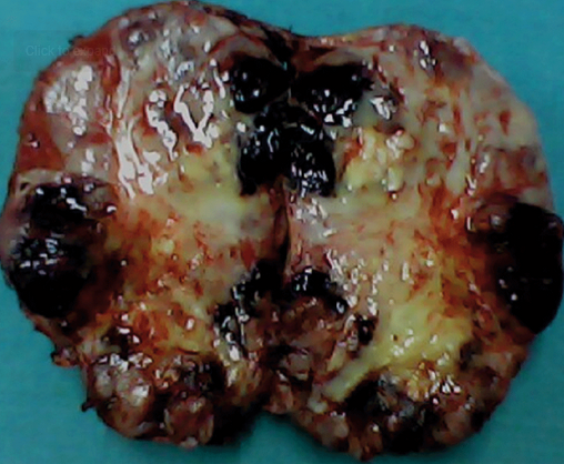
Pheochromocytomas are tumors arising from chromaffin cells of the adrenal medulla (see Image. Gross Specimen of Giant Pheochromocytoma). The clinical manifestations of these tumors are primarily related to the excessive secretion of catecholamines. Similar tumors that arise from extra-adrenal chromaffin cells have been referred to as paragangliomas.[1] These tumors are predominantly benign but can be malignant in a minority of cases.[2]

Etiology

The tumors can be sporadic or familial in origin. According to a recent study, as many as 35% of the cases may be related to germline disease-causing mutations.[3] Familial syndromes known to be associated with pheochromocytoma include Von-Hippel Lindau (VHL), multiple endocrine neoplasia type 2 (MEN2), and neurofibromatosis type 1 (NF1).

Epidemiology

As with many secondary causes of hypertension, pheochromocytomas appear to be underdiagnosed. An autopsy study identified undiagnosed pheochromocytomas in 0.05% of the autopsies.[4] 

In a single-center study of 4180 patients from Brooklyn, pheochromocytoma was found in 0.2% of patients with hypertension. The study calculated an average annual incidence rate of 0.5 per 100,000 person-years.[5]

Pathophysiology

Catecholamines are produced in the chromaffin cells starting with the rate-limiting step of tyrosine hydroxylase (TH), regulating the conversion of tyrosine to dihydroxyphenylalanine (DOPA). Di-hydroxyphenylalanine is subsequently converted into dopamine by the action of dopa decarboxylase, which is further converted into norepinephrine by the action of dopamine β-hydroxylase. PNMT(phenylethanolamine-N-methyltransferase) enzyme methylates the norepinephrine to epinephrine. These catecholamines are sequestered in storage vesicles and released into circulation, and cardiac manifestations are mediated through the adrenoreceptors.[6]

Pheochromocytomas release these catecholamines in various patterns ranging from paroxysmal, continuous, and mixed patterns. Norepinephrine is released continuously and can result in persistent hypertension, while epinephrine is released in a paroxysmal pattern resulting in tachyarrhythmias.[7][8] Alpha(1,2) and beta(1,2) adrenoreceptors bind epinephrine and norepinephrine with varying affinities. These adrenoceptors are influenced by glucocorticoids and thyroid hormones and can result in either an increased number of adrenoceptors or affect their affinity.[9][10][11]

The heart has Beta-1 adrenoceptors, and stimulation of these receptors leads to the activation of adenylate cyclase through the guanine triphosphate protein-coupled receptor(Gs). Activating adenylate cyclase (AC) can convert adenosine triphosphate to cyclic adenosine monophosphate (cAMP). This results in the activation of hyperpolarization-activated cyclic nucleotide-gated(HCN) channels and protein kinase (PKA). This cascade of activities can result in increased ionotropy in SA nodal cells and increased dromotropy in AV nodal cells.[12]

Ryanodine-2 receptors (RyR2) present on the sarcoplasmic reticulum of cardiac myocytes are also phosphorylated by protein kinase A and cause calcium efflux into the cytosol.[13] Calcium binding to the troponin-tropomyosin complex escalates cross-bridge cycling and inotropy by revealing myosin binding sites on actin.[14]

Beta-2 adrenoceptors are present in the peripheral blood vessels and can cause vaso-dilation when activated by epinephrine and norepinephrine. Alpha(1)-adrenoreceptors are present in the vascular smooth muscle cells and can lead to hypertension induced by vasoconstriction after being activated by norepinephrine and epinephrine. Synaptic nerve terminals have Alpha-2 adrenoceptors which inhibit the release of norepinephrine.

Histopathology

Histology of the lesion characteristically shows zellballen nests of chromaffin cells with strong positivity to chromogranin, synaptophysin, CD56, and focally to S100.[15][16] The presence of chromaffin cells in the extra-adrenal tissue is the only pathognomonic characteristic of the malignant form of the entity.[15]

History and Physical

In pheochromocytoma, episodes of hypertension tend to be paroxysmal and are described in the literature as associated with headaches, tachycardia, and sweating. However, it is not uncommon for patients to present with sustained resistant hypertension. The presence of hypertension resistant to multiple antihypertensive medications therapies should raise suspicion for pheochromocytoma.

Episodes of severe hypertension may be precipitated by dopamine receptor antagonists, beta-blockers (typically non-selective), tricyclic antidepressants, corticosteroids, sympathomimetics, and neuromuscular agents; or may be noted in the setting of surgery or induction of anesthesia.[1] 

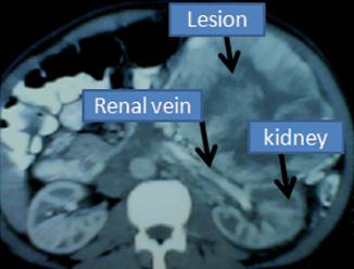
When associated with familial syndromes associated with pheochromocytoma, the condition may be diagnosed during screening tests (after diagnosis has already been established); many such patients do not have hypertension at diagnosis. Giant lesions can paradoxically present only as abdominal lumps with the paucity of the characteristic symptoms of pheochromocytoma owing to tumoral necrosis, comparatively higher volume of interstitial tissue compared to chromaffin cells, and encapsulation by the connective tissues (see Image. Giant Pheochromocytoma).[15] Incidentally detected adrenal masses on CT scans may also result in the diagnosis of pheochromocytoma.

Familial Pheochromocytomas

Pheochromocytomas are known to occur in multiple hereditary syndromes: von Hippel Lindau (VHL) syndrome, multiple endocrine neoplasia type 2 (MEN2), and neurofibromatosis type 1 (NF1). All these are inherited in an autosomal dominant fashion.

VHL disease: Typical phenotype would include hemangioblastomas (brain and spine), retinal angiomas, clear cell renal cell carcinomas, pheochromocytomas, paragangliomas, pancreatic neuroendocrine tumors, endolymphatic sac tumors of the middle ear and cystadenomas (pancreas, epididymis, and broad ligament). The disease is usually caused by mutations in the VHL tumor suppressor gene.[17] VHL type I patients are at lower risk of developing pheochromocytomas than VHL type II patients.[18] VHL patients tend to have higher levels of norepinephrine metabolite normetanephrine than MEN2 patients.[19] 

MEN2: Typical phenotype includes medullary thyroid cancer, pheochromocytomas, primary hyperparathyroidism, mucocutaneous neuromas, skeletal deformities, intestinal ganglioneuromas, and cutaneous lichen amyloidosis. This syndrome is caused by mutations in RET proto-oncogene and is inherited in an autosomal dominant fashion.[20] MEN2 patients tend to have elevated levels of metanephrines (epinephrine metabolite) and be more symptomatic with a higher incidence of hypertension than VHL patients.[19]

NF1: This is caused by mutations in the NF1 tumor suppressor gene and is also inherited in an autosomal dominant fashion.[21] Diagnosis is typically made clinically, and typical characteristics include café-au-lait macules, axillary freckling, Lisch nodules, optic gliomas, osseous lesions, and neurofibromas.[22] Pheochromocytomas are present in a small proportion of these patients, and one study identified these in 0.1 to 5.7% of patients with NF1.[23] 

Subclinical Presentation

Pheochromocytoma can have varied clinical presentations depending on tumor secretory patterns. Dopamine-secreting tumors and tumors with scant hormone secretion can result in subclinical & mild disease presentation. Dopamine-secreting tumors tend to secrete noradrenaline simultaneously; these hormones exert counteractive effects on vessels and attenuate the development of clinical signs and symptoms.[24]

Secretory paragangliomas (secretory tumors outside the adrenal gland) can have varied clinical presentations depending on the location and size of the tumor. Head and neck region paragangliomas (HNPGL) are parasympathetic in origin compared to pheochromocytomas and other secretory paragangliomas, which are sympathetic in origin.[25][26] HNPGL usually produces symptoms by compressing the surrounding local tissues and nerves and can have subclinical presentation when smaller in size.[27][28] 

The recent increase in genetic screening of family members has also resulted in increased detection of patients with subclinical diseases. Succinate dehydrogenase subunit B (SDHB) mutation in tumors have been reported to have undifferentiated catecholamine biosynthetic phenotype containing low concentrations of catecholamine and can have a subclinical presentation before it reaches a larger size to produce signs and symptoms.[29] 

Catecholamine secretory patterns can give rise to a different clinical picture. Adrenaline-secreting tumors usually lead to paroxysmal episodes of hypertensive crisis and otherwise can have a silent course, whereas noradrenaline-secreting tumors usually lead to essential hypertension. Chronically elevated levels of noradrenaline can result in the down-regulation of adrenoreceptors and lead to mild clinical presentation.[30][31]

Patients with subclinical disease also appear to have significant cardiovascular risk. Some cases reports of sudden hypertensive crisis associated with sudden death have been described.[32][33] Mechanical factors, including palpation of the abdomen, sexual intercourse, cough, sneezing, defecation, pain, extreme emotions, and cold, can precipitate a hypertensive crisis in catecholamine-secreting tumors that were otherwise silent. Annual screening with plasma or urinary metanephrines is recommended for genetic carriers to detect disease development earlier.

Another important aspect of the subclinical presentation is incidentalomas discovered on CT scanning. These findings are prevalent in 6% of autopsy studies and 4% of CT scans.[34][35] Adrenals develop nodules with advancing age and are reported to have a 7% prevalence after age 70. In a large series of incidentalomas, more than 4% were reported to be pheochromocytomas.[36] Screening with plasma or urinary metanephrines is recommended before any surgery in cases of adrenal incidentalomas.

In summary, examination findings may include the following:

  • Hypertension
  • Tachycardia
  • Anxiety
  • Diaphoresis
  • Subcutaneous neurofibromas
  • Cafe-au-lait macules
  • Thyroid mass
  • Axillary freckling
  • Lisch nodules on the iris
  • Retinal angiomas
  • Abdominal mass

Evaluation

In the appropriate clinical scenario, diagnosis of pheochromocytoma can be established by biochemical confirmation of hypersecretion of metanephrines and catecholamines. Per the latest endocrine society guidelines, initial biochemical evaluation should include plasma-fractionated metanephrines or urinary-fractionated metanephrines.[1]

Plasma fractionated metanephrines: It has been noted in several studies that plasma metanephrine concentrations tend to be higher in the seated position when compared to the supine position; this results in a decrease in specificity and an increase in false positive results.[37][38][39] 

Guidelines recommend that blood be drawn in the supine position after the patient has been fully recumbent for at least 30 minutes before sampling.[1] In many laboratories where this is often not feasible due to logistical constraints, positive results from samples drawn in a seated position should be repeated in the supine position. Reference intervals established for the same position in which the blood is drawn should be used.

Patient 24-hour urine fractionated metanephrines and catecholamines: This is also a good screening test for biochemical evaluation of pheochromocytoma. Per guidelines, urinary creatinine should be measured to verify the completeness of the collection.[1]

The next step in evaluation after biochemical tests suggest pheochromocytoma is to locate the tumor using imaging studies. Per guidelines, a CT scan of the abdomen and pelvis is the recommended initial imaging test to locate the tumor. MRI is an acceptable alternative to CT scans, particularly when avoidance of radiation or contrast is desired, although its spatial resolution is lower. For metastatic disease, 18F-FDG PET scanning is recommended. The 123I-MIBG scan is an alternative when radiotherapy with 123I-MIBG is planned.[1]

Genetic testing: Since as many as 35% of the cases may be related to germline disease-causing mutations, due consideration should be given to genetic testing in all patients diagnosed with pheochromocytoma.[3] Bilateral tumors and tumors presenting at younger ages are more commonly associated with hereditary syndromes.

Once the diagnosis of pheochromocytoma is confirmed, careful clinical evaluation and family history may reveal characteristic symptoms and signs of hereditary syndromes associated with pheochromocytomas. Targeted genetic testing for VHL, MEN2, and NF1 syndromes should be performed in these cases. After identifying a pathogenic mutation, genetic screening of first-degree family relatives should be considered.

Typical sporadic cases are unilateral, associated with negative family history and absence of symptoms/signs of VHL, MEN2, and NF1 syndromes. These individuals should be clinically monitored for the development of characteristic signs of these syndromes. Genetic testing should be considered in sporadic cases depending on clinical suspicion. A study from Germany reported the detection of characteristic mutations in 24% of patients with apparently sporadic pheochromocytoma.[40]

Several genes associated with the development of pheochromocytomas have been identified and have been categorized into three clusters based on the mechanism of tumorigenesis:

  1. The pseudohypoxia pathway (Cluster 1) has been associated with mutations in EGLN1, EGLN2, DLST, FH, IDH3B, MDH2, SDHA, SDHAF2, SDHB, SDHC, SDHD or VHL,  EPAS1, IDH1, and IDH2 genes,
  2. The kinase-signaling pathway (Cluster 2) has been associated with mutations in NF1, MAX, MERTK, MET, MYCN, RET, or TMEM127 genes, and
  3. Cluster 3 is a somatic cluster associated with alterations in the Wnt signaling pathway.[41]

Treatment / Management

The definitive treatment of pheochromocytoma is surgical resection.

Unilateral pheochromocytomas: Most sporadic tumors are unilateral.

Minimally invasive adrenalectomy: Per endocrine society guidelines, minimally invasive adrenalectomy (laparoscopic) is the preferred treatment for most unilateral adrenal pheochromocytomas.

Open adrenalectomy: This can be considered in larger adrenal pheochromocytomas or in cases where the tumor is considered invasive.[1]

Bilateral pheochromocytomas: These are most commonly associated with hereditary cases, particularly multiple endocrine neoplasia type 2 (MEN2) and von Hippel-Lindau (VHL) disease.[42] In these cases, bilateral total adrenalectomy is associated with a lifelong need for steroid replacement with long-term side effects.[43] 

Bilateral partial or cortical sparing adrenalectomy (open or laparoscopic) is an option in these cases and has demonstrated similar survival despite tumor recurrence and preservation of adrenocortical function with reduced need for lifelong glucocorticoid replacement therapy.[43][44]

Preoperative Preparation

No randomized studies are available to guide approaches for medical optimization before surgery. It is, however, well recognized that initiation of medical therapy in the preoperative period is associated with a lower risk of uncontrolled hypertension (including hypertensive crises), tachycardia, and volume expansion in the perioperative period.

Per endocrine society guidelines, the initial drugs of choice for preoperative preparation are alpha-adrenergic receptor blockers.[1] A typical regimen would include initiating phenoxybenzamine 7 to 14 days before surgery at a dose of 10 mg orally twice daily, carefully up-titrated to a maximum amount of 1 mg/kg/day. Due to a better side effect profile, doxazosin (alpha-1 selective agent) can be used alternatively.

At least 3 to 4 days after initiation of alpha-blockers, beta-blockers (propranolol, atenolol, metoprolol) are initiated to control tachycardia. These agents should not be initiated without prior alpha blockade because of the risk of precipitating hypertensive crisis due to unopposed alpha-receptor stimulation.[1]

Calcium channel blockers (amlodipine and nifedipine) are alternative/add-on agents that effectively control hypertension in the preoperative period.[45]

Volume contraction associated with catecholamine excess in pheochromocytoma patients can be managed with the initiation of a high-sodium diet a few days after the initiation of an alpha blockade. This can also decrease the risk of hypotension after surgery.[7]

The cornerstones in perioperative management include:

  1. Preventing catecholamine surge through minimal handling of the lesion, preventing spillage of the tumor content (especially in cystic lesions), and early control of the adrenal vein.
  2. Managing sudden decrease in peripheral vascular resistance (hypotension) following surgical removal of the lesion.[15]

Differential Diagnosis

  • Hyperthyroidism
  • Anxiety disorder
  • Panic attacks
  • Renal artery stenosis
  • Hyperaldosteronism
  • Migraine
  • Pre-eclampsia
  • Cardiomyopathy
  • Postural tachycardia syndrome (POTS)
  • Drug-induced hypertension
  • Cushing syndrome
  • Carcinoid
  • Acrodynia

Surgical Oncology

Metastatic Pheochromocytomas

Approximately 10% of pheochromocytomas are estimated to be malignant, as defined by the presence of metastasis. There are no definitively known histological or biochemical features that would enable the differentiation of a malignant pheochromocytoma from a benign pheochromocytoma.[46] The presence of chromaffin cells in the extra-adrenal tissue is the only pathognomonic characteristic of the malignant form of the entity.[15]

Pheochromocytoma of the adrenal gland scaled score (PASS) score has been applied to differentiate the benign lesions from their malignant counterparts.[15][16] The following are the available treatment options for a malignant pheochromocytoma:

Surgery

Resection of primary and metastatic disease should be considered if technically feasible; however, it will likely not result in a cure for the disease. The utilized approach to surgery has to be individualized depending on the location of the metastases and can include open or laparoscopic modalities. Larger tumors are preferably treated with an open approach since it may be associated with a lower risk of tumor rupture and may allow for the simultaneous removal of metastases.[46][47]

Debulking of the primary and metastatic lesions may result in the improvement of symptoms from the catecholamine surge.[46][47]

The reported five-year survival is only 50%.[15]

Medical Oncology

External Beam Radiation Therapy

This can be useful for the control of primary and metastatic disease at a variety of sites. Based on data from smaller studies, these treatments can result in symptom relief and debulking of disease.[48][49]

Systemic Chemotherapy

This can be considered in patients with extensive metastatic disease and those with unresectable disease. Studies reporting on treatment outcomes with chemotherapy in the setting of metastatic pheochromocytoma utilized regimens involving cyclophosphamide, doxorubicin, dacarbazine, and vincristine.[50][51] According to a meta-analysis, approximately 37% of the patients responded to chemotherapy.[52] Patients generally do not have complete responses.[46] Chemotherapy can reduce tumor size and improve blood pressure control.[50][53]

Radionuclide Therapy

MIBG: This treatment can only be considered for tumors that can take up MIBG (meta-iodobenzylguanidine) and utilized in unresectable diseases or cases with high tumor burden. MIBG scintigraphy can be used for the localization of catecholamine-secreting tissues because of its structural similarity with noradrenaline.[54] Therapy with Iobenguane I-131 can achieve symptomatic improvement in a significant number of patients and tumor size reduction in a considerable proportion (24 to 45%) of patients.[55][56][57][58]

Novel Therapies

A significant new development in the treatment of metastatic pheochromocytomas includes the utilization of agents that can inhibit angiogenesis and proliferative signaling in the tumor cells. Many such activities in tumor cells are mediated by the interaction of several growth factors (vascular endothelial growth factors [VEGF], platelet-derived growth factors [PDGF], and others) with tyrosine kinase receptors.[46]

Sunitinib: This medication blocks VEGF1, VEGF2, VEGF3, PDGF-alpha, PDGF-beta, c-kit, fms-related tyrosine kinase 3, and RET protooncogene receptors and has the potential to reduce angiogenesis and cell growth.[46][59] The drug has been evaluated in small studies where disease control rates (including stable disease and partial response) have ranged between 57 to 83%.[60][61] Median progression-free survival ranged between 4 and 13 months.[60][61] Sunitinib can cause hypertension; antihypertensive therapy may need to be adjusted to allow for adequate BP control during treatment.

Axitinib: This medication is an inhibitor of VEGFR2 (vascular endothelial growth factor receptor 2). This is especially important in metastatic pheochromocytomas, which frequently exhibit a tumor environment of pseudohypoxia which stimulates the synthesis of VEGF by the tumor cells and results in enhanced angiogenesis.[46] This treatment has been evaluated in phase 2 clinical trials where a partial response was noted in 36% of the patients. Dose adjustments for hypertension are frequently needed as this medication can worsen blood pressure control in a significant proportion of treated patients.[46]

Other Investigational Targeted Therapies

Other therapies currently undergoing investigation include cabozantinib (VEGFR2 and c-MET receptor inhibitor) and hypoxia-inducible factor 2 alpha (HIF2A) inhibitors.

Prognosis

It is essential to recognize the risk of recurrence of pheochromocytomas. This risk is present in sporadic as well as familial cases. In a study of 192 patients with pheochromocytomas and paragangliomas, the recurrence risk was higher in familial, right adrenal, and extra-adrenal tumors.[62] 

As estimated by a recent meta-analysis, the recurrence rate after curative surgery appears to be low at 3% at a mean follow-up of 77 months.[63] Endocrine Society guidelines recommend lifelong annual biochemical testing to assess for recurrent or metastatic disease.[1]

Complications

  • Myocardial infarction
  • Cardiogenic shock
  • Cerebrovascular accident
  • Renal Failure
  • Pulmonary edema
  • Acute respiratory distress syndrome (ARDS)
  • Cardiac arrhythmias
  • Lactic acidosis
  • Hypertensive retinopathy
  • Hypertensive encephalopathy
  • Seizures (children)
  • Polydipsia (children)
  • Polyuria (children)
  • Cerebral vasculitis
  • Ischemic enterocolitis
  • Renal infarction
  • Anxiety
  • Depression

Deterrence and Patient Education

Patient education is critical to the management of pheochromocytomas. It is important for them to understand the episodic nature of symptoms prevalent in the condition. They should be informed that episodes of severe hypertension may be precipitated by medications like dopamine receptor antagonists, beta-blockers (typically non-selective), tricyclic antidepressants, corticosteroids, sympathomimetics, and neuromuscular agents, or may be noted in the setting of surgery or induction of anesthesia. Common episodic symptoms would include:

  • Headaches
  • Sweating
  • Heart racing
  • Shortness of breath
  • Chest pain
  • Anxiety

All patients diagnosed with pheochromocytoma, especially those with a known family history of von Hippel Lindau syndrome, multiple endocrine neoplasia type 2, and neurofibromatosis type 1, should be advised of the need for genetic screening of their relatives.

Several blood, urine, and imaging tests are available to assist medical professionals in diagnosing pheochromocytoma. Options for treatment include medications to control blood pressure and other symptoms. Options for surgical removal of the tumor are also available depending on several factors.

Pearls and Other Issues

Pheochromocytomas and Pregnancy

It is a rare but anxiety-provoking clinical condition in pregnant patients. Various studies have reported the prevalence anywhere between 1 per 15000 and 1 per 54000 pregnancies.[64][65] The clinical course appears to be unpredictable. Increased secretion of catecholamines into maternal circulation can cause severe hypertension, uteroplacental ischemia, arrhythmia, and death. Due to the rare occurrence of the condition, there are no established guidelines to guide management in this setting.

In a large international multicenter retrospective cohort study, authors provided constructive evidence for helping the clinicians.[66] The authors concluded that unrecognized and sub-optimally treated PPGL (pheochromocytoma paraganglioma) was associated with maternal or fetal death in 33 out of (14%) 230 pregnancies. Undiagnosed or untreated patients with PPGLs had a significantly increased risk of maternal and fetal complications with an odds ratio of 27·0 (95% CI 3·5–3473·1) when compared with PPGLs identified before pregnancy, providing compelling evidence to start adrenoceptor blockade after the diagnosis of tumor.

Authors reported that severe maternal complications occurred in 7% of and death occurred in 1% of pregnancies associated with untreated pheochromocytoma paragangliomas (PPGL). Overall fetal mortality of 9% was reported. These rates of complications appeared significantly lower when compared to previously published literature. Abdominal and pelvic PPGL were associated with higher adverse events either due to compression by the uterus or higher catecholamine release. Tumor size was not reported to have higher complications at delivery time. The type of delivery was not found to be associated with adverse outcomes.

Antepartum surgery was not associated with better outcomes; adverse events occurred in 8% of such pregnancies. In case of surgery has to be performed, 2nd trimester is considered safer when compared to the first trimester. Metastatic PPGL was not associated with adverse outcomes; these were thought to be likely related to closer monitoring and treatment. Authors reported lower adverse outcomes in syndromic PPGL; this was thought to be secondary to earlier diagnosis and treatment.

In conclusion, undiagnosed and untreated PPGL was associated with a several-fold higher risk of maternal and fetal complications compared to excellent outcomes among patients treated with alpha-adrenergic blockade agents, including phenoxybenzamine and doxazosin during pregnancy. The transperitoneal approach is recommended for laparoscopic resection instead of extraperitoneal because prone positioning is contraindicated in pregnancy.[67]

Enhancing Healthcare Team Outcomes

Given the involvement of multiple organ systems, as detailed above, pheochromocytoma is a complex condition that requires close multidisciplinary coordination to ensure that the patients receive appropriate care. Timely communication between endocrinologists, surgeons, oncologists, obstetricians, cardiologists, palliative care, and primary care physicians is imperative and cannot be overemphasized.

This is especially important when patients require surgical or obstetric interventions. Adequate support from allied health professionals (pharmacists, nursing, and social workers) and working as part of an interprofessional team is also essential to optimize care delivery and improve patient outcomes.


(Click Image to Enlarge)
<p>Giant Pheochromocytoma</p>

Giant Pheochromocytoma


Contributed by Sunil Munakomi, MD


(Click Image to Enlarge)
<p>Gross Specimen of Giant Pheochromocytoma</p>

Gross Specimen of Giant Pheochromocytoma


Contributed by Sunil Munakomi, MD

Details

Editor:

Bharat Marwaha

Updated:

3/5/2023 3:04:14 PM

References


[1]

Lenders JW, Duh QY, Eisenhofer G, Gimenez-Roqueplo AP, Grebe SK, Murad MH, Naruse M, Pacak K, Young WF Jr, Endocrine Society. Pheochromocytoma and paraganglioma: an endocrine society clinical practice guideline. The Journal of clinical endocrinology and metabolism. 2014 Jun:99(6):1915-42. doi: 10.1210/jc.2014-1498. Epub     [PubMed PMID: 24893135]

Level 1 (high-level) evidence

[2]

Plouin PF, Chatellier G, Fofol I, Corvol P. Tumor recurrence and hypertension persistence after successful pheochromocytoma operation. Hypertension (Dallas, Tex. : 1979). 1997 May:29(5):1133-9     [PubMed PMID: 9149678]


[3]

Neumann HP, Young WF Jr, Krauss T, Bayley JP, Schiavi F, Opocher G, Boedeker CC, Tirosh A, Castinetti F, Ruf J, Beltsevich D, Walz M, Groeben HT, von Dobschuetz E, Gimm O, Wohllk N, Pfeifer M, Lourenço DM Jr, Peczkowska M, Patocs A, Ngeow J, Makay Ö, Shah NS, Tischler A, Leijon H, Pennelli G, Villar Gómez de Las Heras K, Links TP, Bausch B, Eng C. 65 YEARS OF THE DOUBLE HELIX: Genetics informs precision practice in the diagnosis and management of pheochromocytoma. Endocrine-related cancer. 2018 Aug:25(8):T201-T219. doi: 10.1530/ERC-18-0085. Epub 2018 May 24     [PubMed PMID: 29794110]


[4]

Lo CY, Lam KY, Wat MS, Lam KS. Adrenal pheochromocytoma remains a frequently overlooked diagnosis. American journal of surgery. 2000 Mar:179(3):212-5     [PubMed PMID: 10827323]


[5]

Ariton M, Juan CS, AvRuskin TW. Pheochromocytoma: clinical observations from a Brooklyn tertiary hospital. Endocrine practice : official journal of the American College of Endocrinology and the American Association of Clinical Endocrinologists. 2000 May-Jun:6(3):249-52     [PubMed PMID: 11421540]


[6]

Mariani-Costantini R, Shen Y, Cheng L. Biochemical Diagnosis of Pheochromocytoma and Paraganglioma. Paraganglioma: A Multidisciplinary Approach. 2019 Jul 2:():     [PubMed PMID: 31294941]


[7]

Pacak K. Preoperative management of the pheochromocytoma patient. The Journal of clinical endocrinology and metabolism. 2007 Nov:92(11):4069-79     [PubMed PMID: 17989126]


[8]

Gupta G, Pacak K, AACE Adrenal Scientific Committee. PRECISION MEDICINE: AN UPDATE ON GENOTYPE/BIOCHEMICAL PHENOTYPE RELATIONSHIPS IN PHEOCHROMOCYTOMA/PARAGANGLIOMA PATIENTS. Endocrine practice : official journal of the American College of Endocrinology and the American Association of Clinical Endocrinologists. 2017 Jun:23(6):690-704. doi: 10.4158/EP161718.RA. Epub 2017 Mar 23     [PubMed PMID: 28332883]


[9]

Sundaresan PR, Banerjee SP. Differential regulation of beta-adrenergic receptor-coupled adenylate cyclase by thyroid hormones in rat liver and heart: possible role of corticosteroids. Hormone research. 1987:27(2):109-18     [PubMed PMID: 2820855]


[10]

Motulsky HJ, Insel PA. Adrenergic receptors in man: direct identification, physiologic regulation, and clinical alterations. The New England journal of medicine. 1982 Jul 1:307(1):18-29     [PubMed PMID: 6123082]


[11]

Ciaraldi TP, Marinetti GV. Hormone action at the membrane level. VIII. Adrenergic receptors in rat heart and adipocytes and their modulation by thyroxine. Biochimica et biophysica acta. 1978 Jul 3:541(3):334-46     [PubMed PMID: 149563]


[12]

Nazari MA, Rosenblum JS, Haigney MC, Rosing DR, Pacak K. Pathophysiology and Acute Management of Tachyarrhythmias in Pheochromocytoma: JACC Review Topic of the Week. Journal of the American College of Cardiology. 2020 Jul 28:76(4):451-464. doi: 10.1016/j.jacc.2020.04.080. Epub     [PubMed PMID: 32703516]


[13]

Kushnir A, Marks AR. The ryanodine receptor in cardiac physiology and disease. Advances in pharmacology (San Diego, Calif.). 2010:59():1-30. doi: 10.1016/S1054-3589(10)59001-X. Epub     [PubMed PMID: 20933197]

Level 3 (low-level) evidence

[14]

Wakabayashi T. Mechanism of the calcium-regulation of muscle contraction--in pursuit of its structural basis. Proceedings of the Japan Academy. Series B, Physical and biological sciences. 2015:91(7):321-50. doi: 10.2183/pjab.91.321. Epub     [PubMed PMID: 26194856]


[15]

Munakomi S, Rajbanshi S, Adhikary PS. Case Report: A giant but silent adrenal pheochromocytoma - a rare entity. F1000Research. 2016:5():290     [PubMed PMID: 27785358]

Level 3 (low-level) evidence

[16]

Cajipe KM, Gonzalez G, Kaushik D. Giant cystic pheochromocytoma. BMJ case reports. 2017 Nov 8:2017():. pii: bcr-2017-222264. doi: 10.1136/bcr-2017-222264. Epub 2017 Nov 8     [PubMed PMID: 29122903]

Level 3 (low-level) evidence

[17]

Lonser RR, Glenn GM, Walther M, Chew EY, Libutti SK, Linehan WM, Oldfield EH. von Hippel-Lindau disease. Lancet (London, England). 2003 Jun 14:361(9374):2059-67     [PubMed PMID: 12814730]


[18]

Zbar B, Kishida T, Chen F, Schmidt L, Maher ER, Richards FM, Crossey PA, Webster AR, Affara NA, Ferguson-Smith MA, Brauch H, Glavac D, Neumann HP, Tisherman S, Mulvihill JJ, Gross DJ, Shuin T, Whaley J, Seizinger B, Kley N, Olschwang S, Boisson C, Richard S, Lips CH, Lerman M. Germline mutations in the Von Hippel-Lindau disease (VHL) gene in families from North America, Europe, and Japan. Human mutation. 1996:8(4):348-57     [PubMed PMID: 8956040]


[19]

Eisenhofer G, Walther MM, Huynh TT, Li ST, Bornstein SR, Vortmeyer A, Mannelli M, Goldstein DS, Linehan WM, Lenders JW, Pacak K. Pheochromocytomas in von Hippel-Lindau syndrome and multiple endocrine neoplasia type 2 display distinct biochemical and clinical phenotypes. The Journal of clinical endocrinology and metabolism. 2001 May:86(5):1999-2008     [PubMed PMID: 11344198]


[20]

Frank-Raue K, Rondot S, Raue F. Molecular genetics and phenomics of RET mutations: Impact on prognosis of MTC. Molecular and cellular endocrinology. 2010 Jun 30:322(1-2):2-7. doi: 10.1016/j.mce.2010.01.012. Epub 2010 Jan 18     [PubMed PMID: 20083156]


[21]

Gutmann DH, Ferner RE, Listernick RH, Korf BR, Wolters PL, Johnson KJ. Neurofibromatosis type 1. Nature reviews. Disease primers. 2017 Feb 23:3():17004. doi: 10.1038/nrdp.2017.4. Epub 2017 Feb 23     [PubMed PMID: 28230061]


[22]

DeBella K, Szudek J, Friedman JM. Use of the national institutes of health criteria for diagnosis of neurofibromatosis 1 in children. Pediatrics. 2000 Mar:105(3 Pt 1):608-14     [PubMed PMID: 10699117]


[23]

Walther MM, Herring J, Enquist E, Keiser HR, Linehan WM. von Recklinghausen's disease and pheochromocytomas. The Journal of urology. 1999 Nov:162(5):1582-6     [PubMed PMID: 10524872]


[24]

Mannelli M, Lenders JW, Pacak K, Parenti G, Eisenhofer G. Subclinical phaeochromocytoma. Best practice & research. Clinical endocrinology & metabolism. 2012 Aug:26(4):507-15. doi: 10.1016/j.beem.2011.10.008. Epub 2012 May 22     [PubMed PMID: 22863392]


[25]

Kuhweide R, Lanser MJ, Fisch U. Catecholamine-secreting paragangliomas at the skull base. Skull base surgery. 1996:6(1):35-45     [PubMed PMID: 17170951]


[26]

van Duinen N, Steenvoorden D, Kema IP, Jansen JC, Vriends AH, Bayley JP, Smit JW, Romijn JA, Corssmit EP. Increased urinary excretion of 3-methoxytyramine in patients with head and neck paragangliomas. The Journal of clinical endocrinology and metabolism. 2010 Jan:95(1):209-14. doi: 10.1210/jc.2009-1632. Epub 2009 Nov 6     [PubMed PMID: 19897674]


[27]

Pellitteri PK, Rinaldo A, Myssiorek D, Gary Jackson C, Bradley PJ, Devaney KO, Shaha AR, Netterville JL, Manni JJ, Ferlito A. Paragangliomas of the head and neck. Oral oncology. 2004 Jul:40(6):563-75     [PubMed PMID: 15063383]


[28]

Papaspyrou K, Mewes T, Rossmann H, Fottner C, Schneider-Raetzke B, Bartsch O, Schreckenberger M, Lackner KJ, Amedee RG, Mann WJ. Head and neck paragangliomas: Report of 175 patients (1989-2010). Head & neck. 2012 May:34(5):632-7. doi: 10.1002/hed.21790. Epub 2011 Jun 20     [PubMed PMID: 21692132]


[29]

Timmers HJ, Kozupa A, Eisenhofer G, Raygada M, Adams KT, Solis D, Lenders JW, Pacak K. Clinical presentations, biochemical phenotypes, and genotype-phenotype correlations in patients with succinate dehydrogenase subunit B-associated pheochromocytomas and paragangliomas. The Journal of clinical endocrinology and metabolism. 2007 Mar:92(3):779-86     [PubMed PMID: 17200167]


[30]

Tsujimoto G, Manger WM, Hoffman BB. Desensitization of beta-adrenergic receptors by pheochromocytoma. Endocrinology. 1984 Apr:114(4):1272-8     [PubMed PMID: 6323140]


[31]

Streeten DH, Anderson GH Jr. Mechanisms of orthostatic hypotension and tachycardia in patients with pheochromocytoma. American journal of hypertension. 1996 Aug:9(8):760-9     [PubMed PMID: 8862222]


[32]

Song G, Joe BN, Yeh BM, Meng MV, Westphalen AC, Coakley FV. Risk of catecholamine crisis in patients undergoing resection of unsuspected pheochromocytoma. International braz j urol : official journal of the Brazilian Society of Urology. 2011 Jan-Feb:37(1):35-40;discussion 40-1     [PubMed PMID: 21385478]


[33]

Shen SJ, Cheng HM, Chiu AW, Chou CW, Chen JY. Perioperative hypertensive crisis in clinically silent pheochromocytomas: report of four cases. Chang Gung medical journal. 2005 Jan:28(1):44-50     [PubMed PMID: 15804148]

Level 3 (low-level) evidence

[34]

Kloos RT, Gross MD, Francis IR, Korobkin M, Shapiro B. Incidentally discovered adrenal masses. Endocrine reviews. 1995 Aug:16(4):460-84     [PubMed PMID: 8521790]


[35]

Glazer HS, Weyman PJ, Sagel SS, Levitt RG, McClennan BL. Nonfunctioning adrenal masses: incidental discovery on computed tomography. AJR. American journal of roentgenology. 1982 Jul:139(1):81-5     [PubMed PMID: 6979870]


[36]

Mantero F, Terzolo M, Arnaldi G, Osella G, Masini AM, Alì A, Giovagnetti M, Opocher G, Angeli A. A survey on adrenal incidentaloma in Italy. Study Group on Adrenal Tumors of the Italian Society of Endocrinology. The Journal of clinical endocrinology and metabolism. 2000 Feb:85(2):637-44     [PubMed PMID: 10690869]

Level 3 (low-level) evidence

[37]

Lenders JW, Willemsen JJ, Eisenhofer G, Ross HA, Pacak K, Timmers HJ, Sweep CG. Is supine rest necessary before blood sampling for plasma metanephrines? Clinical chemistry. 2007 Feb:53(2):352-4     [PubMed PMID: 17200132]


[38]

de Jong WH, Eisenhofer G, Post WJ, Muskiet FA, de Vries EG, Kema IP. Dietary influences on plasma and urinary metanephrines: implications for diagnosis of catecholamine-producing tumors. The Journal of clinical endocrinology and metabolism. 2009 Aug:94(8):2841-9. doi: 10.1210/jc.2009-0303. Epub 2009 Jun 30     [PubMed PMID: 19567530]


[39]

Deutschbein T, Unger N, Jaeger A, Broecker-Preuss M, Mann K, Petersenn S. Influence of various confounding variables and storage conditions on metanephrine and normetanephrine levels in plasma. Clinical endocrinology. 2010 Aug:73(2):153-60. doi: 10.1111/j.1365-2265.2009.03761.x. Epub 2009 Dec 18     [PubMed PMID: 20039892]


[40]

Neumann HP, Bausch B, McWhinney SR, Bender BU, Gimm O, Franke G, Schipper J, Klisch J, Altehoefer C, Zerres K, Januszewicz A, Eng C, Smith WM, Munk R, Manz T, Glaesker S, Apel TW, Treier M, Reineke M, Walz MK, Hoang-Vu C, Brauckhoff M, Klein-Franke A, Klose P, Schmidt H, Maier-Woelfle M, Peçzkowska M, Szmigielski C, Eng C, Freiburg-Warsaw-Columbus Pheochromocytoma Study Group. Germ-line mutations in nonsyndromic pheochromocytoma. The New England journal of medicine. 2002 May 9:346(19):1459-66     [PubMed PMID: 12000816]


[41]

Sarkadi B, Saskoi E, Butz H, Patocs A. Genetics of Pheochromocytomas and Paragangliomas Determine the Therapeutical Approach. International journal of molecular sciences. 2022 Jan 27:23(3):. doi: 10.3390/ijms23031450. Epub 2022 Jan 27     [PubMed PMID: 35163370]


[42]

Lee JE, Curley SA, Gagel RF, Evans DB, Hickey RC. Cortical-sparing adrenalectomy for patients with bilateral pheochromocytoma. Surgery. 1996 Dec:120(6):1064-70; discussion 1070-1     [PubMed PMID: 8957496]


[43]

Neumann HP, Reincke M, Bender BU, Elsner R, Janetschek G. Preserved adrenocortical function after laparoscopic bilateral adrenal sparing surgery for hereditary pheochromocytoma. The Journal of clinical endocrinology and metabolism. 1999 Aug:84(8):2608-10     [PubMed PMID: 10443647]


[44]

Neumann HPH, Tsoy U, Bancos I, Amodru V, Walz MK, Tirosh A, Kaur RJ, McKenzie T, Qi X, Bandgar T, Petrov R, Yukina MY, Roslyakova A, van der Horst-Schrivers ANA, Berends AMA, Hoff AO, Castroneves LA, Ferrara AM, Rizzati S, Mian C, Dvorakova S, Hasse-Lazar K, Kvachenyuk A, Peczkowska M, Loli P, Erenler F, Krauss T, Almeida MQ, Liu L, Zhu F, Recasens M, Wohllk N, Corssmit EPM, Shafigullina Z, Calissendorff J, Grozinsky-Glasberg S, Kunavisarut T, Schalin-Jäntti C, Castinetti F, Vlcek P, Beltsevich D, Egorov VI, Schiavi F, Links TP, Lechan RM, Bausch B, Young WF Jr, Eng C, International Bilateral-Pheochromocytoma-Registry Group. Comparison of Pheochromocytoma-Specific Morbidity and Mortality Among Adults With Bilateral Pheochromocytomas Undergoing Total Adrenalectomy vs Cortical-Sparing Adrenalectomy. JAMA network open. 2019 Aug 2:2(8):e198898. doi: 10.1001/jamanetworkopen.2019.8898. Epub 2019 Aug 2     [PubMed PMID: 31397861]

Level 2 (mid-level) evidence

[45]

Ulchaker JC, Goldfarb DA, Bravo EL, Novick AC. Successful outcomes in pheochromocytoma surgery in the modern era. The Journal of urology. 1999 Mar:161(3):764-7     [PubMed PMID: 10022680]


[46]

Jimenez C. Treatment for Patients With Malignant Pheochromocytomas and Paragangliomas: A Perspective From the Hallmarks of Cancer. Frontiers in endocrinology. 2018:9():277. doi: 10.3389/fendo.2018.00277. Epub 2018 May 28     [PubMed PMID: 29892268]

Level 3 (low-level) evidence

[47]

Roman-Gonzalez A, Zhou S, Ayala-Ramirez M, Shen C, Waguespack SG, Habra MA, Karam JA, Perrier N, Wood CG, Jimenez C. Impact of Surgical Resection of the Primary Tumor on Overall Survival in Patients With Metastatic Pheochromocytoma or Sympathetic Paraganglioma. Annals of surgery. 2018 Jul:268(1):172-178. doi: 10.1097/SLA.0000000000002195. Epub     [PubMed PMID: 28257320]


[48]

Breen W, Bancos I, Young WF Jr, Bible KC, Laack NN, Foote RL, Hallemeier CL. External beam radiation therapy for advanced/unresectable malignant paraganglioma and pheochromocytoma. Advances in radiation oncology. 2018 Jan-Mar:3(1):25-29. doi: 10.1016/j.adro.2017.11.002. Epub 2017 Nov 22     [PubMed PMID: 29556576]

Level 3 (low-level) evidence

[49]

Fishbein L, Bonner L, Torigian DA, Nathanson KL, Cohen DL, Pryma D, Cengel KA. External beam radiation therapy (EBRT) for patients with malignant pheochromocytoma and non-head and -neck paraganglioma: combination with 131I-MIBG. Hormone and metabolic research = Hormon- und Stoffwechselforschung = Hormones et metabolisme. 2012 May:44(5):405-10. doi: 10.1055/s-0032-1308992. Epub 2012 May 7     [PubMed PMID: 22566196]


[50]

Ayala-Ramirez M, Feng L, Habra MA, Rich T, Dickson PV, Perrier N, Phan A, Waguespack S, Patel S, Jimenez C. Clinical benefits of systemic chemotherapy for patients with metastatic pheochromocytomas or sympathetic extra-adrenal paragangliomas: insights from the largest single-institutional experience. Cancer. 2012 Jun 1:118(11):2804-12. doi: 10.1002/cncr.26577. Epub 2011 Oct 17     [PubMed PMID: 22006217]


[51]

Tanabe A, Naruse M, Nomura K, Tsuiki M, Tsumagari A, Ichihara A. Combination chemotherapy with cyclophosphamide, vincristine, and dacarbazine in patients with malignant pheochromocytoma and paraganglioma. Hormones & cancer. 2013 Apr:4(2):103-10. doi: 10.1007/s12672-013-0133-2. Epub 2013 Jan 30     [PubMed PMID: 23361939]


[52]

Niemeijer ND, Alblas G, van Hulsteijn LT, Dekkers OM, Corssmit EP. Chemotherapy with cyclophosphamide, vincristine and dacarbazine for malignant paraganglioma and pheochromocytoma: systematic review and meta-analysis. Clinical endocrinology. 2014 Nov:81(5):642-51. doi: 10.1111/cen.12542. Epub 2014 Jul 30     [PubMed PMID: 25041164]

Level 1 (high-level) evidence

[53]

Huang H, Abraham J, Hung E, Averbuch S, Merino M, Steinberg SM, Pacak K, Fojo T. Treatment of malignant pheochromocytoma/paraganglioma with cyclophosphamide, vincristine, and dacarbazine: recommendation from a 22-year follow-up of 18 patients. Cancer. 2008 Oct 15:113(8):2020-8. doi: 10.1002/cncr.23812. Epub     [PubMed PMID: 18780317]


[54]

van der Harst E, de Herder WW, Bruining HA, Bonjer HJ, de Krijger RR, Lamberts SW, van de Meiracker AH, Boomsma F, Stijnen T, Krenning EP, Bosman FT, Kwekkeboom DJ. [(123)I]metaiodobenzylguanidine and [(111)In]octreotide uptake in begnign and malignant pheochromocytomas. The Journal of clinical endocrinology and metabolism. 2001 Feb:86(2):685-93     [PubMed PMID: 11158032]


[55]

Loh KC, Fitzgerald PA, Matthay KK, Yeo PP, Price DC. The treatment of malignant pheochromocytoma with iodine-131 metaiodobenzylguanidine (131I-MIBG): a comprehensive review of 116 reported patients. Journal of endocrinological investigation. 1997 Dec:20(11):648-58     [PubMed PMID: 9492103]


[56]

Mukherjee JJ, Kaltsas GA, Islam N, Plowman PN, Foley R, Hikmat J, Britton KE, Jenkins PJ, Chew SL, Monson JP, Besser GM, Grossman AB. Treatment of metastatic carcinoid tumours, phaeochromocytoma, paraganglioma and medullary carcinoma of the thyroid with (131)I-meta-iodobenzylguanidine [(131)I-mIBG]. Clinical endocrinology. 2001 Jul:55(1):47-60     [PubMed PMID: 11453952]


[57]

Troncone L, Rufini V. Nuclear medicine therapy of pheochromocytoma and paraganglioma. The quarterly journal of nuclear medicine : official publication of the Italian Association of Nuclear Medicine (AIMN) [and] the International Association of Radiopharmacology (IAR). 1999 Dec:43(4):344-55     [PubMed PMID: 10731785]


[58]

Sisson JC. Radiopharmaceutical treatment of pheochromocytomas. Annals of the New York Academy of Sciences. 2002 Sep:970():54-60     [PubMed PMID: 12381541]


[59]

Hao Z, Sadek I. Sunitinib: the antiangiogenic effects and beyond. OncoTargets and therapy. 2016:9():5495-505. doi: 10.2147/OTT.S112242. Epub 2016 Sep 8     [PubMed PMID: 27660467]


[60]

O'Kane GM, Ezzat S, Joshua AM, Bourdeau I, Leibowitz-Amit R, Olney HJ, Krzyzanowska M, Reuther D, Chin S, Wang L, Brooks K, Hansen AR, Asa SL, Knox JJ. A phase 2 trial of sunitinib in patients with progressive paraganglioma or pheochromocytoma: the SNIPP trial. British journal of cancer. 2019 Jun:120(12):1113-1119. doi: 10.1038/s41416-019-0474-x. Epub 2019 May 20     [PubMed PMID: 31105270]


[61]

Ayala-Ramirez M, Chougnet CN, Habra MA, Palmer JL, Leboulleux S, Cabanillas ME, Caramella C, Anderson P, Al Ghuzlan A, Waguespack SG, Deandreis D, Baudin E, Jimenez C. Treatment with sunitinib for patients with progressive metastatic pheochromocytomas and sympathetic paragangliomas. The Journal of clinical endocrinology and metabolism. 2012 Nov:97(11):4040-50. doi: 10.1210/jc.2012-2356. Epub 2012 Sep 10     [PubMed PMID: 22965939]


[62]

Amar L, Servais A, Gimenez-Roqueplo AP, Zinzindohoue F, Chatellier G, Plouin PF. Year of diagnosis, features at presentation, and risk of recurrence in patients with pheochromocytoma or secreting paraganglioma. The Journal of clinical endocrinology and metabolism. 2005 Apr:90(4):2110-6     [PubMed PMID: 15644401]


[63]

Holscher I, van den Berg TJ, Dreijerink KMA, Engelsman AF, Nieveen van Dijkum EJM. Recurrence Rate of Sporadic Pheochromocytomas After Curative Adrenalectomy: A Systematic Review and Meta-analysis. The Journal of clinical endocrinology and metabolism. 2021 Jan 23:106(2):588-597. doi: 10.1210/clinem/dgaa794. Epub     [PubMed PMID: 33125073]

Level 1 (high-level) evidence

[64]

Harrington JL, Farley DR, van Heerden JA, Ramin KD. Adrenal tumors and pregnancy. World journal of surgery. 1999 Feb:23(2):182-6     [PubMed PMID: 9880429]


[65]

Lenders JWM, Langton K, Langenhuijsen JF, Eisenhofer G. Pheochromocytoma and Pregnancy. Endocrinology and metabolism clinics of North America. 2019 Sep:48(3):605-617. doi: 10.1016/j.ecl.2019.05.006. Epub 2019 Jun 13     [PubMed PMID: 31345526]


[66]

Bancos I, Atkinson E, Eng C, Young WF Jr, Neumann HPH, International Pheochromocytoma and Pregnancy Study Group. Maternal and fetal outcomes in phaeochromocytoma and pregnancy: a multicentre retrospective cohort study and systematic review of literature. The lancet. Diabetes & endocrinology. 2021 Jan:9(1):13-21. doi: 10.1016/S2213-8587(20)30363-6. Epub 2020 Nov 26     [PubMed PMID: 33248478]

Level 2 (mid-level) evidence

[67]

van der Weerd K, van Noord C, Loeve M, Knapen MFCM, Visser W, de Herder WW, Franssen G, van der Marel CD, Feelders RA. ENDOCRINOLOGY IN PREGNANCY: Pheochromocytoma in pregnancy: case series and review of literature. European journal of endocrinology. 2017 Aug:177(2):R49-R58. doi: 10.1530/EJE-16-0920. Epub 2017 Apr 5     [PubMed PMID: 28381449]

Level 2 (mid-level) evidence