Definition/Introduction
Polarization of light is a fundamental optical phenomenon with significant implications in ophthalmology and optometry, enhancing diagnostic and therapeutic techniques for various ocular conditions.
Polarization of light is a fundamental optical phenomenon with significant implications in ophthalmology and optometry, enhancing diagnostic and therapeutic techniques for various ocular conditions.
Polarization of light refers to the process by which the oscillations of the electric field vector within an electromagnetic (EM) wave become restricted to a single plane.[1] The plane of polarization is determined by the direction of propagation and the orientation of the electric-field oscillations. Different types of polarization, such as linear, circular, and elliptical, exhibit distinct characteristics and behaviors.[2]
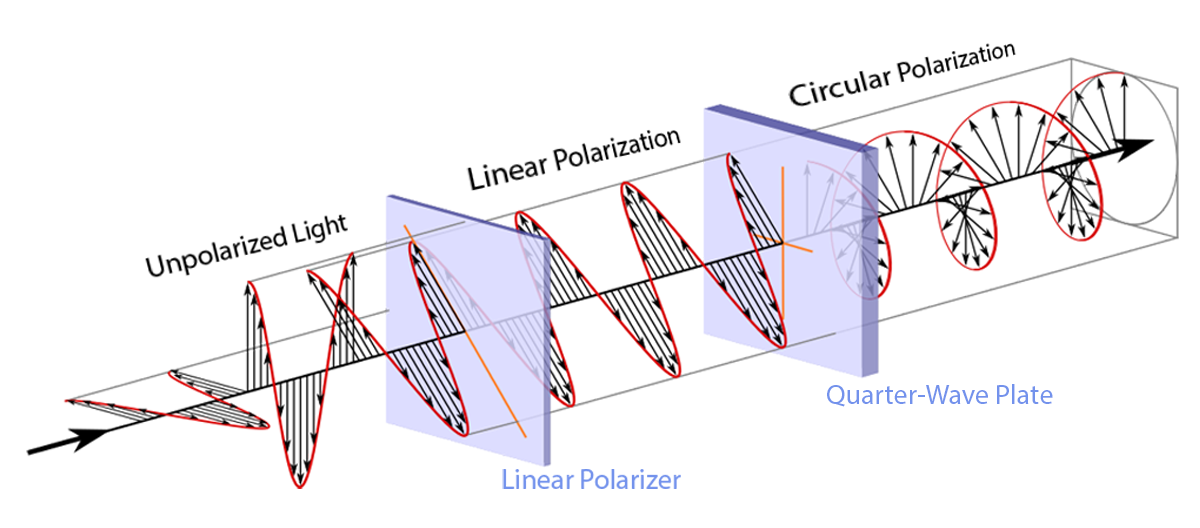
All light possesses polarization. Light, commonly termed "unpolarized," lacks organized polarization, instead exhibiting randomized polarization. Randomly polarized light is a type of light in which the electric field vector oscillates in random directions perpendicular to the direction of the light's propagation. In other words, the light waves are not aligned in any particular direction and are constantly changing orientation.[3] As a result, the light cannot be blocked or transmitted by a polarizer, which is a material that only allows light waves of a specific polarization orientation to pass through.
Linearly polarized light occurs when the electric field vector oscillates in a single plane perpendicular to the direction of the light's propagation. The direction of polarization is typically denoted as either vertical or horizontal but can be at any angle relative to the viewer. Light can be linearly polarized with a polarizer that selectively transmits light waves in a desired polarization direction while blocking others.[4] Various methods can be employed to generate polarized light, including reflection, refraction, scattering, and absorption.[5]
Circularly polarized light occurs when the electric field vector rotates around the direction of the light's propagation in a circle, with a constant amplitude and frequency; it can be thought of as a combination of two linearly polarized waves of equal amplitude and perpendicular polarization directions, with a phase difference of 90 degrees between them.[6] Circularly polarized light can be generated by passing linearly polarized light through a quarter-wave plate or a circular polarizer.[7]
Elliptically polarized light is a type in which the electric field vector traces out an ellipse as it propagates, with varying amplitude and phase. This type of polarization can be considered a combination of two linearly polarized waves of unequal amplitude and perpendicular polarization directions, with a phase difference between them that is not necessarily 90 degrees.[8] Elliptically polarized light can be generated by passing linearly polarized light through a material that induces a phase shift between the two perpendicular polarization directions, such as a birefringent crystal.[9]
Polarized light is utilized in various diagnostic and therapeutic techniques in ophthalmology, such as anterior segment imaging, retinal imaging, photodynamic therapy, low-level light therapy, and laser surgery. The unique properties of polarized light offer numerous benefits for enhancing patient care, from improving visual performance to facilitating the diagnosis and management of various ocular conditions.
Diagnostic Applications
Birefringence refers to the phenomenon where a material causes the polarization state of light to change as it passes through. This occurs because the material has different refractive indices along different axes, causing the components of the light wave polarized parallel to each axis to travel at different speeds, resulting in a phase shift. Several ocular structures exhibit birefringence due to their cellular arrangement.
The birefringent properties of these structures enable various diagnostic techniques, as described below.
Corneal Examination
The use of polarized light in corneal examination allows for the visualization of corneal stress patterns, which can indicate structural changes in the cornea.[15] These patterns can help clinicians diagnose and monitor conditions such as keratoconus, post-LASIK ectasia, and other corneal thinning disorders.[16] Additionally, evaluating corneal stress patterns can help assess the success of corneal cross-linking procedures, which strengthen and stabilize the cornea in patients with progressive corneal ectasia.[17]
Anterior Segment Imaging
Polarized light is particularly valuable in enhancing the contrast of anterior segment structures, making it easier to visualize and diagnose various ocular conditions. For instance, polarized light can improve the visualization of the trabecular meshwork in gonioscopy.[18] Similarly, AS-OCT and Scheimpflug imaging can use polarized light to provide high-resolution images of the cornea, iris, and anterior chamber, enabling clinicians to detect and monitor conditions like corneal dystrophies, pterygium, and iridocorneal endothelial syndromes.[19]
Retinal Imaging
Scanning Laser Polarimetry (SLP)
SLP is a noninvasive imaging technique that utilizes polarized light to measure the thickness of the retinal nerve fiber layer (RNFL). The RNFL consists of axons of retinal ganglion cells responsible for transmitting visual information from the retina to the brain. The change in polarization as light moves through the layer is directly proportional to the thickness of the RNFL. SLP is beneficial as it is a rapid and quantitative assessment of the RNFL in high resolution. However, ocular media opacities, such as cataracts or corneal irregularities, can affect SLP, which may influence the accuracy of RNFL thickness measurements. In addition, the birefringence of the RNFL may vary among individuals, which can impact the accuracy of SLP measurements.[20]
Glaucoma Diagnostics and Variable Corneal Compensation
The glaucoma diagnostics (GDx) test utilizes SLP to measure RNFL thickness, allowing for the detection of glaucomatous damage and monitoring of disease progression. The GDx test has two main iterations: the GDx Nerve Fiber Analyzer and the GDx VCC (variable corneal compensation). The GDx Nerve Fiber Analyzer was the first-generation device that used a fixed corneal compensation algorithm to account for corneal birefringence. However, this fixed compensation could not accurately account for the variability in corneal birefringence between individuals, leading to potential measurement errors. To address this limitation, the GDx VCC was developed, incorporating a variable corneal compensation algorithm that individually measures and compensates for the corneal birefringence of each patient. This improvement allows for more accurate RNFL measurements, making the GDx VCC a valuable tool in glaucoma assessment.[21]
Polarization-Sensitive Optical Coherence Tomography (PS-OCT)
PS-OCT combines the principles of optical coherence tomography (OCT) with polarization-sensitive detection. PS-OCT provides depth-resolved, high-resolution images of the retina, choroid, and optic nerve head, along with information on their polarization properties. This enables the assessment of structural changes and the birefringence of the tissues, which can indicate various pathological conditions.[13] Compared with OCT, PS-OCT offers improved contrast between different retinal layers and structures and can evaluate birefringent tissues, such as the RNFL, Henle fiber layer, and RPE. Compared with SLP, PS-OCT allows for a comprehensive evaluation of the retina, choroid, and optic nerve head, making it useful for diagnosing and monitoring a range of ocular conditions.[22]
Use of Polarizing Filters
Circular polarization filters are incorporated into many slit lamps and ophthalmoscopes. Corneal reflections can hinder the clinician's ability to view ocular structures, especially when examining the anterior segment. When circularly polarized light interacts with the cornea, the reflected light changes its polarization state. A circular polarizing filter placed in the observation pathway, oriented perpendicular to the first filter on the light source, effectively filters out the altered polarization state of the corneal reflections. This process results in a clearer and more detailed view of other ocular structures.[23]
Stereo Testing
Stereo testing, also known as stereopsis or depth perception testing, is a critical element in the comprehensive eye examinations conducted in optometry and ophthalmology.[24] Stereopsis is the visual perception of depth and three-dimensional structure. This perception is primarily due to the slight differences in the images projected onto the retinas of the two eyes, a concept known as binocular disparity.[25]
In stereo testing, the role of polarization is significant. Polarized glasses are employed to deliver different images to each eye, simulating the binocular disparity that is inherent in natural vision. This process effectively tests the patient's ability to perceive depth and integrate binocular visual information. Polarized glasses used in these tests have differently oriented filters for each eye, allowing different images to be presented to each eye simultaneously.
Titmus Fly stereotest, Randot stereotest, and Lang stereotest are among the commonly used stereo tests in clinical practice.[26][27] These tests use polarized glasses to present a series of images or patterns with different disparities, requiring the integration of visual information from both eyes to correctly perceive the depth or three-dimensional structure in the images or patterns. In the Titmus Fly Stereotest, for example, patients wearing polarized glasses are asked to identify a three-dimensional fly. Similarly, in the Randot stereotest, patients must identify three-dimensional shapes among two-dimensional images.
Therapeutic Applications
Polarized Eyewear
Polarized eyewear reduces glare, enhances visual acuity and contrast sensitivity, and improves overall visual comfort. Applications of polarized light in eyewear extend beyond sunglasses to include various types of eyewear, such as swimming and ski goggles. Polarization is also applied to non-eyewear surfaces such as windshields and windows.
While polarized lenses are generally beneficial in reducing glare and enhancing visual comfort, they can interfere with the visibility of certain displays, such as computer and liquid crystal display (LCD) screens, that use polarization to display images. This is due to the orientation of the polarizing filters in the lenses, which may block or distort the light emitted from these screens. Users may experience a darkening or color shift when viewing computer monitors, smartphones, tablets, or other devices with LCD screens while wearing polarized eyewear.[28] Additionally, polarized lenses can interfere with the visibility of certain instruments or displays in vehicles and aircraft, such as GPS systems or heads-up displays, potentially affecting the performance of these devices.
Photodynamic Therapy (PDT)
In PDT, polarized light activates photosensitizing agents that selectively target pathological tissues, such as neovascular membranes in age-related macular degeneration (AMD) or tumor cells in ocular malignancies. The photosensitizing agents absorb the polarized light, which triggers a series of chemical reactions that produce reactive oxygen species (ROS). The ROS then cause localized cellular damage and death, ultimately destroying the targeted tissue. Polarized light in PDT ensures that the light is precisely delivered to the target tissue, minimizing damage to surrounding healthy structures.[29]
Low-Level Light Therapy (LLLT)
LLLT uses polarized light at specific wavelengths to modulate cellular processes, such as mitochondrial respiration, and promote tissue healing. This noninvasive therapy has shown promising results in managing ocular conditions, such as dry eye syndrome, by reducing inflammation and stimulating the production of tear film components. Additionally, LLLT has been explored as a potential treatment for retinal diseases, such as diabetic retinopathy and AMD, by promoting cellular repair mechanisms and reducing oxidative stress.[30]
Laser Surgery
Ophthalmic laser systems increasingly utilize polarized light to ensure precise tissue ablation or photocoagulation during various surgical procedures. The controlled delivery of polarized light in these surgeries improves the accuracy and precision of tissue targeting. Some of the key ophthalmic procedures that involve the use of polarized light in laser surgery include:
Polarization of light has been an area of interest in ophthalmology and optometry due to its potential diagnostic and therapeutic applications. The unique properties of polarized light enable various applications, such as eyewear with contrast sensitivity and glare reduction, advanced imaging techniques, and enhanced treatment options.
The development of specialized instrumentation and devices has facilitated the utilization of polarized light in clinical practice, allowing for assessing and managing various ocular conditions. Further advancements in polarized light-based technologies have the potential to contribute to the evolution of patient care, offering more accurate and noninvasive diagnostic modalities, as well as innovative treatment approaches.
As our understanding of the principles underlying light polarization and its applications in ophthalmology and optometry continues to grow, healthcare professionals must remain informed of the latest developments and consider integrating these technologies into their clinical practice when appropriate. Interprofessional collaboration and education are essential in leveraging the benefits of polarized light to enhance the patient experience and contribute to the ongoing advancement of the fields of ophthalmology and optometry.
Light polarization plays a vital role for the interprofessional healthcare team to ensure the best patient outcomes and satisfaction with their vision. The understanding and practical application of polarized light requires collaborative efforts from ophthalmologists, optometrists, opticians, and optical technologists or nurses. Opticians and optical technologists play a key role in assisting patients with the selection of polarized eyewear. These lenses can reduce glare and improve visual comfort, particularly in bright conditions or during outdoor activities.
Opticians need to understand light polarization to provide the best advice on eyewear selection. The advantages of polarized lenses can be substantial for patients with specific needs, such as those involved in particular sports or professions. Different lens materials affect the polarization of light in varying ways. Knowledge of this is essential when recommending lens materials or coatings. For instance, some lenses may have a polarizing filter or coating that can reduce glare and increase visual comfort.[35]
The interprofessional team should be prepared to explain these benefits to patients, assisting them in making informed decisions about their eyewear. Further, using polarized light in advanced imaging techniques such as optical coherence tomography (OCT) is essential. OCT is a non-invasive imaging test that uses light waves to take cross-section pictures of the retina.
Trained technicians and nurses must be knowledgeable about operating these machines and obtaining quality results to provide the most accurate diagnostic information for the patient's condition. The interprofessional team's comprehensive understanding of light polarization can significantly improve patient outcomes and satisfaction. Effective coordination among ophthalmologists, optometrists, opticians, and optical technologists or nurses ensures patients receive the most appropriate and beneficial ocular care.
Cronin TW, Marshall J. Patterns and properties of polarized light in air and water. Philosophical transactions of the Royal Society of London. Series B, Biological sciences. 2011 Mar 12:366(1565):619-26. doi: 10.1098/rstb.2010.0201. Epub [PubMed PMID: 21282165]
Sparks WB, Hough J, Germer TA, Chen F, DasSarma S, DasSarma P, Robb FT, Manset N, Kolokolova L, Reid N, Macchetto FD, Martin W. Detection of circular polarization in light scattered from photosynthetic microbes. Proceedings of the National Academy of Sciences of the United States of America. 2009 May 12:106(19):7816-21. doi: 10.1073/pnas.0810215106. Epub 2009 Apr 28 [PubMed PMID: 19416893]
Duan J, Qu Y, Fu Q, Yu T, Yang Y, Zhang S, Zhan J, Bai X. Polarized light transmission characteristics in a smoky ellipsoidal particle medium. Applied optics. 2023 Apr 1:62(10):2510-2521. doi: 10.1364/AO.480857. Epub [PubMed PMID: 37132799]
Sedov ES, Rubo YG, Kavokin AV. Polariton polarization rectifier. Light, science & applications. 2019:8():79. doi: 10.1038/s41377-019-0189-z. Epub 2019 Aug 28 [PubMed PMID: 31645925]
Li X, Chen B, He L, Gao X. Optimal configurations for different incident polarization states in linear polarization calibration. Applied optics. 2020 Oct 20:59(30):9520-9531. doi: 10.1364/AO.403647. Epub [PubMed PMID: 33104672]
Xu X, Zhang Y, Song H, Lin X, Huang Z, Kuroda K, Tan X. Generation of circular polarization with an arbitrarily polarized reading wave. Optics express. 2021 Jan 18:29(2):2613-2623. doi: 10.1364/OE.414531. Epub [PubMed PMID: 33726453]
Zheng J, He X, Beckett P, Sun X, Cai Z, Zhang W, Liu X, Hao X. Dichroic Circular Polarizers Based on Plasmonics for Polarization Imaging Applications. Nanomaterials (Basel, Switzerland). 2021 Aug 23:11(8):. doi: 10.3390/nano11082145. Epub 2021 Aug 23 [PubMed PMID: 34443976]
Park S, Kim M, Kim I, Taylor RA, Song J, Kyhm K. Elliptical Polarization of Localized States in an Anisotropic Single GaAs Quantum Ring. Nanomaterials (Basel, Switzerland). 2022 Dec 31:13(1):. doi: 10.3390/nano13010184. Epub 2022 Dec 31 [PubMed PMID: 36616094]
Herne CM, Cartwright NA, Cattani MT. Determining elliptical polarization of light from rotation of calcite crystals. Optics express. 2017 May 1:25(9):10044-10050. doi: 10.1364/OE.25.010044. Epub [PubMed PMID: 28468379]
Bharti K, den Hollander AI, Lakkaraju A, Sinha D, Williams DS, Finnemann SC, Bowes-Rickman C, Malek G, D'Amore PA. Cell culture models to study retinal pigment epithelium-related pathogenesis in age-related macular degeneration. Experimental eye research. 2022 Sep:222():109170. doi: 10.1016/j.exer.2022.109170. Epub 2022 Jul 11 [PubMed PMID: 35835183]
Kesim C, Bektas SN, Kulali Z, Yildiz E, Ersoz MG, Sahin A, Gunduz-Demir C, Hasanreisoglu M. HENLE FIBER LAYER MAPPING WITH DIRECTIONAL OPTICAL COHERENCE TOMOGRAPHY. Retina (Philadelphia, Pa.). 2022 Sep 1:42(9):1780-1787. doi: 10.1097/IAE.0000000000003514. Epub [PubMed PMID: 35504010]
Eghrari AO, Riazuddin SA, Gottsch JD. Overview of the Cornea: Structure, Function, and Development. Progress in molecular biology and translational science. 2015:134():7-23. doi: 10.1016/bs.pmbts.2015.04.001. Epub 2015 Jun 4 [PubMed PMID: 26310146]
Level 3 (low-level) evidenceAzuma S, Makita S, Kasaragod D, Sugiyama S, Miura M, Yasuno Y. Clinical multi-functional OCT for retinal imaging. Biomedical optics express. 2019 Nov 1:10(11):5724-5743. doi: 10.1364/BOE.10.005724. Epub 2019 Oct 14 [PubMed PMID: 31799043]
Hejtmancik JF, Shiels A. Overview of the Lens. Progress in molecular biology and translational science. 2015:134():119-27. doi: 10.1016/bs.pmbts.2015.04.006. Epub 2015 May 27 [PubMed PMID: 26310153]
Level 3 (low-level) evidenceAvetisov SE, Bubnova IA, Novikov IA, Antonov AA, Siplivyi VI. Experimental study on the mechanical strain of corneal collagen. Journal of biomechanics. 2013 Jun 21:46(10):1648-54. doi: 10.1016/j.jbiomech.2013.04.008. Epub 2013 May 13 [PubMed PMID: 23680349]
Joseph Antony S. Imaging shear stress distribution and evaluating the stress concentration factor of the human eye. Scientific reports. 2015 Mar 10:5():8899. doi: 10.1038/srep08899. Epub 2015 Mar 10 [PubMed PMID: 25754336]
Gulzar A, Yıldız E, Kaleli HN, Nazeer MA, Zibandeh N, Malik AN, Taş AY, Lazoğlu I, Şahin A, Kizilel S. Ruthenium-induced corneal collagen crosslinking under visible light. Acta biomaterialia. 2022 Jul 15:147():198-208. doi: 10.1016/j.actbio.2022.05.040. Epub 2022 May 25 [PubMed PMID: 35643198]
Carmichael-Martins A, Gast TJ, Burns SA, Walker BR, King BJ. Characterization of the human iridocorneal angle in vivo using a custom design goniolens with OCT gonioscopy. Biomedical optics express. 2022 Sep 1:13(9):4652-4667. doi: 10.1364/BOE.465317. Epub 2022 Aug 10 [PubMed PMID: 36187241]
Gupta N, Varshney A, Ramappa M, Basu S, Romano V, Acharya M, Gaur A, Kapur N, Singh A, Shah G, Chaudhary I, Patel N, Tiwari A, Kate A, Sangwan V, Mathur U. Role of AS-OCT in Managing Corneal Disorders. Diagnostics (Basel, Switzerland). 2022 Apr 7:12(4):. doi: 10.3390/diagnostics12040918. Epub 2022 Apr 7 [PubMed PMID: 35453966]
Lever M, Halfwassen C, Unterlauft JD, Bechrakis NE, Manthey A, Böhm MRR. Retinal nerve fibre layer thickness measurements in childhood glaucoma: the role of scanning laser polarimetry and optical coherence tomography. Graefe's archive for clinical and experimental ophthalmology = Albrecht von Graefes Archiv fur klinische und experimentelle Ophthalmologie. 2021 Dec:259(12):3777-3786. doi: 10.1007/s00417-021-05276-z. Epub 2021 Jun 26 [PubMed PMID: 34173881]
Karvonen E, Stoor K, Luodonpää M, Hägg P, Lintonen T, Liinamaa J, Tuulonen A, Saarela V. Diagnostic performance of modern imaging instruments in glaucoma screening. The British journal of ophthalmology. 2020 Oct:104(10):1399-1405. doi: 10.1136/bjophthalmol-2019-314795. Epub 2020 Jan 16 [PubMed PMID: 31949097]
Bille JF, Aumann S, Donner S, Fischer J, Müller F. Optical Coherence Tomography (OCT): Principle and Technical Realization. High Resolution Imaging in Microscopy and Ophthalmology: New Frontiers in Biomedical Optics. 2019:(): [PubMed PMID: 32091846]
OʼSullivan R, Tom LM, Bunya VY, Nyberg WC, Massaro-Giordano M, Daniel E, Smith E, Brainard DH, Gee J, Maguire MG, Stone RA. Use of Crossed Polarizers to Enhance Images of the Eyelids. Cornea. 2017 May:36(5):631-635. doi: 10.1097/ICO.0000000000001157. Epub [PubMed PMID: 28257379]
Levi DM. Learning to see in depth. Vision research. 2022 Nov:200():108082. doi: 10.1016/j.visres.2022.108082. Epub 2022 Jul 14 [PubMed PMID: 35841717]
Cumming BG, DeAngelis GC. The physiology of stereopsis. Annual review of neuroscience. 2001:24():203-38 [PubMed PMID: 11283310]
Furr BA, Musch DC, Andrews CA, Schumann R. Testability Study of the Titmus V3 Test in Pre-school Children. Optometry and vision science : official publication of the American Academy of Optometry. 2018 Jul:95(7):588-593. doi: 10.1097/OPX.0000000000001240. Epub [PubMed PMID: 29957735]
Ohlsson J, Villarreal G, Abrahamsson M, Cavazos H, Sjöström A, Sjöstrand J. Screening merits of the Lang II, Frisby, Randot, Titmus, and TNO stereo tests. Journal of AAPOS : the official publication of the American Association for Pediatric Ophthalmology and Strabismus. 2001 Oct:5(5):316-22 [PubMed PMID: 11641643]
Sasaki S, Udono M, Koike Y. Real-color displays realized by randomized polarization. Applied optics. 2021 Apr 10:60(11):3108-3113. doi: 10.1364/AO.420403. Epub [PubMed PMID: 33983207]
Level 1 (high-level) evidenceKwiatkowski S, Knap B, Przystupski D, Saczko J, Kędzierska E, Knap-Czop K, Kotlińska J, Michel O, Kotowski K, Kulbacka J. Photodynamic therapy - mechanisms, photosensitizers and combinations. Biomedicine & pharmacotherapy = Biomedecine & pharmacotherapie. 2018 Oct:106():1098-1107. doi: 10.1016/j.biopha.2018.07.049. Epub 2018 Jul 17 [PubMed PMID: 30119176]
Glass GE. Photobiomodulation: The Clinical Applications of Low-Level Light Therapy. Aesthetic surgery journal. 2021 May 18:41(6):723-738. doi: 10.1093/asj/sjab025. Epub [PubMed PMID: 33471046]
Bashir ZS, Ali MH, Anwar A, Ayub MH, Butt NH. Femto-lasik: The recent innovation in laser assisted refractive surgery. JPMA. The Journal of the Pakistan Medical Association. 2017 Apr:67(4):609-615 [PubMed PMID: 28420926]
Landers J. Selective laser trabeculoplasty: A review. Clinical & experimental ophthalmology. 2021 Dec:49(9):1102-1110. doi: 10.1111/ceo.13979. Epub 2021 Aug 16 [PubMed PMID: 34331388]
Hassanpoor N, Ahoor M, Latifi A, Niyousha M. Conventional and Pattern Scanning Pan-Retinal Photocoagulation Laser in Diabetic Patients' Visual Field. Journal of lasers in medical sciences. 2022:13():e40. doi: 10.34172/jlms.2022.40. Epub 2022 Sep 26 [PubMed PMID: 36743140]
Kobashi H, Rong SS. Corneal Collagen Cross-Linking for Keratoconus: Systematic Review. BioMed research international. 2017:2017():8145651. doi: 10.1155/2017/8145651. Epub 2017 Jun 11 [PubMed PMID: 28691035]
Level 1 (high-level) evidenceLacherez P, Saeri AK, Wood JM, Atchison DA, Horswill MS. A yellow filter improves response times to low-contrast targets and traffic hazards. Optometry and vision science : official publication of the American Academy of Optometry. 2013 Mar:90(3):242-8. doi: 10.1097/OPX.0b013e3182815783. Epub [PubMed PMID: 23400022]