Traumatic Cataract

Earn CME/CE in your profession:


Continuing Education Activity

The formation of traumatic cataracts is common after blunt or penetrating ocular trauma. Up to 65% of eye traumas lead to cataract formation, resulting in significant short- and long-term vision loss. Most cases of ocular trauma result in some degree of lenticular swelling. The severity of the trauma and the integrity of the capsular bag determine the type of cataract developed and the clinical course. Traumatic cataracts may occur acutely or develop slowly over time, as in the case of a concussion cataract.

Surgery for traumatic cataracts is often more complex than standard age-related cataracts due to associated damage to the lens capsule and zonules, synechiae, and reduced media clarity. A thorough preoperative assessment and planning are crucial to achieve successful outcomes and optimal vision.[

This activity reviews the etiology, epidemiology, evaluation, management, procedural techniques, complications, deterrence, and age-related specifics of traumatic cataracts while highlighting the role of the interprofessional team in improving outcomes for patients after ocular trauma.

Objectives:

  • Identify patients at risk for developing traumatic cataracts based on their clinical history.
  • Assess patients with ocular trauma for globe injury and traumatic cataracts.
  • Apply best practices when treating traumatic cataracts to reduce the risk of complications and facilitate optimal visual acuity.
  • Develop and implement effective interprofessional team strategies to improve outcomes for patients with traumatic cataracts.

Introduction

Ocular trauma is a significant cause of vision loss, and as many as 1.6 million people lose sight yearly due to traumatic cataracts.[1] Eye injuries occur in approximately one-fifth of adults, with men and young people being the most commonly affected.[2] There are an estimated 55 million eye injuries annually, with developed countries experiencing a high incidence of one-sided blindness.[3] Thorough assessment and management of oculofacial trauma are crucial, and guidelines are available to determine the visual prognosis.[4] Factors such as initial visual acuity, pupillary reflex response, and the severity of the trauma are essential in this assessment.[5] This activity provides a comprehensive guide to managing lens injuries, particularly traumatic cataracts, including aseptic and antiseptic procedures before surgery and surgical indications and timing. Patients can receive the appropriate treatment and care with this approach, leading to better visual outcomes after ocular trauma.

Etiology

Ocular trauma can occur via many mechanisms, affecting the crystalline lens in various ways, including perforating or blunt trauma, electric shock, ultraviolet and ionizing radiation, and chemical injuries.[6][7] 

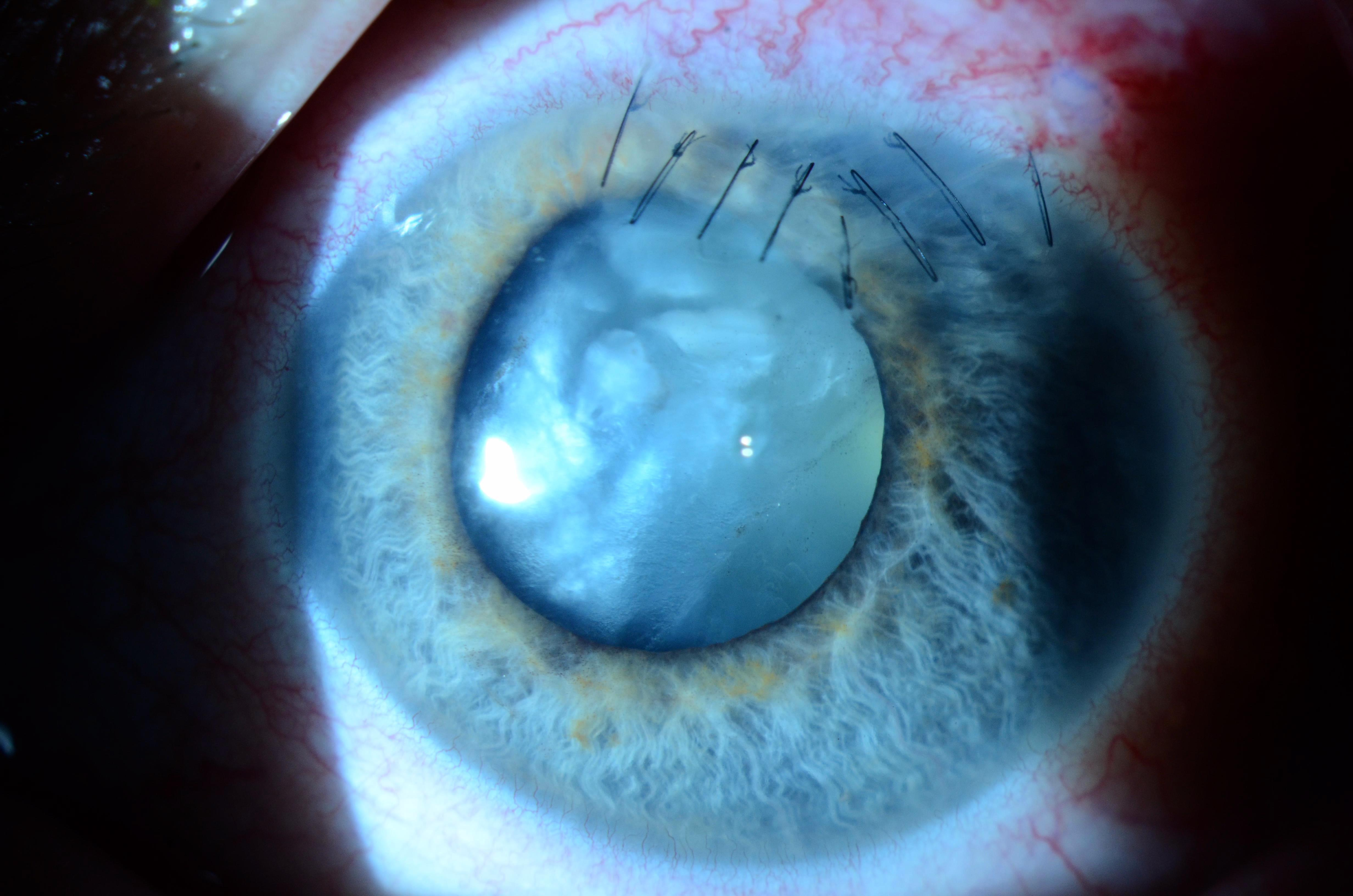
In penetrating trauma, where an object with a sharp edge, such as glass, wood, or metal, pierces the eye, a traumatic cataract may develop immediately if the object reaches the lens after passing through the cornea.[4][8] The lens may also be damaged or completely ruptured, leading to partial or complete cataracts and blindness (see Image. Traumatic Cataract Secondary to Penetrating Ocular Injury). Chemical trauma may occur when a foreign substance enters the eye and alters the composition of the lens fibers, leading to traumatic cataracts.[7][9] Radiation exposure can also damage and rupture the lens, resulting in traumatic cataracts over time.[10] Radiation-induced ocular trauma is often seen in children.[11] 

Opacification of the lens may occur immediately after an injury or years later; the type of cataract formed will depend on the nature and extent of the ocular trauma.[11] Penetrating trauma may result in a cataract proportional to the size of the opening in the lens capsule. No morphologic grouping exists for cataracts due to penetrating trauma.[12][4][13] 

In contrast, cataracts secondary to blunt trauma often exhibit a distinctive rosette- or flower-shaped appearance.[14] Larger capsular openings can cause the entire lens to become cataractous, while smaller openings may produce only a focal opacity.[8] Additionally, blunt trauma can cause cataract formation without a loss of capsular integrity due to the forces of the original trauma or subsequent inflammation.[15] Ocular trauma may also lead to subcapsular cataracts.[16] Electric shock can cause diffuse milky-white opacification or multiple snowflake-like opacities. Ultraviolet radiation can induce true exfoliation of the anterior lens capsule with subsequent cataract development. The ionizing radiation used to treat ocular tumors or during cardiac interventions may cause posterior subcapsular opacities. Finally, chemical injuries to the lens can result from various sources, including naphthalene, thallium, lactose, and galactose.[17]

Epidemiology

The prevalence of cataract formation after ocular trauma is approximately 65%.[2] Traumatic cataract formation contributes significantly to short-term and long-term vision impairment on a global scale.[18] Eye injuries occur in approximately one-fifth of adults, with men and young people being the most commonly affected.[19][20] There are an estimated 55 million eye injuries annually, with up to 1.6 million people losing sight yearly due to traumatic cataracts.[21] 

The incidence of traumatic cataract formation varies globally, influenced by age, gender, environment, and socioeconomic factors.[22] While males are more prone to ocular injuries from outdoor activities, gender does not significantly affect visual outcomes.[22] Younger individuals are more susceptible to ocular injuries but respond well to treatment.[21][22] The incidence of ocular injuries is higher in rural areas, but the visual outcome does not differ significantly from urban counterparts. The etiology of the traumatic incident varies with geography and socioeconomic status, but the results do not differ significantly. 

Pathophysiology

Disruption of the lens fibers after blunt or penetrating ocular trauma commonly leads to a traumatic cataract.[23][24][25] The traumatic mechanism and the integrity of the capsular bag dictate the morphology of the cataract and the clinical course.[26] Trauma disrupts and injures the lens fibers, leading to lens swelling.[23][27] Worldwide, traumatic cataract formation is observed in 24% of patients with globe contusions. Concussion cataracts, another type of traumatic cataract, occur due to blunt trauma. Although the lens capsule is not extensively damaged, it becomes progressively opaque over time. The pathophysiology of traumatic cataracts occurs through direct rupture and distortion of the capsule or coup and equatorial expansion due to various forces transferring the traumatic energy to the other side of the eye.[28] Traumatic cataracts typically present as rosette or stellate subtypes.[9][29] 

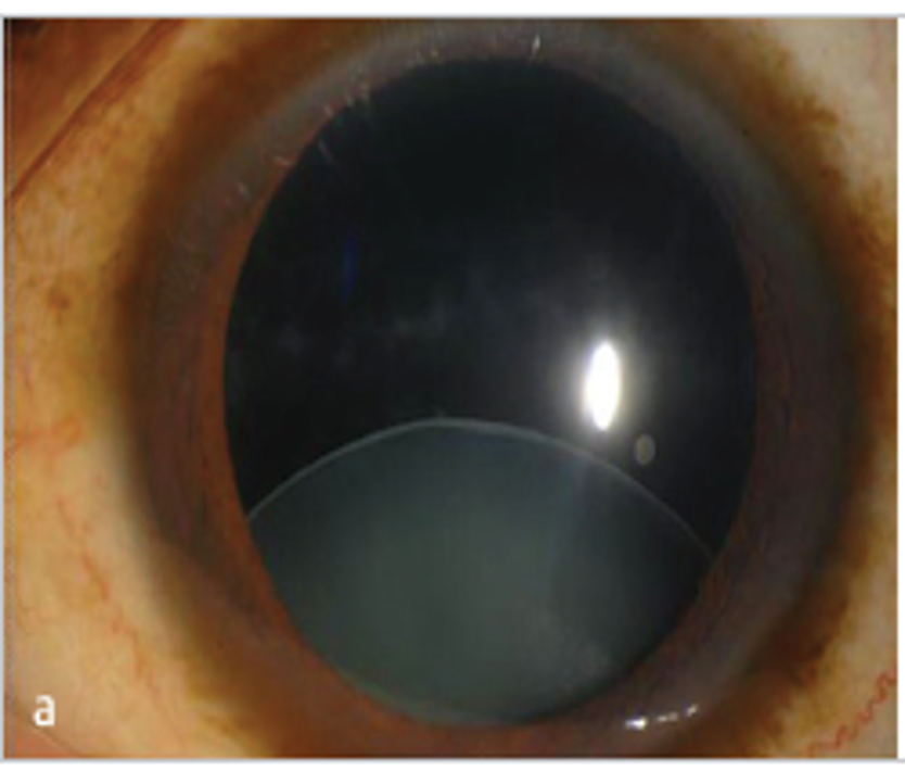
The lens comprises the cortex and the nucleus, and the lens capsule is an uninterrupted basement membrane of modified epithelial cells. Denaturation and coagulation of lens proteins can lead to cataract formation through loss of transparency due to degenerative processes.[28][30] Various disturbances can cause these degenerative processes. Disturbances during lens growth and formation lead to congenital cataracts. Fibrous changes in the lens epithelium result in subcapsular cataracts, cortical hydration between lens fibers induces cortical cataracts, and the deposition of pigments such as urochrome leads to nuclear cataracts.[30] Trauma can also cause partial (subluxated) or complete (luxated) displacement of the lens, leading to ocular disorders such as phacomorphic glaucoma, in which the lens diameter swells and occludes the iridocorneal angle. (see Image. Posttraumatic Crystalline Lens Subluxation) Trauma may also induce lens-particle glaucoma, where lens proteins clog the trabecular meshwork, or an inflammatory response within the anterior chamber leading to the development of phacoantigenic glaucoma.[7][28][30][31][32][33]

Histopathology

Traumatic cataracts lack a standardized morphologic classification system. Cataract morphology is dictated by the type of injury and the time interval between injury and intervention.[13][34] Cataracts are categorized by lenticular opacity and are total, white soft, or membranous. A total cataract is characterized by a lack of visible lens matter between the capsule and nucleus. The fusion of both capsules is characteristic of a membranous cataract with minimal cortical material. White soft cataracts have loose cortical material in the anterior chamber with a ruptured lens capsule. Additionally, a lens with a rosette opacity pattern indicates a traumatic cataract caused by blunt trauma.[14]

Ultrastructural findings of traumatic white cataracts suggest that dysfunction of the lens epithelium can lead to edema of superficial cortical lens fibers, resulting in degeneration and localized vacuolization.[23][28][35] Gap junctions and desmosomes interconnect lens epithelial cells and contain scattered microfilaments.[36] A break in the capsule can cause a change in tension on the underlying epithelium, which may be transmitted along the points of strong attachment from cell to cell. Capsulorhexis may be turned to the periphery at a site where these changes are most pronounced, especially if the inner pressure in the lens is high.[37] These findings may increase the rate of capsular breaks during surgery, indicating a potential correlation between histopathology, morphology, and clinical behavior of the anterior lens capsule in traumatic white cataracts.

History and Physical

The first step in evaluating ocular trauma is determining if an acute ocular emergency is present. The second step is determining if the injury is due to blunt or penetrating trauma.[38][5][39][38] If the intraocular pressure is significantly low, there may be an open globe injury, which requires caution when using topical medication or eye devices. Pediatric patients may be unable to recount their ocular trauma accurately, and an increased level of suspicion of an open globe injury or intraocular foreign body is imperative.

A comprehensive medical history should be obtained, as specific comorbidities, such as poorly controlled diabetes or hypertension, can increase the risk of complications, particularly expulsive hemorrhage or infection.[40] Particular attention should be paid to the ocular history to estimate vision potential accurately. A prior history of intraocular surgery may affect the planning of cataract extraction. The date of the most recent tetanus vaccination should be ascertained.

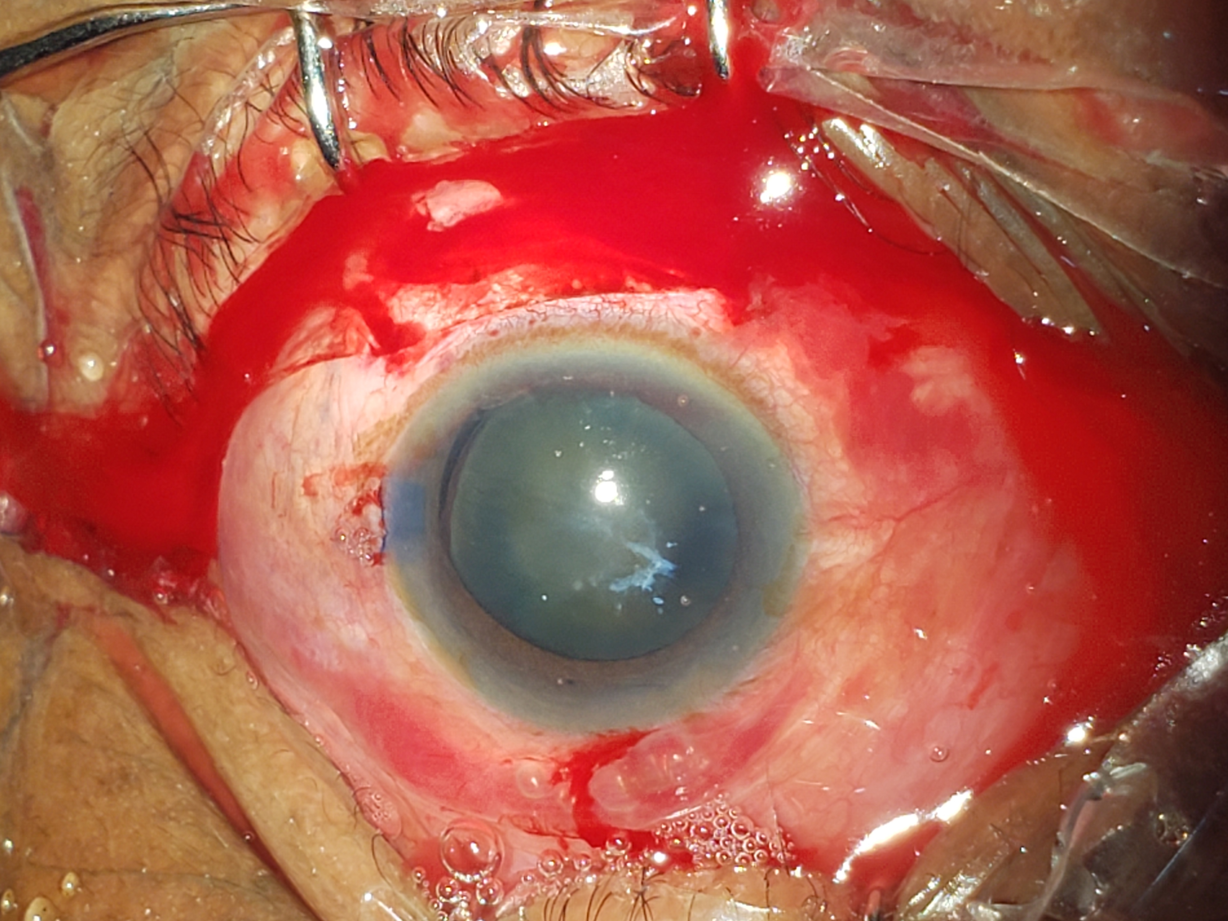
The physical evaluation of a traumatic cataract aims to determine if surgery is necessary and, if so, which procedure will be safest and most effective.[20][41][42] When assessing acute ocular trauma, a thorough evaluation is essential to determine the severity of the eye injury.[2][20] The physical examination should carefully evaluate visual acuity, pupillary function, and intraocular pressure. Slit lamp biomicroscopy and a dilated fundus examination should be performed. Identifying signs of zonular damage, such as phacodonesis, focal iridodonesis, vitreous prolapse, and lens subluxation, is crucial though these signs may not always be present (see Image. Traumatic Cataract).[20][43] Subtle signs of lens injury include seeing the lens equator during eccentric gaze, a decentered nucleus in the primary position, an iridolenticular gap, or changes in the lens periphery contour. The formation of a cataract within minutes to hours after ocular trauma may indicate a violation of the anterior lens capsule.[44]

Evaluation

Surgery for traumatic cataracts is a complex procedure that requires careful consideration.[5][19][45][19][46] The lens capsule and zonules are often damaged, increasing intraoperative risks, such as intraoperative lens dislocation, capsular rupture, and vitreous loss.[45][47]

Preoperative assessment and planning are essential for successful surgical outcomes. In cases where corneal or other media opacities obscure visualization, computed tomography (CT) imaging may facilitate the identification of a traumatic cataract.[48] Capsular tears can occur simultaneously or separately, and traditional B-scan ocular ultrasonography can detect ocular pathology but lacks resolution for posterior capsule or zonular structures.[49][50] Instead, ocular echography with a 20-MHz frequency is an effective imaging method for detecting occult posterior lens capsular rupture.[51][52][53]

Ultrasound biomicroscopy is also an effective method for identifying occult zonular damage in patients with anterior segment trauma.[51][54][55] Ocular CT of the anterior segment and Scheimpflug imaging have utility in determining the presence and extent of posterior capsular rupture and zonular integrity.[55][56] Accurate intraocular lens (IOL) calculations may not be possible in the acute setting. However, options include using data from the traumatized or fellow eye or deferring IOL placement to a secondary procedure.[43] Studies indicate that acceptable IOL calculations can be obtained from the fellow eye in most cases.[57][58]

Treatment / Management

The anesthesia choice should be tailored to the individual based on age, overall health, and estimated procedure duration.[59] General anesthesia is frequently used for open globe injuries, uncooperative patients, complex procedures, and pediatric patients.[60] 

Aseptic and antiseptic procedures should be performed with care to prevent eye damage or the introduction of bacteria. Preoperative cleansing with diluted povidone-iodine 5% in the conjunctival sac is the most effective method for reducing the risk of endophthalmitis.

Initial Management Considerations

Surgery to remove traumatic cataracts is typically not recommended in the acute setting. The standard approach is first to perform primary globe closure for penetrating or perforating injuries, followed by a secondary procedure to remove the cataract and place an intraocular lens.[18][61][62] This approach offers the benefit of a thorough assessment of associated injuries to adjacent ocular structures, which is crucial for preoperative planning. Additionally, cataracts may not be severe enough to affect vision immediately following the injury. Poor intraoperative visualization and limited ability to assess retinal or optic nerve damage during primary procedures can make cataract surgery more challenging in the acute setting. Nevertheless, some studies have demonstrated that earlier surgical intervention can enhance visual outcomes, and immediate cataract removal can alleviate inflammation and pressure elevation.[62][4] While deciding to place an IOL during primary surgery is debatable, small case series have reported favorable outcomes.[63][64] Primary extraction is imperative for patients with lens vitreous admixture to avoid further complications.[63]

Considerations in the Pediatric Population

Eye injuries in children are severe in about one-third of cases. In cases of acquired cataracts in children with vague trauma histories, it is essential to consider possible child abuse. To prevent stimulus deprivation amblyopia in children aged 5 years or younger, the timing of clearing the visual axis must be considered.[63][39] Cataract surgery should be performed within 1 year of ocular trauma; delay can increase amblyopia risk. Prompt cataract extraction is necessary to prevent phacomorphic glaucoma in pediatric eyes caused by a swollen cataractous lens causing the pupillary block.[65] Primary posterior capsulectomy and vitrectomy should be considered for children with traumatic cataracts to slow the opacification of the posterior capsule.[66][67]

Epilenticular IOL implantation is an option that may avoid common complications such as intraoperative high positive pressure and postoperative fibrinous uveitis, resulting in clear visual pathways and improved visual acuity. However, using contact lenses for unilateral aphakia is not feasible for many children worldwide due to cost, inadequate sanitation, and lack of availability. A study in sub-Saharan Africa supports using posterior chamber IOLs as the standard of care for children older than 2 years, as it produces the best visual acuity results. IOLs placed in the capsular bag are much less likely to require future capsulotomy, reducing amblyopia risk from developing posterior capsule opacification.[5] In cases from India where the posterior segment was not involved, extracapsular cataract extraction with posterior chamber IOL implantation resulted in better visual acuity in children following blunt trauma than penetrating trauma.

Concurrent Injury to the Cornea or Anterior Capsule

In cases where a traumatic corneal injury has occurred, there may be laceration or loss of corneal tissue. The anterior chamber must be sealed to prevent fluid leakage during cataract surgery. Corneal wounds may be closed using 10-0 nylon sutures during the same surgical session as the cataract extraction. Sutures should be placed perpendicularly when closing an irregular wound, starting from the wound edges.[68][69] The elasticity of the cornea decreases with age. Wound closure under higher tension to prevent leakage of the aqueous solution during and after the cataract extraction procedure is preferred in pediatric patients. Obtaining an accurate keratometry measurement may be challenging when the cornea is lacerated or traumatized, severely compromising the IOL calculation.[57][70][71] IOL calculation may be based on the uninjured fellow eye or an average keratometry value of 44 D.

A capsular dye such as trypan blue can be used to assess the condition of the anterior capsule. A standard continuous curvilinear capsulorhexis (CCC) may be performed if the anterior capsule is intact. If the anterior capsule is ruptured, bridges should be incised with Vannas scissors to avoid pulling with the phacoemulsification handpiece or irrigation-and-aspiration tip. In the presence of zonular weakness or dialysis, the bag for the cataract extraction may be secured by placing iris or capsular bag hooks. Capsular tension rings are indicated if zonular dialysis is present but contraindicated without a CCC or if posterior capsule rupture is suspected.[72][73][74]

Traumatic Cataract with Preexisting Ocular Comorbidities

Numerous factors must be considered when choosing a surgical approach to a traumatic cataract. Surgical expertise and preference play a role, as does the condition of the capsular bag, the quality of zonular support, and the presence of synechiae or vitreous prolapse.[73][75] In a study of 120 patients with traumatic cataracts, the inclusion of posterior capsulectomy and anterior vitrectomy during the primary procedure led to better visual outcomes. A pars plana lensectomy may be utilized in posterior segment injuries to remove the cataract while concurrently addressing the posterior pathology.[74] However, this technique has been associated with a higher risk of complications, such as macular pucker, cystoid macular edema, and secondary glaucoma.

Preexisting tears in the anterior capsule can be easily identified with trypan blue staining. Capsulorhexis may be performed in routine cataract surgery; a complete circular capsulorhexis is preferred to a can-opener opening. If a thick fibrotic capsule is present, Vannas scissors may be required. It is imperative to avoid damaging the zonules; using flexible iris retractors to anchor the capsule or placing a fixated capsular tension ring is helpful. In some traumatic settings, femtosecond laser-assisted capsulorhexis has been used successfully, achieving a more predictable capsulorhexis and reducing zonular stress.

Managing preexisting posterior tears requires a tailored approach dictated by the type and size of the tear.[76] Type 1 tears have fibrosed margins less likely to extend during irrigation. Type 2 tears have thin, transparent margins that can enlarge during irrigation-aspiration. The use of IOLs is not supported for tears larger than 6 mm. Both anterior and posterior approaches to posterior tears have reported successful outcomes; exercising precautionary measures when using an anterior approach is crucial to prevent further complications. The literature thoroughly explains these measures, including preventing lens matter from mixing with the vitreous, establishing a semi-closed system, using dry aspiration techniques, meticulously controlling infusion, and performing anterior vitrectomy.

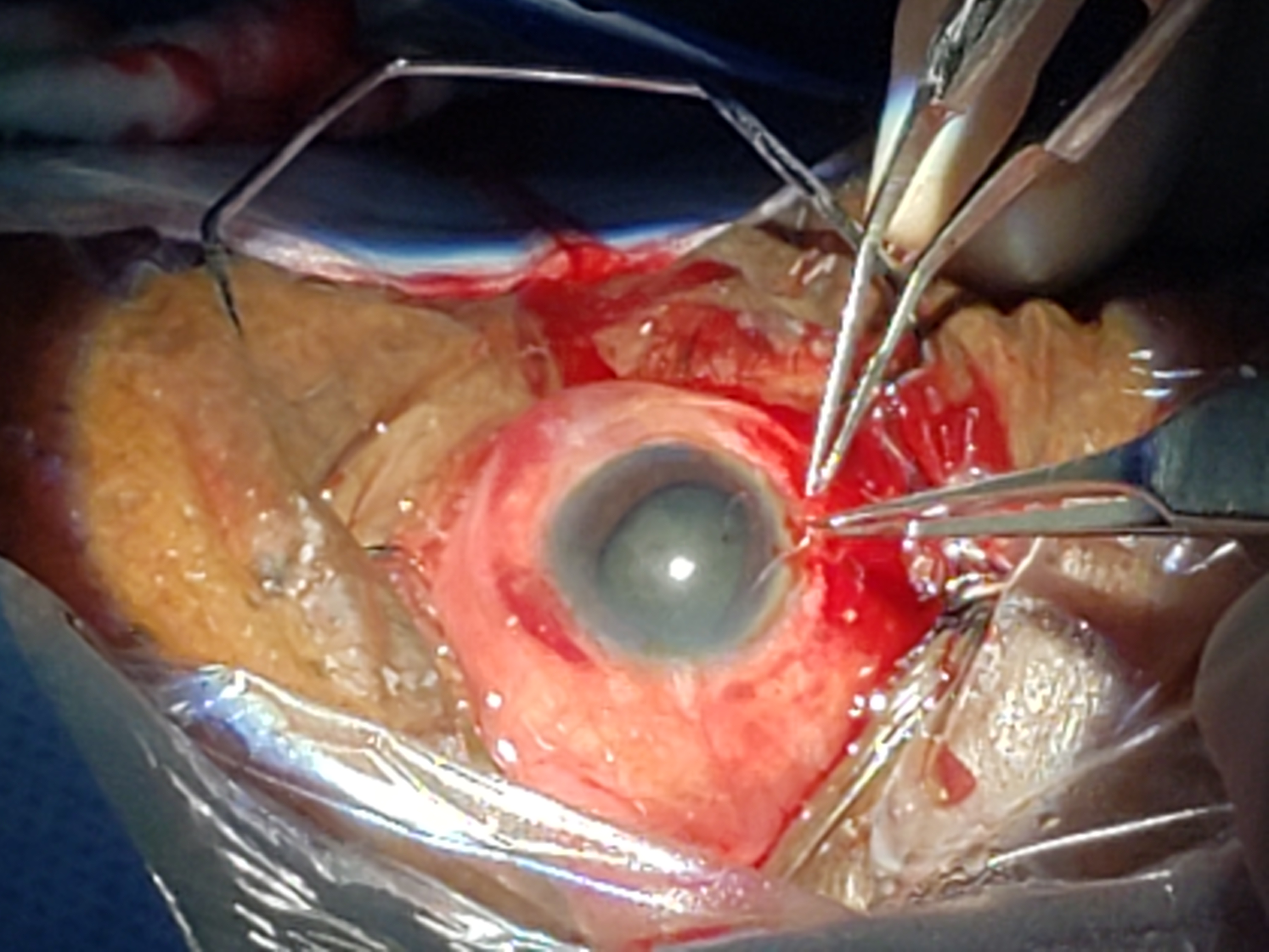
Phacoemulsification requires the use of low flow rates and ultrasound settings. Triamcinolone staining can identify and remove the prolapsed vitreous in the anterior chamber.[77] Cases of compromised zonular support may be inadequate for an in-the-bag IOL. However, capsular tension rings (CTR) can be used during surgery as a support tool or long-term implant device for IOL fixation (Image. Traumatic Cataract Repair).[74] Traumatic subluxation can be addressed by preserving the capsular bag using fixated CTR.[75] The key surgical steps of capsulorhexis and cortical removal can worsen preexisting zonular compromise. Nonetheless, the use of CTR can effectively address this issue.

When there is a small tear in the posterior capsule, placing a foldable IOL in the capsular bag is a viable solution if there is enough support.[76] A three-piece IOL can also be placed in the ciliary sulcus with sufficient capsular support.[78] However, studies have demonstrated that visual acuity is superior when the IOL is placed in the capsular bag rather than the ciliary sulcus due to decreased inflammation. Other alternatives, such as an anterior chamber IOL and iris and scleral suture fixated IOLs, can be considered if there is inadequate support.[78][79] Furthermore, newer techniques for glued sutureless scleral-fixated IOLs are promising. Light-adjustable IOLs may also improve refractive outcomes in difficult cases of ocular trauma.

Procedural Considerations

Either a Venturi or peristaltic pump may be employed during phacoemulsification surgery. Keeping the parameters low is recommended, and the divide-and-conquer technique is preferred. If the nucleus is visible, the stop-and-chop method can be employed using an Akahoshi pre-chopper after sculpting a central groove. Pushing down the nucleus during chopping should be avoided. The irrigation and aspiration tip can easily aspirate the lens nucleus. Beginning with the nucleus and finishing with the cortex is the best approach to prevent damaging the posterior capsule.

During phacoemulsification surgery, it is recommended to utilize conservative settings.[73][80] A low bottle height of 60 to 75 cm, a low aspiration rate of 18 to 20 mL/min, and a low vacuum pressure of 180 to 200 mm Hg can be used initially and adjusted based on the specific device and clinical situation. The amount of ultrasound power utilized will depend on the hardness of the cataract.[81] 

A Simcoe cannula is another option to aspirate a soft cataract and can be inserted through a small incision measuring 2.8 to 3.0 mm. If both lens components are present, the hardness of the cataract should be assessed before surgery. Hydrodissection can separate the nucleus from the cortex, protecting the posterior capsule during phacoemulsification.

The selection and placement of the IOL are entirely dependent on the clinical situation, with consideration given to visibility.[63] Occasionally, placing the IOL in a later secondary procedure may be recommended.[63] It is not advised to use an anterior chamber lens for young patients or those with a lacerated cornea. After the surgery, cefuroxime is injected into the anterior chamber for antibiotic prophylaxis. Tamponade with dispersive viscoelastic or intracameral epinephrine injection for vasoconstriction may be utilized to control bleeding during cataract extraction.

Differential Diagnosis

Traumatic cataract always occurs in the setting of acute or remote ocular trauma. However, the following conditions must be excluded:

  • Acute angle-closure glaucoma
  • Angle-recession glaucoma 
  • Choroidal rupture
  • Retinal detachment
  • Laceration of the corneoscleral complex
  • Iridodialysis
  • Iridocyclitis
  • Ectopia Lentis (subluxation and luxation)
  • Hyphema 
  • Vitreous Hemorrhage 
  • Age-related or senile cataract
  • Sudden vision loss

Prognosis

Blindness caused by injury poses a significant burden on society and individuals. There remains a pressing need for standardized classification, investigations, and treatment guidelines to enhance outcomes.[4][13][40] The Birmingham Eye Trauma Terminology (BETT) was established in 1996 to standardize ocular trauma documentation. The BETT facilitates the investigation of visual outcomes following traumatic cataract surgery.[4][13] However, many issues and controversies surrounding the management of traumatic cataracts remain. Although a controlled, prospective clinical trial is not feasible, studies have been conducted to forecast visual outcomes in cases of traumatic cataracts. Predicting visual outcomes is complicated by multiple factors beyond just the lens.[82] 

Predicting visual outcomes in patients with traumatic cataracts is a complex process requiring consideration of various factors.[2][4][19][83] The Ocular Trauma Score (OTS), developed in the early 2000s, is a validated tool for predicting visual acuity after ocular trauma.[4][20][84] The variables used in the OTS are initial vision, rupture, endophthalmitis, perforating injury, retinal detachment, and afferent pupillary defect. A retrospective study of over 300 children has demonstrated the effectiveness of OTS in predicting visual outcomes in pediatric patients with traumatic cataracts. However, further research is needed to improve the accuracy of OTS in children. Two recent studies evaluating the value of the OTS in 2-year-old pediatric patients reached conflicting conclusions, leading to some controversy.[85][86][87] A new pediatric OTS has been developed to enhance accuracy in children with uncertain initial vision.[83][87] 

The morphology of cataracts plays a significant role in determining the surgical technique and visual outcome.[4][88][89] However, there is no consensus on whether primary or secondary cataract extraction is optimal.

Infection and endophthalmitis following an open globe injury are common.[90] Infection is also common with a retained intraocular foreign body.[91] Using plants with antimicrobial and antifungal properties may effectively prevent infection following penetrating injuries caused by a wooden object.[90][92] According to a study of patients with open or closed globe injuries with similar characteristics, a satisfactory grade of vision after managing traumatic cataracts was significantly better in the eyes with open injuries.[40] 

Complications

The complications of traumatic cataracts include phacoanaphylactic uveitis, retinal detachment, choroidal rupture, hyphema, retrobulbar hemorrhage, traumatic optic neuropathy, globe rupture, and phacolytic, phacomorphic, pupillary block, or angle-recession glaucoma.[2][62][93][94][32][95][96][97][98][99][100][101][102] 

Preventing retinal detachment requires removing the prolapsed vitreous in the anterior chamber through a lateral incision and performing an anterior vitrectomy using a second instrument.[103] Using the phacoemulsification handpiece to pull or heat the vitreous or removing it through the wound with a sponge must be avoided. If bleeding occurs in the anterior chamber, prompt removal and irrigation are essential to prevent hematocornea. During cataract extraction, dispersive viscoelastic or intracameral epinephrine can be utilized to control spontaneous bleeding.

Postoperative and Rehabilitation Care

Progress after cataract surgery is monitored during examinations performed 1 day, 1 week, and 1 month after the procedure.[5][104] Patients are prescribed ophthalmic antibiotics and anti-inflammatory medications. If complications occur, ophthalmic steroids or IOP-lowering medications may be prescribed.

Each postoperative examination should include a slit-lamp examination and an assessment of visual acuity.[105]

Pediatric patients at risk for postoperative amblyopia may benefit from patching the good eye during postoperative visual rehabilitation. Pediatric patients risk developing posterior capsular opacification that can lead to amblyopia if left untreated.[106][107] Inflammatory responses in young patients may cause fibrinous uveitis, which can be managed with aggressive perioperative steroid regimens.[108]

Deterrence and Patient Education

Educating patients about the risks of developing traumatic cataracts and their sequelae is crucial.[109][110]. Patients should be advised to seek immediate care following any ocular trauma. Additionally, patients should be educated on the benefits of wearing sunglasses to shield their eyes from harmful ultraviolet rays and the proper use of eye protection during high-risk activities. 

Enhancing Healthcare Team Outcomes

Managing traumatic cataracts requires an interprofessional team of healthcare professionals, including nurses, laboratory technologists, pharmacists, and clinicians of various specialties.[5][111] Without proper management, traumatic cataracts can lead to devastating outcomes, blindness, and poor quality of life.[38][112] Risk factors, including smoking, excessive alcohol consumption, extended exposure to sunlight without protective sunglasses, diabetes, severe eye or head injuries, other eye conditions, prolonged use of systemic steroids, and radiation treatment for cancer or other diseases, can contribute to traumatic cataracts.[113] Taking proper precautions to prevent eye trauma, such as using protective eyewear like glasses and eye shields, limiting time spent in hazardous situations, and avoiding exposure to infrared or ultraviolet rays, is something every patient should be educated about.[114]


(Click Image to Enlarge)
Traumatic Cataract Secondary to Penetrating Ocular Injury
Traumatic Cataract Secondary to Penetrating Ocular Injury. Corneal sutures are visualized at the site of the ruptured globe repair.
Contributed by CJ Rapuano, MD

(Click Image to Enlarge)
Posttraumatic Crystalline Lens Subluxation
Contributed by F Woreta, MD, MPH

(Click Image to Enlarge)
Traumatic Cataract. A traumatic cataract associated with zonular dialysis and anterior capsular fibrosis.
Contributed by Dalil Bonabe, MD

(Click Image to Enlarge)
Traumatic Cataract Repair
Traumatic Cataract Repair. A traumatic cataract with zonular dialysis secondary to remote penetrating ocular injury managed with a capsular tension ring and 3-piece intraocular lens.
Contributed by Dalil Bonabe, MD
Details

Editor:

Bharat Gurnani

Updated:

8/27/2023 5:27:59 PM

References


[1]

Khokhar S, Gupta S, Yogi R, Gogia V, Agarwal T. Epidemiology and intermediate-term outcomes of open- and closed-globe injuries in traumatic childhood cataract. European journal of ophthalmology. 2014 Jan-Feb:24(1):124-30. doi: 10.5301/ejo.5000342. Epub 2013 Jul 26     [PubMed PMID: 23918072]


[2]

Peleja MB, da Cunha FBS, Peleja MB, Rohr JTD. Epidemiology and prognosis factors in open globe injuries in the Federal District of Brazil. BMC ophthalmology. 2022 Mar 9:22(1):111. doi: 10.1186/s12886-021-02183-z. Epub 2022 Mar 9     [PubMed PMID: 35264122]


[3]

Baur ID, Auffarth GU, Łabuz G, Khoramnia R. [Implantation of a Toric IOL with Enhanced Depth of Focus for Unilateral Traumatic Cataract]. Klinische Monatsblatter fur Augenheilkunde. 2023 Jun:240(6):819-823. doi: 10.1055/a-1809-5187. Epub 2022 May 17     [PubMed PMID: 35580622]


[4]

Chaudhary A, Singh R, Singh SP. Prognostic value of Ocular Trauma Score and pediatric Penetrating Ocular Trauma Score in predicting the visual prognosis following ocular injury. Romanian journal of ophthalmology. 2022 Apr-Jun:66(2):146-152. doi: 10.22336/rjo.2022.29. Epub     [PubMed PMID: 35935081]


[5]

Zhu AY, Kraus CL. Practice Patterns in the Surgical Management of Pediatric Traumatic Cataracts. Journal of pediatric ophthalmology and strabismus. 2020 May 1:57(3):190-198. doi: 10.3928/01913913-20200304-01. Epub     [PubMed PMID: 32453853]


[6]

Gupta VB, Rajagopala M, Ravishankar B. Etiopathogenesis of cataract: an appraisal. Indian journal of ophthalmology. 2014 Feb:62(2):103-10. doi: 10.4103/0301-4738.121141. Epub     [PubMed PMID: 24618482]


[7]

Shanbagh S, Matalia J, Kannan R, Shetty R, Panmand P, Muthu SO, Chaurasia SS, Deshpande V, Bhattacharya SS, Gopalakrishnan AV, Ghosh A. Distinct gene expression profiles underlie morphological and etiological differences in pediatric cataracts. Indian journal of ophthalmology. 2023 May:71(5):2143-2151. doi: 10.4103/IJO.IJO_3269_22. Epub     [PubMed PMID: 37203095]


[8]

Sen P, Sreelakshmi K, Bhende P, Gopal L, Rishi P, Rishi E, Susvar P, Attiku Y. Outcome of Sutured Scleral-Fixated Intraocular Lens in Blunt and Penetrating Trauma in Children. Ophthalmic surgery, lasers & imaging retina. 2018 Oct 1:49(10):757-764. doi: 10.3928/23258160-20181002-03. Epub     [PubMed PMID: 30395661]


[9]

Stepp MA, Menko AS. Immune responses to injury and their links to eye disease. Translational research : the journal of laboratory and clinical medicine. 2021 Oct:236():52-71. doi: 10.1016/j.trsl.2021.05.005. Epub 2021 May 27     [PubMed PMID: 34051364]


[10]

Hilely A, Leiba H, Achiron A, Hecht I, Parness-Yossifon R. Traumatic Cataracts in Children, Long-Term Follow-up in an Israeli Population: A Retrospective Study. The Israel Medical Association journal : IMAJ. 2019 Sep:21(9):599-602     [PubMed PMID: 31542904]

Level 2 (mid-level) evidence

[11]

Tartarella MB, Britez-Colombi GF, Milhomem S, Lopes MC, Fortes Filho JB. Pediatric cataracts: clinical aspects, frequency of strabismus and chronological, etiological, and morphological features. Arquivos brasileiros de oftalmologia. 2014 May-Jun:77(3):143-7     [PubMed PMID: 25295898]


[12]

Agrawal R, Ho SW, Teoh S. Pre-operative variables affecting final vision outcome with a critical review of ocular trauma classification for posterior open globe (zone III) injury. Indian journal of ophthalmology. 2013 Oct:61(10):541-5. doi: 10.4103/0301-4738.121066. Epub     [PubMed PMID: 24212303]


[13]

Shah M, Shah S, Upadhyay P, Agrawal R. Controversies in traumatic cataract classification and management: a review. Canadian journal of ophthalmology. Journal canadien d'ophtalmologie. 2013 Aug:48(4):251-8. doi: 10.1016/j.jcjo.2013.03.010. Epub     [PubMed PMID: 23931462]


[14]

Asano N, Schlötzer-Schrehardt U, Dörfler S, Naumann GO. Ultrastructure of contusion cataract. Archives of ophthalmology (Chicago, Ill. : 1960). 1995 Feb:113(2):210-5     [PubMed PMID: 7864754]


[15]

El Kaissoumi L, Mrini B. [Neglected post-traumatic ruptured cataract]. The Pan African medical journal. 2022:42():3. doi: 10.11604/pamj.2022.42.3.33130. Epub 2022 May 4     [PubMed PMID: 35685383]


[16]

Fagerholm PP, Philipson BT. Human traumatic cataract. A quantitative microradiographic and electron microscopic study. Acta ophthalmologica. 1979 Feb:57(1):20-32     [PubMed PMID: 419973]


[17]

Bendeddouche K, Assaf E, Emadisson H, Forestier F, Salvanet-Bouccara A. [Air bags and eye injuries: chemical burns and major traumatic ocular lesions--a case study]. Journal francais d'ophtalmologie. 2003 Oct:26(8):819-23     [PubMed PMID: 14586223]

Level 3 (low-level) evidence

[18]

Rodricks D, Loya A, Mohamed M, Al-Mohtaseb Z. Visual outcomes of open globe injury patients with traumatic cataracts. International ophthalmology. 2022 Jul:42(7):2039-2046. doi: 10.1007/s10792-021-02195-0. Epub 2022 Feb 8     [PubMed PMID: 35133577]


[19]

Khatri A, Shrestha SM, Kuhn F, Subramanian P, Hoskin AK, Pradhan E, Agrawal R. Ophthalmic Trauma Correlation Matrix (OTCM): a potential novel tool for evaluation of concomitant ocular tissue damage in open globe injuries. Graefe's archive for clinical and experimental ophthalmology = Albrecht von Graefes Archiv fur klinische und experimentelle Ophthalmologie. 2022 May:260(5):1773-1778. doi: 10.1007/s00417-021-05491-8. Epub 2021 Nov 18     [PubMed PMID: 34792638]


[20]

Doğan E, Çelik E, Gündoğdu KÖ, Alagöz G. Characteristics of pediatric traumatic cataract and factors affecting visual outcomes. Injury. 2023 Jan:54(1):168-172. doi: 10.1016/j.injury.2022.09.034. Epub 2022 Sep 21     [PubMed PMID: 36167690]


[21]

Goldblum D, Körner F, Garweg JG. [Pharmacological therapy approaches in ocular traumatology]. Therapeutische Umschau. Revue therapeutique. 2001 Jan:58(1):8-12     [PubMed PMID: 11217490]


[22]

Memon MN, Narsani AK, Nizamani NB. Visual outcome of unilateral traumatic cataract. Journal of the College of Physicians and Surgeons--Pakistan : JCPSP. 2012 Aug:22(8):497-500     [PubMed PMID: 22868014]


[23]

Mangan MS, Arıcı C, Tuncer İ, Yetik H. Isolated Anterior Lens Capsule Rupture Secondary to Blunt Trauma: Pathophysiology and Treatment. Turkish journal of ophthalmology. 2016 Aug:46(4):197-199. doi: 10.4274/tjo.85547. Epub 2016 Aug 15     [PubMed PMID: 28058159]


[24]

Elksnis Ē, Vanags J, Elksne E, Gertners O, Laganovska G. Isolated posterior capsule rupture after blunt eye injury. Clinical case reports. 2021 Apr:9(4):2105-2108. doi: 10.1002/ccr3.3956. Epub 2021 Feb 18     [PubMed PMID: 33936647]

Level 3 (low-level) evidence

[25]

Gulati S, Hanebrink KA, Henry M, Munro M, Chan RVP, Edward DP. Open globe injury and intraocular foreign body following crossbow-related penetrating ocular trauma. American journal of ophthalmology case reports. 2022 Jun:26():101441. doi: 10.1016/j.ajoc.2022.101441. Epub 2022 Feb 23     [PubMed PMID: 35252625]

Level 3 (low-level) evidence

[26]

Smith MP, Colyer MH, Weichel ED, Stutzman RD. Traumatic cataracts secondary to combat ocular trauma. Journal of cataract and refractive surgery. 2015 Aug:41(8):1693-8. doi: 10.1016/j.jcrs.2014.12.059. Epub     [PubMed PMID: 26432127]


[27]

Liu X, Wang L, Du C, Li D, Fan Y. Mechanism of lens capsular rupture following blunt trauma: a finite element study. Computer methods in biomechanics and biomedical engineering. 2015:18(8):914-21. doi: 10.1080/10255842.2014.975798. Epub     [PubMed PMID: 25427212]


[28]

Inanc M, Tekin K, Erol YO, Sargon MF, Koc M, Budakoglu O, Yılmazbas P. The ultrastructural alterations in the lens capsule and epithelium in eyes with traumatic white cataract. International ophthalmology. 2019 Jan:39(1):47-53. doi: 10.1007/s10792-017-0783-0. Epub 2017 Nov 30     [PubMed PMID: 29189944]


[29]

Singh RB, Thakur S, Ichhpujani P. Traumatic rosette cataract. BMJ case reports. 2018 Nov 28:11(1):. doi: 10.1136/bcr-2018-227465. Epub 2018 Nov 28     [PubMed PMID: 30567140]

Level 3 (low-level) evidence

[30]

Marcantonio JM, Syam PP, Liu CS, Duncan G. Epithelial transdifferentiation and cataract in the human lens. Experimental eye research. 2003 Sep:77(3):339-46     [PubMed PMID: 12907166]


[31]

Kuriyan AE, Flynn HW Jr, Yoo SH. Subluxed traumatic cataract: optical coherence tomography findings and clinical management. Clinical ophthalmology (Auckland, N.Z.). 2012:6():1997-9. doi: 10.2147/OPTH.S37393. Epub 2012 Dec 4     [PubMed PMID: 23271877]


[32]

Leung VC, Fung SSM, Muni R, Ali A. Phacomorphic Angle-closure Following Silicone Oil Tamponade in a Pediatric Patient. Journal of glaucoma. 2018 Jun:27(6):e106-e109. doi: 10.1097/IJG.0000000000000957. Epub     [PubMed PMID: 29613981]


[33]

Fagerholm PP. The response of the lens to trauma. Transactions of the ophthalmological societies of the United Kingdom. 1982:102 Pt 3():369-74     [PubMed PMID: 6964283]


[34]

Shah MA, Shah SM, Applewar A, Patel C, Shah S, Patel U. OcularTrauma Score: a useful predictor of visual outcome at six weeks in patients with traumatic cataract. Ophthalmology. 2012 Jul:119(7):1336-41. doi: 10.1016/j.ophtha.2012.01.020. Epub 2012 Mar 27     [PubMed PMID: 22459803]


[35]

Walker NJ, Foster A, Apel AJ. Traumatic expulsive iridodialysis after small-incision sutureless cataract surgery. Journal of cataract and refractive surgery. 2004 Oct:30(10):2223-4     [PubMed PMID: 15474840]


[36]

Rafferty NS, Goossens W. Ultrastructure of traumatic cataractogenesis in the frog: a comparison with mouse and human lens. The American journal of anatomy. 1977 Mar:148(3):385-407     [PubMed PMID: 300987]


[37]

Klysik A, Kaszuba-Bartkowiak K, Jurowski P. Axial Length of the Eyeball Is Important in Secondary Dislocation of the Intraocular Lens, Capsular Bag, and Capsular Tension Ring Complex. Journal of ophthalmology. 2016:2016():6431438. doi: 10.1155/2016/6431438. Epub 2016 Mar 16     [PubMed PMID: 27069675]


[38]

Jones WL. Traumatic injury to the lens. Optometry clinics : the official publication of the Prentice Society. 1991:1(2):125-42     [PubMed PMID: 1799823]


[39]

Shah MA, Shah SM, Shah SB, Patel UA. Effect of interval between time of injury and timing of intervention on final visual outcome in cases of traumatic cataract. European journal of ophthalmology. 2011 Nov-Dec:21(6):760-5. doi: 10.5301/EJO.2011.6482. Epub     [PubMed PMID: 21445838]

Level 3 (low-level) evidence

[40]

Batchelor A, Lacy M, Hunt M, Lu R, Lee AY, Lee CS, Saraf SS, Chee YE, IRIS Registry Analytic Center Consortium. Predictors of Long-term Ophthalmic Complications after Closed Globe Injuries Using the Intelligent Research in Sight (IRIS®) Registry. Ophthalmology science. 2023 Mar:3(1):100237. doi: 10.1016/j.xops.2022.100237. Epub 2022 Oct 28     [PubMed PMID: 36561352]


[41]

Sharma AK, Aslami AN, Srivastava JP, Iqbal J. Visual Outcome of Traumatic Cataract at a Tertiary Eye Care Centre in North India: A Prospective Study. Journal of clinical and diagnostic research : JCDR. 2016 Jan:10(1):NC05-8. doi: 10.7860/JCDR/2016/17216.7049. Epub 2016 Jan 1     [PubMed PMID: 26894101]


[42]

Singh RK, Singh S. Management of partially absorbed white soft cataract post penetrating injury to eye. Indian journal of ophthalmology. 2023 Jan:71(1):321. doi: 10.4103/ijo.IJO_1586_22. Epub     [PubMed PMID: 36588276]


[43]

Wu B, Yuan X, Chen S. Comperative analysis of accuracy between low-frequency ultrasound biomicroscopy and 14-MHz ultrasonography with tissue harmonic imaging for the evaluation of the posterior lens capsule in traumatic cataracts. BMC ophthalmology. 2021 Oct 22:21(1):375. doi: 10.1186/s12886-021-02094-z. Epub 2021 Oct 22     [PubMed PMID: 34686169]


[44]

Dezhagah H. Circular anterior lens capsule rupture caused by blunt ocular trauma. Middle East African journal of ophthalmology. 2010 Jan:17(1):103-5. doi: 10.4103/0974-9233.61227. Epub     [PubMed PMID: 20543947]


[45]

Eze KC, Enock ME, Eluehike SU. Ultrasonic evaluation of orbito-ocular trauma in Benin-City, Nigeria. The Nigerian postgraduate medical journal. 2009 Sep:16(3):198-202     [PubMed PMID: 19767906]


[46]

Agrawal R, Shah M, Mireskandari K, Yong GK. Controversies in ocular trauma classification and management: review. International ophthalmology. 2013 Aug:33(4):435-45. doi: 10.1007/s10792-012-9698-y. Epub 2013 Jan 22     [PubMed PMID: 23338232]


[47]

Xiong S, Xia X. Risk factors and surgical outcomes for the concurrence of intraocular lens dislocation with vitreoretinal diseases. Zhong nan da xue xue bao. Yi xue ban = Journal of Central South University. Medical sciences. 2022 Jul 28:47(7):881-887. doi: 10.11817/j.issn.1672-7347.2022.220264. Epub     [PubMed PMID: 36039584]


[48]

Segev Y, Goldstein M, Lazar M, Reider-Groswasser I. CT appearance of a traumatic cataract. AJNR. American journal of neuroradiology. 1995 May:16(5):1174-5     [PubMed PMID: 7639149]


[49]

Chaniyara MH, Pujari A, Aron N, Sharma N. Optimising the surgical outcome in a case of post-traumatic cataract using ultrasound biomicroscopy. BMJ case reports. 2017 Jul 26:2017():. pii: bcr-2017-220579. doi: 10.1136/bcr-2017-220579. Epub 2017 Jul 26     [PubMed PMID: 28751510]

Level 3 (low-level) evidence

[50]

Wang K, Liu J, Chen M. Role of B-scan ultrasonography in the localization of intraocular foreign bodies in the anterior segment: a report of three cases. BMC ophthalmology. 2015 Aug 14:15():102. doi: 10.1186/s12886-015-0076-1. Epub 2015 Aug 14     [PubMed PMID: 26268356]

Level 3 (low-level) evidence

[51]

Tabatabaei SA, Soleimani M, Etesali H, Naderan M. Accuracy of Swept-Source Optical Coherence Tomography and Ultrasound Biomicroscopy for Evaluation of Posterior Lens Capsule in Traumatic Cataract. American journal of ophthalmology. 2020 Aug:216():55-58. doi: 10.1016/j.ajo.2020.03.030. Epub 2020 Apr 2     [PubMed PMID: 32247777]


[52]

Mansour AM, Jaroudi MO, Hamam RN, Maalouf FC. Isolated posterior capsular rupture following blunt head trauma. Clinical ophthalmology (Auckland, N.Z.). 2014:8():2403-7. doi: 10.2147/OPTH.S73990. Epub 2014 Nov 28     [PubMed PMID: 25506201]


[53]

Pujari A, Sharma N. The Emerging Role of Anterior Segment Optical Coherence Tomography in Cataract Surgery: Current Role and Future Perspectives. Clinical ophthalmology (Auckland, N.Z.). 2021:15():389-401. doi: 10.2147/OPTH.S286996. Epub 2021 Feb 3     [PubMed PMID: 33568893]

Level 3 (low-level) evidence

[54]

Załecki K. [Diagnostic B-scan ultrasound of the lens before cataract surgery with intraocular artificial lens implantation]. Klinika oczna. 1995 Jun:97(6):192-9     [PubMed PMID: 7643563]


[55]

Tabatabaei A, Hasanlou N, Kheirkhah A, Mansouri M, Faghihi H, Jafari H, Arefzadeh A, Moghimi S. Accuracy of 3 imaging modalities for evaluation of the posterior lens capsule in traumatic cataract. Journal of cataract and refractive surgery. 2014 Jul:40(7):1092-6. doi: 10.1016/j.jcrs.2013.10.048. Epub 2014 May 15     [PubMed PMID: 24836968]


[56]

Limpas Y, Rodarie C, Wary P. [Anterior segment examination of a post-traumatic, multioperated eye with Visante OCT. A case report]. Journal francais d'ophtalmologie. 2009 Nov:32(9):669-72. doi: 10.1016/j.jfo.2009.09.008. Epub 2009 Oct 29     [PubMed PMID: 19879017]

Level 3 (low-level) evidence

[57]

Qi Y, Zhang YF, Zhu Y, Wan MG, Du SS, Yue ZZ. Prognostic Factors for Visual Outcome in Traumatic Cataract Patients. Journal of ophthalmology. 2016:2016():1748583. doi: 10.1155/2016/1748583. Epub 2016 Aug 9     [PubMed PMID: 27595014]

Level 2 (mid-level) evidence

[58]

Cohen KL. Inaccuracy of intraocular lens power calculation after traumatic corneal laceration and cataract. Journal of cataract and refractive surgery. 2001 Sep:27(9):1519-22     [PubMed PMID: 11566543]


[59]

Giles K, Christelle D, Yannick B, Fricke OH, Wiedemann P. Cataract surgery with intraocular lens implantation in children aged 5-15 in local anaesthesia: visual outcomes and complications. The Pan African medical journal. 2016:24():200     [PubMed PMID: 27795795]


[60]

Navaleza JS, Pendse SJ, Blecher MH. Choosing anesthesia for cataract surgery. Ophthalmology clinics of North America. 2006 Jun:19(2):233-7     [PubMed PMID: 16701160]


[61]

Sen P, Shah C, Sen A, Jain E, Mohan A. Primary versus secondary intraocular lens implantation in traumatic cataract after open-globe injury in pediatric patients. Journal of cataract and refractive surgery. 2018 Dec:44(12):1446-1453. doi: 10.1016/j.jcrs.2018.07.061. Epub 2018 Oct 5     [PubMed PMID: 30297231]


[62]

Özbilen KT, Altınkurt E. Impact of the possible prognostic factors for visual outcomes of traumatic cataract surgery. International ophthalmology. 2020 Nov:40(11):3163-3173. doi: 10.1007/s10792-020-01502-5. Epub 2020 Jul 10     [PubMed PMID: 32651906]


[63]

Rumelt S, Rehany U. The influence of surgery and intraocular lens implantation timing on visual outcome in traumatic cataract. Graefe's archive for clinical and experimental ophthalmology = Albrecht von Graefes Archiv fur klinische und experimentelle Ophthalmologie. 2010 Sep:248(9):1293-7. doi: 10.1007/s00417-010-1378-x. Epub 2010 Jun 29     [PubMed PMID: 20585800]


[64]

Bowe T, Serina A, Armstrong M, Welcher JE, Adebona O, Gore C, Staffa SJ, Zurakowski D, Shah AS. Timing of Ocular Hypertension After Pediatric Closed-Globe Traumatic Hyphema: Implications for Surveillance. American journal of ophthalmology. 2022 Jan:233():135-143. doi: 10.1016/j.ajo.2021.04.033. Epub 2021 May 13     [PubMed PMID: 33991515]


[65]

Lim Z, Rubab S, Chan YH, Levin AV. Management and outcomes of cataract in children: the Toronto experience. Journal of AAPOS : the official publication of the American Association for Pediatric Ophthalmology and Strabismus. 2012 Jun:16(3):249-54. doi: 10.1016/j.jaapos.2011.12.158. Epub     [PubMed PMID: 22681941]


[66]

Günaydın NT, Oral AYA. Pediatric traumatic cataracts: 10-year experience of a tertiary referral center. BMC ophthalmology. 2022 May 2:22(1):199. doi: 10.1186/s12886-022-02427-6. Epub 2022 May 2     [PubMed PMID: 35501774]


[67]

Writing Committee for the Pediatric Eye Disease Investigator Group (PEDIG), Bothun ED, Repka MX, Dean TW, Gray ME, Lenhart PD, Li Z, Morrison DG, Wallace DK, Kraker RT, Cotter SA, Holmes JM. Visual Outcomes and Complications After Lensectomy for Traumatic Cataract in Children. JAMA ophthalmology. 2021 Jun 1:139(6):647-653. doi: 10.1001/jamaophthalmol.2021.0980. Epub     [PubMed PMID: 33956055]


[68]

Hamad JB, Sivaraman KR, Snyder ME. Lamellar Dissection Technique for Traumatic Cataract With Corneal Incarceration. Cornea. 2021 Mar 1:40(3):393-397. doi: 10.1097/ICO.0000000000002544. Epub     [PubMed PMID: 33214414]


[69]

Nagra PK, Raber IM. Epithelial ingrowth in a phakic corneal transplant patient after traumatic wound dehiscence. Cornea. 2003 Mar:22(2):184-6     [PubMed PMID: 12605060]


[70]

Titiyal JS, Kaur M, Falera R. Intraoperative optical coherence tomography in anterior segment surgeries. Indian journal of ophthalmology. 2017 Feb:65(2):116-121. doi: 10.4103/ijo.IJO_868_16. Epub     [PubMed PMID: 28345566]


[71]

Marmur RK, Skripnichenko ZM, Iakimenko SA. [Evaluation of the state of the anterior part of the eye in patients with traumatic cataracts using the method of ultrasonic echography]. Oftalmologicheskii zhurnal. 1970:25(3):192-8     [PubMed PMID: 5469359]


[72]

Allapitchai F, Ravindran M. Management of pediatric subluxated cataract and different methods to face the challenges. Indian journal of ophthalmology. 2023 Feb:71(2):673. doi: 10.4103/ijo.IJO_2057_22. Epub     [PubMed PMID: 36727391]


[73]

Oudjani N, Renault D, Courrier E, Malek Y. Phacoemulsification And Zonular Weakness: Contribution Of The Capsular Tension Ring With A Thread. Clinical ophthalmology (Auckland, N.Z.). 2019:13():2301-2304. doi: 10.2147/OPTH.S212063. Epub 2019 Dec 11     [PubMed PMID: 31849440]


[74]

Rai G, Sahai A, Kumar PR. Outcome of Capsular Tension Ring (CTR) Implant in Complicated Cataracts. Journal of clinical and diagnostic research : JCDR. 2015 Dec:9(12):NC05-7. doi: 10.7860/JCDR/2015/10425.6999. Epub 2015 Dec 1     [PubMed PMID: 26816928]


[75]

Ma X, Li Z. Capsular tension ring implantation after lens extraction for management of subluxated cataracts. International journal of clinical and experimental pathology. 2014:7(7):3733-8     [PubMed PMID: 25120749]


[76]

Vajpayee RB, Sharma N, Dada T, Gupta V, Kumar A, Dada VK. Management of posterior capsule tears. Survey of ophthalmology. 2001 May-Jun:45(6):473-88     [PubMed PMID: 11425354]

Level 3 (low-level) evidence

[77]

Kazem MA, Behbehani JH, Uboweja AK, Paramasivam RB. Traumatic cataract surgery assisted by trypan blue. Ophthalmic surgery, lasers & imaging : the official journal of the International Society for Imaging in the Eye. 2007 Mar-Apr:38(2):160-3. doi: 10.3928/15428877-20070301-14. Epub     [PubMed PMID: 17396700]


[78]

Faria MY, Ferreira NP, Canastro M. Management of dislocated intraocular lenses with iris suture. European journal of ophthalmology. 2017 Jan 19:27(1):45-48. doi: 10.5301/ejo.5000823. Epub 2016 Jun 23     [PubMed PMID: 27338117]


[79]

Kaynak S, Ozbek Z, Pasa E, Oner FH, Cingil G. Transscleral fixation of foldable intraocular lenses. Journal of cataract and refractive surgery. 2004 Apr:30(4):854-7     [PubMed PMID: 15093650]


[80]

Devgan U. Surgical techniques in phacoemulsification. Current opinion in ophthalmology. 2007 Feb:18(1):19-22     [PubMed PMID: 17159442]

Level 3 (low-level) evidence

[81]

Cotlier E, Rose M. Cataract extraction by the intracapsular methods and by phacoemulsification: the results of surgeons in training. Transactions. Section on Ophthalmology. American Academy of Ophthalmology and Otolaryngology. 1976 Jan-Feb:81(1):OP163-82     [PubMed PMID: 1274036]


[82]

Shah M, Shah S, Shah S, Prasad V, Parikh A. Visual recovery and predictors of visual prognosis after managing traumatic cataracts in 555 patients. Indian journal of ophthalmology. 2011 May-Jun:59(3):217-22. doi: 10.4103/0301-4738.81043. Epub     [PubMed PMID: 21586844]


[83]

Shah S, Shah M, Gunay R, Kataria A, Makhloga S, Vaghela M. New model for the prediction of visual outcomes in young children with mechanical ocular conditions and comparison with other models. Indian journal of ophthalmology. 2022 Aug:70(8):3045-3049. doi: 10.4103/ijo.IJO_3144_21. Epub     [PubMed PMID: 35918970]


[84]

Irawati Y, Ardiani LS, Gondhowiardjo TD, Hoskin AK. Predictive value and applicability of ocular trauma scores and pediatric ocular trauma scores in pediatric globe injuries. International journal of ophthalmology. 2022:15(8):1352-1356. doi: 10.18240/ijo.2022.08.19. Epub 2022 Aug 18     [PubMed PMID: 36017051]


[85]

Schörkhuber MM, Wackernagel W, Riedl R, Schneider MR, Wedrich A. Ocular trauma scores in paediatric open globe injuries. The British journal of ophthalmology. 2014 May:98(5):664-8. doi: 10.1136/bjophthalmol-2013-304469. Epub 2014 Feb 11     [PubMed PMID: 24518079]


[86]

Shah MA, Agrawal R, Teoh R, Shah SM, Patel K, Gupta S, Gosai S. Pediatric ocular trauma score as a prognostic tool in the management of pediatric traumatic cataracts. Graefe's archive for clinical and experimental ophthalmology = Albrecht von Graefes Archiv fur klinische und experimentelle Ophthalmologie. 2017 May:255(5):1027-1036. doi: 10.1007/s00417-017-3616-y. Epub 2017 Feb 22     [PubMed PMID: 28224290]


[87]

Shah MA, Shah SM, Applewar A, Patel C, Patel K. Ocular Trauma Score as a predictor of final visual outcomes in traumatic cataract cases in pediatric patients. Journal of cataract and refractive surgery. 2012 Jun:38(6):959-65. doi: 10.1016/j.jcrs.2011.12.032. Epub     [PubMed PMID: 22624894]

Level 3 (low-level) evidence

[88]

Luo XJ, Cao K, Liu J, Duan QY, Chen SY, Zhang Y, Huang T, Mao XN, Li CG, Chen YS. [Gene analysis and clinical features of MYH9-related disease]. Zhonghua er ke za zhi = Chinese journal of pediatrics. 2021 Nov 2:59(11):957-962. doi: 10.3760/cma.j.cn112140-20210507-00389. Epub     [PubMed PMID: 34711031]


[89]

Ben Zina Z, Trigui A, Feki J, Ellouze S, Dhouib I, Charfi N, Chaabouni M. [Traumatic cataracts. Epidemiology, treatment, and prognosis (report of 60 cases)]. La Tunisie medicale. 1998 Aug-Sep:76(8-9):254-7     [PubMed PMID: 9810862]

Level 3 (low-level) evidence

[90]

Bohrani Sefidan B, Tabatabaei SA, Soleimani M, Ahmadraji A, Shahriari M, Daraby M, Dehghani Sanij A, Mehrakizadeh A, Ramezani B, Cheraqpour K. Epidemiological characteristics and prognostic factors of post-traumatic endophthalmitis. The Journal of international medical research. 2022 Feb:50(2):3000605211070754. doi: 10.1177/03000605211070754. Epub     [PubMed PMID: 35114823]

Level 2 (mid-level) evidence

[91]

Li X, Zarbin MA, Bhagat N. Pediatric open globe injury: A review of the literature. Journal of emergencies, trauma, and shock. 2015 Oct-Dec:8(4):216-23. doi: 10.4103/0974-2700.166663. Epub     [PubMed PMID: 26604528]


[92]

Uppuluri A, Zarbin MA, Bhagat N. Risk Factors for Post-Open-Globe Injury Endophthalmitis. Journal of vitreoretinal diseases. 2020 Sep-Oct:4(5):353-359. doi: 10.1177/2474126420932322. Epub 2020 Jul 16     [PubMed PMID: 37008290]


[93]

Yeung L, Chen TL, Kuo YH, Chao AN, Wu WC, Chen KJ, Hwang YS, Chen Y, Lai CC. Severe vitreous hemorrhage associated with closed-globe injury. Graefe's archive for clinical and experimental ophthalmology = Albrecht von Graefes Archiv fur klinische und experimentelle Ophthalmologie. 2006 Jan:244(1):52-7     [PubMed PMID: 16044322]


[94]

Fau R. [Phacolytic glaucoma on a preceding traumatic cataract]. Bulletin des societes d'ophtalmologie de France. 1968 Nov:68(11):901-5     [PubMed PMID: 5746613]


[95]

Netland KE, Martinez J, LaCour OJ 3rd, Netland PA. Traumatic anterior lens dislocation: a case report. The Journal of emergency medicine. 1999 Jul-Aug:17(4):637-9     [PubMed PMID: 10431953]

Level 3 (low-level) evidence

[96]

Jan S, Khan S, Mohammad S. Hyphaema due to blunt trauma. Journal of the College of Physicians and Surgeons--Pakistan : JCPSP. 2003 Jul:13(7):398-401     [PubMed PMID: 12887842]


[97]

Mowatt L, Chambers C. Ocular morbidity of traumatic hyphema in a Jamaican hospital. European journal of ophthalmology. 2010 May-Jun:20(3):584-9     [PubMed PMID: 19967662]


[98]

Sridhar U, Tripathy K. Lens-Induced Inflammation. StatPearls. 2023 Jan:():     [PubMed PMID: 35015464]


[99]

Samanta R, Jayaraj S, Sood G, Agrawal A. Post-traumatic posterior giant retinal tear and macular hole associated retinal detachment. Indian journal of ophthalmology. 2022 May:70(5):1869. doi: 10.4103/ijo.IJO_1017_22. Epub     [PubMed PMID: 35502118]


[100]

Park JH, Jang JW, Kim SJ, Lee YJ. Traumatic optic neuropathy accompanying orbital grease gun injury. Korean journal of ophthalmology : KJO. 2010 Apr:24(2):134-8. doi: 10.3341/kjo.2010.24.2.134. Epub 2010 Apr 6     [PubMed PMID: 20379466]


[101]

Chung IY, Seo SW, Han YS, Kim E, Jung JM. Penetrating retrobulbar orbital foreign body: a transcranial approach. Yonsei medical journal. 2007 Apr 30:48(2):328-30     [PubMed PMID: 17461536]


[102]

Thakker MM, Ray S. Vision-limiting complications in open-globe injuries. Canadian journal of ophthalmology. Journal canadien d'ophtalmologie. 2006 Feb:41(1):86-92     [PubMed PMID: 16462880]


[103]

Sheng YH. [Vitreous prolapse during cataract surgery]. [Zhonghua yan ke za zhi] Chinese journal of ophthalmology. 1993 Jan:29(1):27-9     [PubMed PMID: 8334906]


[104]

Khodzhaev NS, Sobolev NP, Mushkova IA, Izmaylova SB, Karimova AN. [Visual rehabilitation of patients with large post-traumatic defects of the anterior eye segment through iris-lens diaphragm implantation]. Vestnik oftalmologii. 2017:133(6):23-29. doi: 10.17116/oftalma2017133623-29. Epub     [PubMed PMID: 29319666]


[105]

Eriksen JR, Bronsard A, Mosha M, Carmichael D, Hall A, Courtright P. Predictors of poor follow-up in children that had cataract surgery. Ophthalmic epidemiology. 2006 Aug:13(4):237-43     [PubMed PMID: 16877282]


[106]

Trivedi RH, Wilson ME. Posterior capsule opacification in pediatric eyes with and without traumatic cataract. Journal of cataract and refractive surgery. 2015 Jul:41(7):1461-4. doi: 10.1016/j.jcrs.2014.10.034. Epub 2015 Jul 23     [PubMed PMID: 26210053]


[107]

Verma N, Ram J, Sukhija J, Pandav SS, Gupta A. Outcome of in-the-bag implanted square-edge polymethyl methacrylate intraocular lenses with and without primary posterior capsulotomy in pediatric traumatic cataract. Indian journal of ophthalmology. 2011 Sep-Oct:59(5):347-51. doi: 10.4103/0301-4738.83609. Epub     [PubMed PMID: 21836338]


[108]

Baklouti K, Mhiri N, Mghaieth F, El Matri L. [Traumatic cataract: clinical and therapeutic aspects]. Bulletin de la Societe belge d'ophtalmologie. 2005:(298):13-7     [PubMed PMID: 16422217]


[109]

Agrawal R, Wei HS, Teoh S. Prognostic factors for open globe injuries and correlation of ocular trauma score at a tertiary referral eye care centre in Singapore. Indian journal of ophthalmology. 2013 Sep:61(9):502-6. doi: 10.4103/0301-4738.119436. Epub     [PubMed PMID: 24104709]


[110]

Jonas JB, Knorr HL, Budde WM. Prognostic factors in ocular injuries caused by intraocular or retrobulbar foreign bodies. Ophthalmology. 2000 May:107(5):823-8     [PubMed PMID: 10811069]


[111]

Arbisser LB. Managing intraoperative complications in cataract surgery. Current opinion in ophthalmology. 2004 Feb:15(1):33-9     [PubMed PMID: 14743017]

Level 3 (low-level) evidence

[112]

Ajamian PC. Traumatic cataract. Optometry clinics : the official publication of the Prentice Society. 1993:3(2):49-56     [PubMed PMID: 8268696]


[113]

Jinagal J, Gupta G, Gupta PC, Yangzes S, Singh R, Gupta R, Ram J. Visual outcomes of pediatric traumatic cataracts. European journal of ophthalmology. 2019 Jan:29(1):23-27. doi: 10.1177/1120672118757657. Epub 2018 Apr 3     [PubMed PMID: 29609478]


[114]

Guo Y, Liu Y, Xu H, Zhao Z, Gan D. Characteristics of paediatric patients hospitalised for eye trauma in 2007-2015 and factors related to their visual outcomes. Eye (London, England). 2021 Mar:35(3):945-951. doi: 10.1038/s41433-020-1002-1. Epub 2020 Jun 9     [PubMed PMID: 32518396]