Continuing Education Activity
Traumatic dental injuries (TDIs) are common and can have significant long-term consequences for both the hard and soft tissues of the oral cavity. This article outlines the presentation, clinical features, and management of TDIs affecting the primary teeth. As these injuries are common, this article contains information pertinent to clinicians in both primary and secondary care.
Objectives:
- Outline the etiological and risk factors of traumatic dental injury to the primary dentition.
- Summarize the epidemiology of pediatric dental injuries.
- Describe the history and physical examination of traumatic dental injuries in the primary dentition.
- Review the treatment options for primary traumatized teeth.
Introduction
Trauma to the primary dentition and oral cavity are common occurrences in children and young people. A recent international epidemiological study on traumatic dental injuries (TDIs) involving primary teeth revealed a worldwide prevalence of 22.7%.[1] They frequently occur in young children as they learn to crawl, walk, run and embrace their physical environment. These injuries often represent painful, distressing events and may result in adverse long-term physical, aesthetic and psychological consequences for the child.[2]
Children and their caregivers may present to various healthcare settings, including emergency departments, general dental practitioners, community dental services, specialist dental services, and pharmacists. Consequently, each service provider needs to have the appropriate knowledge, skills, and training to care for children with traumatic injuries to their primary dentition.[3]
Etiology
Unintentional falls, collisions, and leisure activities are the most common reasons for TDIs.[3] Falls account for most injuries in the primary dentition. This is unsurprising as it is typical for toddlers to crawl, stagger and fall before being able to walk as part of their development. The anterior teeth are the most commonly injured following trauma, particularly the maxillary central and lateral incisors.[4]
Various studies have examined the risk factors that increase the likelihood of TDIs in the primary dentition. Children with increased overjet and inadequate lip coverage have a higher incidence of dental injuries.[5][6] In primary dentition, the risk of dental trauma is three times greater with an increased overjet greater than 6 mm, whereas children with an overjet less than 3.5 mm are half as likely to suffer from dental trauma. A positive association between anterior open bite and traumatic injuries has also been found.[7]
It is important to remember that traumatic dental injuries and facial trauma can occur in cases of non-accidental (intentional) injuries. The head and neck region is frequently a target of abuse, with injuries occurring in 59-76% of physically abused children.[8]
Clinicians should assess whether the injuries sustained correlate and are consistent with the history of the accident. Repeated injuries, multiple bruises, delayed presentation, or injuries with unclear explanations may raise suspicions of abuse.[9] In scenarios where there is suspicion of abuse, prompt referral following local safeguarding protocols should be followed to investigate the incident thoroughly.
Epidemiology
Traumatic oral injuries account for 5% of all physical injuries in all age groups, and in pre-school children, the proportion increases to 17%.[10] A 2008 literature review concluded that one-third of children with primary dentition sustained a TDI, one-fourth of school-aged children with permanent teeth, and one-third of all adults sustained TDI.[11] However, the prevalence and incidence of TDI vary considerably worldwide, reflecting the socio-economic and behavioral differences and the lack of a standardized classification and registration system for traumatic dental injuries.
A global epidemiological study by Petti estimated worldwide TDI prevalence in primary and permanent dentitions. It concluded that more than one billion living people sustained a traumatic dental injury, which would rank fifth if included in the list of the world’s most frequent acute/chronic diseases and injuries.
History and Physical
A traumatic dental injury to the primary dentition may be the child's first visit to the dentist or emergency department, which can be challenging as a young child is difficult to examine and treat due to fear and lack of understanding of the situation. Early negative dental experiences are linked to significant dental anxiety in children; therefore, we should attempt to minimize anxiety during the initial visit for the child and caregiver. It is essential to adopt a structured and organized approach, conduct a complete clinical history, examine and conduct appropriate investigations, and arrange follow-ups. When examining a young child, the clinician may find knee-to-knee examination advantageous.[3]
Traumatic dental injuries can be classified into soft tissue injuries, hard tissues injuries (e.g., fractures), and periodontal injuries (e.g., luxations).
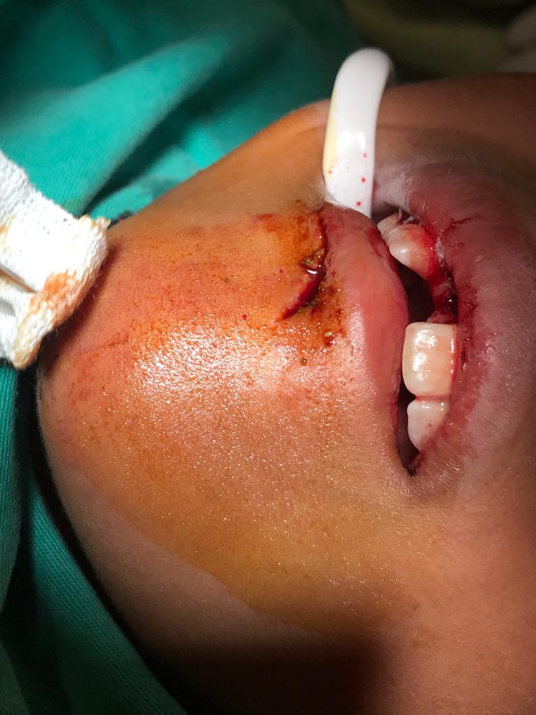
Soft Tissue Injuries
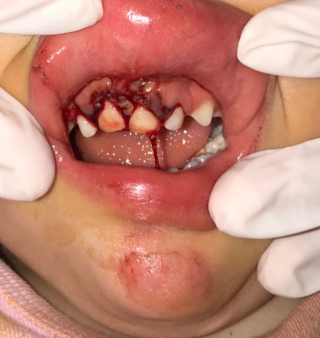
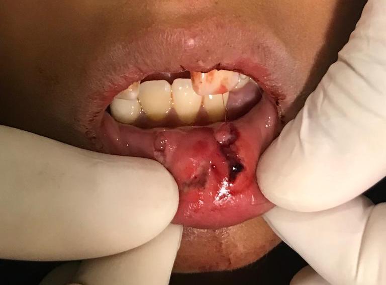
Soft tissue injuries are most commonly found in the 0 to 3 age group.[12] All soft tissue injuries should be identified and recorded with etiology, location, size, and depth of the wound.[13] They may present as abrasions, contusions, or lacerations on the extra-oral and intra-oral soft tissues, including the lips, oral mucosa, gingiva, and frenulum.[14] It is essential to examine the lips for possible embedded tooth fragments. Management of soft tissue injuries, beyond basic first aid, should be provided by those with experience with pediatric orofacial injuries.
Hard Tissue Injuries
Hard tissue injuries are classified into fractures of tooth structure and alveolar bone.
Enamel Fractures
These fractures are limited to the enamel only. No radiographic examination is recommended.
Enamel-Dentine Fractures (uncomplicated crown fracture)
An uncomplicated crown fracture affects the enamel and dentine without exposing the pulp. These fractures generally affect upper central incisors' mesial angles or incisal edges.[14] The missing tooth fragment location should be investigated during history taking. A radiograph of the soft tissues is recommended if the fractured tooth is suspected to be embedded in the lips, cheeks, or tongue.
Crown Fracture With Exposed Pulp (complicated crown fracture)
A complicated crown fracture involves enamel and dentine, plus the pulp is exposed. A periapical or an occlusal X-ray should be taken for diagnostic purposes and to establish a baseline.
Crown-Root Fractures
A crown-root fracture affects the enamel, dentine, and root; the pulp may or may not be exposed (complicated or uncomplicated). Additional findings may include loose but still attached fragments of the tooth. A periapical or an occlusal X-ray should be taken.
Root Fractures
A root fracture affects dentin, cementum, and pulp. They may occur in any direction or orientation and are generally classified as vertical or horizontal root fractures.[15] The coronal fragment may be mobile and displaced, and occlusal interference may be present. A periapical or an occlusal X-ray should be taken.
Alveolar Fractures
An alveolar fracture affects the alveolar bone (labial and palatal/lingual) and may extend to the adjacent bone. In alveolar fractures, the teeth in the affected segment are mobile, usually displaced, and occlusal interference is expected. Intra-oral soft tissue lacerations may also be present.[14][16] A radiographic examination should be performed.
Periodontal Injuries
Concussion
The tooth will be tender to touch, with normal mobility and no gingival sulcular bleeding. No baseline radiographs are recommended for tooth concussions.
Subluxation
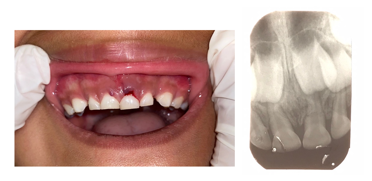
The tooth will show tenderness and increased mobility but no displacement. Bleeding from the gingival crevice may be noted. Radiographs should be taken for diagnostic purposes and to establish a baseline.
Extrusive Luxation
The partial displacement of the tooth out of its socket is known as extrusive luxation. The tooth appears elongated and has increased mobility. Occlusal interference may be present. A radiographic examination is recommended.
Lateral Luxation
The displacement of the tooth in a palatal/lingual or labial direction is known as lateral luxation. The tooth will be immobile and occlusal interference may be present. A radiographic examination is recommended.
Intrusive Luxation
Intrusive luxation is the apical displacement of the tooth, usually through the labial bone plate. The tooth may present as almost or complete disappearance of the tooth into the socket. The tooth can be palpated labially in clinical examination. Radiographic examination is recommended for diagnostic purposes and to determine the orientation of the intrusion.
When the tooth's apex is displaced toward or through the labial bone plate (away from the permanent tooth germ), the apical tip can be seen, and the image of the tooth will appear shorter (foreshortened) than the contralateral tooth. When the tooth's apex is displaced toward the permanent tooth germ, the apical tip cannot be visualized, and the image of the tooth will appear elongated.
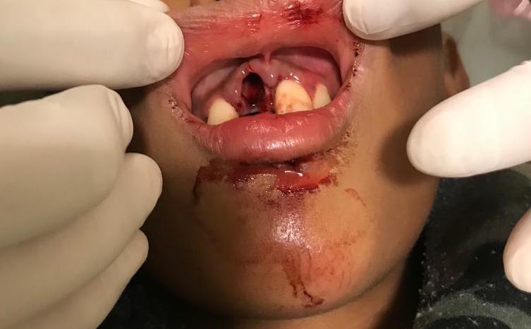
Avulsion
Avulsion means the complete loss of the tooth out of the socket. In cases where the avulsed tooth is missing, a thorough investigation is required. Soft tissue radiographs should be taken if the tooth is suspected to be embedded in the lips, cheeks, or tongue. An intra-oral radiograph must be taken to exclude intrusion. If the avulsed tooth is not found, the child should be referred for medical evaluation to an emergency department for further examination with chest radiography, especially if respiratory symptoms are present.[3]
Evaluation
Tooth mobility, color, tenderness to manual pressure, and the position or displacement of the tooth/teeth should be recorded at the initial assessment. The color of injured and uninjured teeth should also be recorded at every clinic visit. Discoloration is a recognized common complication following luxation injuries. It is usually seen 10 to 14 days after the original injury.[17]
Clinically, teeth with grey discoloration can recover to their original color, become yellowed or remain grey.[18] Teeth with dark discoloration can remain clinically asymptomatic with no radiographic signs of necrosis until natural exfoliation. Root canal treatment is not recommended for discolored teeth unless there are clinical signs of infection of the pulp.[17]
Radiographs are an important diagnostic tool for diagnosing TDIs and serve as a baseline when compared to follow-up appointments. Intra-oral radiographs may include periapical radiographs (size 0 sensor/film, paralleling technique) or occlusal radiographs (size 2 sensor/film). Conventional radiology may not be possible in young children due to compliance, and a clinician may consider alternative views, such as lateral obliques. However, it is important to consider if taking an image and exposing the child to radiation will positively affect the diagnosis or treatment provided. Clinicians must work within the ALARA (As Low As Reasonably Achievable) principles to minimize radiation doses. The use of pulp testing in children is challenging; it is subjective and relies on the child’s understanding and cooperation.[19] False-positive/negative results can be misleading in clinical situations and therefore are not recommended in the IADT guidelines.
Treatment / Management
The management of traumatic dental injuries to the primary dentition aims to prevent damage to the developing permanent tooth germ, alleviate pain, and minimize possible complications, such as infection.[14] There is currently limited evidence to support many of the treatment options of primary dentition.
The child's cooperation in an emergency, the time for natural exfoliation of the tooth, and the impact on the occlusion are all important considerations that influence treatment. A conservative approach involving observation is often the most appropriate option.[20] But, extracting the traumatized tooth is usually required in cases of tooth fracture with pulp involvement, luxation injuries close to the developing permanent tooth, and those that interfere with occlusion.[14] At the initial presentation, a rapid referral should be made to a child-orientated team with expertise in managing TDIs.
There is no evidence for the recommendation of systemic antibiotics to manage luxation injuries in the primary dentition. However, a clinician may wish to consider antibiotics in TDIs associated with soft tissue injuries, where surgical intervention may be necessary, or after considering the child's medical status. A tetanus booster may be needed if environmental contamination of the injury has occurred, and liaison with a medical practitioner would be recommended within 48 hours.
If splint placement is indicated to manage the TDI, The International Association of Dental Traumatology (IADT) guidelines recommend using a flexible splint. A flexible splint (e.g., titanium trauma splint, composite and wire splint) allows functional movement in contrast to a rigid splint (e.g., composite splints) where the injured teeth are immobilized.[21]
It is vital to instruct the caregiver and young patients regarding the care of the injured tooth at home for optimal healing. This includes advice regarding analgesia, a soft diet, meticulous oral hygiene, cleaning the area with antibacterial mouth rinses such as alcohol-free chlorhexidine gluconate 0.12%, and avoidance of further injuries.
Treatment options specific to the type of injury are listed below, in line with The International Association of Dental Traumatology (IADT) guidelines for managing TDIs in primary dentition.
Hard Tissue Injuries
Enamel Fractures
The clinician can consider smoothing any sharp edges. No clinical or radiographic follow-up is recommended.
Enamel-Dentine Fractures (uncomplicated crown fracture)
Managing an enamel dentin fracture with no pulpal exposure involves covering all exposed dentine with glass ionomer or composite. Follow-up clinical examination is recommended after 6 to 8 weeks; a radiographic examination is indicated only when clinical findings suggest pulp infection.
Crown Fractures With Exposed Pulp (complicated crown fracture)
The treatment aims at preserving the pulp by partial pulpotomy. Local anesthesia is required, and a non-setting calcium hydroxide paste is applied over the pulp and covered with glass-ionomer and composite. For more extensive pulpal exposures, a cervical pulpotomy is indicated.
Follow-up clinical examination is recommended after one week, six to eight weeks, and one year, with a radiographic examination at one year following pulpotomy.
Crown-Root Fractures
The immediate management is to remove the loose fragment, often under local anesthesia, and assess if the crown can be restored. If the tooth is restorable and has no pulpal exposure, cover the exposed dentine with glass ionomer. If the tooth is restorable, but the pulp has been exposed, perform a pulpotomy or root canal treatment depending on the root development stage and fracture level.
If the tooth is unrestorable, extract all loose fragments taking care not to damage the permanent successor, leave any firm root fragment in situ, or extract the entire tooth. When the tooth has been retained, a follow-up clinical examination is recommended after one week, six to eight weeks, and one year. A radiographic examination should be done one year following pulpotomy or root canal treatment.
Root Fractures
If the coronal fragment is not displaced, no treatment is required. If the coronal fragment is displaced and is not excessively mobile, leave the coronal fragment to reposition spontaneously, even if occlusal interference is present. A clinical follow-up examination is recommended after one week, six to eight weeks, and one year. Continue clinical follow-up yearly until the eruption of the permanent successor.
If the coronal fragment is displaced, excessively mobile, and interfering with occlusion, there are two options: a) extract the loose coronal fragment only, leaving the apical fragment in place to be reabsorbed; or b) gently reposition the loose coronal fragment. If the fragment is unstable in its new position, stabilize it with a flexible splint attached to adjacent uninjured teeth for four weeks.
If the coronal fragment has been repositioned and splinted, a clinical follow-up examination should be done after one week, four weeks (for splint removal), eight weeks, and one year. A clinical exam after one year is recommended if the coronal fragment has been extracted. Radiographic follow-up is only indicated where clinical findings are suggestive of pathosis.
Alveolar Fractures
Administer a local anesthetic, reposition any displaced segments that are mobile or causing occlusal interference, and stabilize with a flexible splint to the adjacent uninjured teeth for four weeks.
A clinical follow-up examination is recommended after one week, four weeks for splint removal, eight weeks, and one year. Further follow-up is recommended when the child reaches the age of 6 to monitor the permanent tooth eruption.
Radiographic examination is recommended at four weeks and one year to assess the impact on the primary tooth and the permanent tooth germs in the line of the alveolar fracture.
Periodontal Injuries
Concussion Injuries
No treatment is required. A clinical follow-up examination is recommended after one week and 6-8 weeks. Radiographic examination is only required if there are clinical signs of pathosis.
Subluxation Injuries
No treatment is required. A clinical follow-up examination is recommended after one week and six to eight weeks. If an unfavorable outcome is likely, yearly clinical follow-up is recommended until the permanent tooth erupts. Radiographic examination is only required if there are clinical signs of pathosis.
Extrusive Luxation Injuries
Treatment decisions are based on the degree of displacement, mobility, occlusal interferences, root formation, and the child's cooperation.
If the tooth is not interfering with the occlusion, leave it to reposition spontaneously. If the tooth is excessively mobile or extruded >3 mm, extract it under local anesthesia.
A clinical follow-up examination is recommended after one week and six to eight weeks. If an unfavorable outcome is likely, yearly clinical follow-up is recommended until the permanent tooth erupts. Radiographic examination is recommended only if there are clinical signs of pathosis.
Lateral Luxation Injuries
If there is minimal or no occlusal interference, the tooth should be allowed to reposition spontaneously, usually within six months. If the tooth is severely displaced and there is a risk of ingestion or aspiration, the tooth should be extracted under local anesthesia.
Alternatively, for severe displacement, gently reposition the tooth, and if unstable in its new position, apply a flexible splint for four weeks attached to the adjacent uninjured teeth. Clinical examination is recommended after one week (four weeks for splint removal if used), six to eight weeks, six months, and one year.
If an unfavorable outcome is likely, yearly clinical follow-up is recommended until the permanent tooth erupts. Radiographic examination is recommended only if there are clinical signs of pathosis.
Intrusive Luxation Injuries
The tooth should be allowed to reposition spontaneously, irrespective of the direction of the displacement. Spontaneous improvement in the position of the intruded tooth usually occurs within six months but can take up to one year in some cases.
Clinical examination is recommended after one week, six to eight weeks, six months, and one year. Further follow-up when the child reaches the age of six is recommended for a severe intrusion to monitor the eruption of the permanent successor. Radiographic examination is recommended only if there are clinical signs of pathosis.
Avulsion Injuries
The primary tooth should not be replanted. A clinical follow-up examination is recommended after 6-8 weeks and when the child reaches the age of 6 years to monitor the eruption of the permanent tooth.
Radiographic examination is recommended only if there are clinical signs of pathosis. In cases of early loss of an upper central incisor, it is important to consider the impact on the child regarding aesthetics and loss of function. A removable appliance may be indicated if the primary molars have erupted and there is a risk of developing speech disorders or tongue interposition.[22]
Differential Diagnosis
A thorough history and examination usually give an accurate diagnosis. However, the clinician should always be suspicious of non-accidental injury when the history of the accident and the injuries are not consistent. In situations where there is suspicion of abuse, a prompt investigation should be arranged, and referral should follow local protocols.
Treatment Planning
Summary
| Injury Type |
Signs/Symptoms |
Treatment |
| Enamel Fracture |
Fracture of enamel only. |
Consider smoothing any sharp edges of enamel. |
| Uncomplicated crown fracture |
Fracture of enamel & dentine only. |
Cover exposed dentine with GIC or composite. |
| Complicated crown fracture |
Fracture of enamel & dentine with pulpal exposure. |
Partial or complete pulpotomy. |
| Crown-root fracture |
Fracture of enamel, dentine, and root. The pulp may or may not be exposed. |
Extract loose fragments. Restore the tooth if restorable. If unrestorable, leave the remaining tooth in situ if firm, or extract the entire tooth. |
| Root fracture |
Fracture of the root of the tooth. |
If the coronal fragment is not displaced, no treatment is required. If minimally displaced and not mobile, leave for spontaneous repositioning. If displaced, mobile, and interfering with occlusion, either extract the loose coronal fragment or place a flexible splint for 4 weeks. |
| Alveolar fracture |
A fracture involving the alveolar bone: Teeth in the involved segment are mobile and often displaced. Occlusal interference and intra-oral soft tissue lacerations may be present. |
If mobile and/or interfering with occlusion, reposition the displaced segments and apply a flexible splint for 4 weeks. |
| Concussion |
The tooth is tender, has normal mobility, no gingival bleeding. |
No treatment is required. |
| Subluxation |
The tooth is tender, has increased mobility but is not displaced, and may have gingival bleeding. |
No treatment is required. |
| Extrusive luxation |
The tooth will appear elongated and have increased mobility. Occlusal interference may be present. |
If the tooth is not interfering with occlusion, leave for spontaneous repositioning. If the tooth is excessively mobile or extruded >3mm, extract the entire tooth. |
| Lateral luxation |
The tooth is displaced either palatally/lingual or labially. The tooth is immobile and occlusal interference may be present. |
If the tooth is not interfering with occlusion, leave for spontaneous repositioning. If severely displaced and at risk of ingestion or inhalation, either extract the entire tooth or reposition it. If the tooth is unstable after repositioning, place a flexible splint for 4 weeks. |
| Intrusive luxation |
Partial or complete displacement of the tooth into the socket. The tooth is palpated labially. |
Leave and allow for spontaneous repositioning. |
| Avulsion |
There is a complete loss of tooth out of the socket. |
No treatment is necessary. Do not reimplant the avulsed tooth. |
Prognosis
Pulp and periodontal outcomes are influenced by factors related to the injury and subsequent treatment. Favorable outcomes of TDIs in the primary teeth are the absence of symptoms and normal healing of the pulp and periodontal tissues.
Favorable outcomes of pulp healing include a normal color of the crown or transient red/grey or yellow discoloration and pulp canal obliteration. There will be no signs of pulp necrosis and infection, and in immature teeth, root development will continue. Additional favorable outcomes include undisturbed development and eruption of the permanent successor teeth.
Unfavorable outcomes following TDI to primary teeth include the development of symptoms, signs of pulp necrosis such as sinus tract, gingival swelling, abscess, or increased mobility. Persistent dark grey discoloration with an additional sign of infection is an unfavorable outcome.
Various studies have looked at the prognosis of traumatized primary teeth. A study by Soprowski found that 57% of luxated primary teeth showed no post-trauma sequelae, 25% became necrotic, 10% demonstrated calcific degeneration, and 8% became ankylosed.[23]
The clinician should record all favorable and unfavorable outcomes at the initial appointment and subsequent follow-up appointments.
Complications
Due to the close relationship between the apex of the primary tooth root and the underlying permanent tooth germ, there is potential for sequelae to the permanent tooth following orofacial and primary tooth trauma.
Enamel discoloration, enamel hypoplasia, crown or root dilaceration, arrested root formation, and eruption disturbances in the developing permanent dentition are some of the potential complications following primary tooth trauma.[24]
Intrusion and avulsion injuries are the most common type of TDI associated with complications to the permanent dentition.[25]
Deterrence and Patient Education
Given the consequences of traumatic dental injuries on the child and the developing of permanent dentition, there is a need to educate parents, carers, teachers, and healthcare professionals about the need for dental assessment and treatment of injuries to the primary dentition.
Initial first aid management for TDIs in primary dentition may be delivered by pediatricians, nurses, teachers, or caregivers. It is important to stay calm, clean bleeding wounds with saline solution (0.9%) or tap water, stop bleeding wounds by applying pressure with a clean piece of cotton for 5 minutes, and refer to a pediatric dentist for dental injuries.[14]
It is hard to prevent dental injuries; however, management of risk factors, including increased overjet and open bite, should be considered. The cessation of non-nutritive sucking habits should be encouraged before they influence the developing dentition.
Enhancing Healthcare Team Outcomes
The management of TDIs in children is challenging; when possible, acute and follow-up care should be provided by a child-orientated team with experience in managing pediatric orofacial injuries. These teams have access to specialist diagnostic and treatment adjuncts, including sedation and general anesthesia, to minimize pain and anxiety for the child.[3] This collaboration between acute and specialist teams in a multidisciplinary approach will improve patient outcomes.