Hip Osteonecrosis (Archived)

Archived, for historical reference only

Introduction

Osteonecrosis of the hip, commonly known as avascular necrosis (AVN) of the hip, is the death of the femoral head as a result of the vascular disruption. AVN of the hip results in pain around the hip which is insidious in onset. The cause is generally multifactorial and more commonly seen in males compared to females. Furthermore, the age of presentation from symptomatic AVN of the hip is younger than that of osteoarthritis. The treatment of AVN of the hip is controversial, and as such, there are many different treatment options for AVN. The ideal treatment option depends on the severity and stage of the disease. 

Etiology

AVN of the femoral head results from disruption of the vascular supply to the bone. Potential causes of AVN of the femoral head include causes that are clearly known such as when direct trauma has occurred, and in instances where the inciting event is unknown and only can be associated with risk factors that predispose to the disease process. Below are risk factors associated with the development of AVN of the femoral head.

Risk Factors[1][2][3][4][5]

  • Irradiation
  • Trauma
  • Hematologic disease (leukemia, lymphoma)
  • Dysbaric (Caisson disease)
  • Marrow-replacing diseases (Gaucher disease)
  • Sickle cell disease
  • Alcoholism
  • Hypercoagulable states
  • Steroids
  • Systemic lupus erythematosus (SLE)
  • Transplant patient
  • Viral (CMV, hepatitis, HIV, rubella, rubeola, varicella)
  • Protease inhibitors
  • Pancreatitis
  • Vascular insult
  • Subacute bacterial endocarditis
  • Polyarteritis nodosa
  • Rheumatoid Arthritis
  • Giant cell arteritis
  • Sarcoidosis
  • Idiopathic

Previous associations have linked AVN of the femoral head to inherited coagulopathies and genetic polymorphisms that are potentially treatable pathophysiologic conditions. It is believed that AVN of the femoral head is a multifactorial disease associated in some cases with a genetic predisposition and an exposure to one or more risk factors.

Epidemiology

AVN of the femoral head is diagnosed at a rate of 10,000 to 20,000 new cases per year in the United States. The typical age ranges from 35 to 50 years old with the average age of presentation being 36 years old. The prevalence is higher in men than women. There is also a high prevalence of bilateral disease.

Pathophysiology

AVN of the femoral head associated with trauma results from damage to the medial femoral circumflex artery which is the main blood supply to the femoral head. This leads to the death of cells within the bony matrix. The reparative process begins which weakens the subchondral bone, leading to bony collapse and eventually arthritic changes of the joint.[6][7]

AVN in which the direct cause is unknown but likely from the previously mentioned risk factors results from intravascular coagulation. Coagulation of the intraosseous microcirculation results in venous thrombosis. This leads to arterial occlusion due to the lack of venous outflow. The arterial occlusion causes an increase in pressure within the bone which ultimately leads to decreased blood flow to the femoral head and eventual death of the cells. Much like with direct trauma, once the cells die, the reparative process begins and results in bony collapse and arthritic changes.[6] 

History and Physical

It is essential to obtain a full, detailed history pertaining to the symptomatology including onset, location of the pain, duration of symptoms, the characteristics of the pain, alleviating and aggravating symptoms, radiation of symptoms, and timing of symptoms. It is equally important to obtain a detailed medical and surgical history to identify any of the risk factors associated with AVN.

Patients with AVN of the femoral head will present with symptoms of hip pain with insidious onset. Typically, the pain will be associated with standing from a seated position, stairs, inclines, and impact loading. The pain will tend to be more noticeable in the anterior hip or groin as opposed to the buttock or greater trochanter. 

Evaluation

There is no single diagnostic test that is 100% reliable. Proper evaluation of a patient with suspected AVN of the femoral head is similar to the patient that presents with hip pain. The physical exam includes inspection of the hip, including the skin, the position of the lower extremity, and gait evaluation as well as palpation of the surrounding bony structures such as the greater trochanter. The range of motion testing includes flexion, extension, abduction, adduction, and internal and external rotation. A thorough neurovascular exam in needed to evaluate for sensory and motor function. There are helpful special hip tests including the log roll and the Stinchfield test to help determine intra-articular pathology.

Standard imaging includes radiographs of the pelvis and hip, including an anteroposterior view of the pelvis, an anteroposterior, and a frog-leg lateral view of the affected hip. The frog-leg lateral view is the radiographic test to confirm subchondral collapse and make decisions regarding patient management.

If there is a high index of suspicion for AVN of the femoral head despite negative radiographs, an MRI of the hip is indicated. MRI has been shown to be approximately 98% sensitive and specific. Findings consistent with AVN of the femoral head include a “double density” appearance which appears dark on the T1 sequence and bright on the T2 sequence. The presence of a bright signal on the T2 sequence indicates bone marrow edema and is predictive of worsening pain and progression of the disease. 

Treatment / Management

Treatment of AVN of the femoral head is dependent on symptomatology as well as radiographic findings such as evidence of the collapse of the femoral head, size of the necrotic lesion in the femoral head, and involvement of the acetabulum. Treatment options can be broken down into nonoperative and operative categories.

Nonoperative Treatment (Stages 0-2)

  • Bisphosphonates

This is largely limited to small lesions, less than 10% involvement of the femoral head, without evidence of collapse. The evidence is inconclusive on whether or not bisphosphonates prevent femoral head collapse.

Operative Treatment[8][5][9][10][11]

  • Core decompression of the femoral head with or without stem cell injection
  • Vascularized free-fibula grafting
  • Total hip resurfacing
  • Total hip arthroplasty

Core decompression has traditionally been the treatment of choice during precollapse stages, although the evidence is mixed. The idea behind core decompression is to relieve the increase in pressure within the femoral head thus allowing a return in blood flow and healing. There has been some success demonstrated with the injection of stem cells into the necrotic area. Vascularized free-fibula grafting can be performed in patients with or without collapse in stages 0 to 3. The goal of this particular surgery is to remove the necrotic area and use a free-fibular graft to act as a strut to prevent collapse or to tamp up the collapsed area. The long-term results of this surgery have shown consistent and successful results in regards to functional recovery and pain relief. Total hip resurfacing is indicated in select patients with AVN with careful evaluation of the size of the lesion, its location, and the quality of the bone. The resurfacing procedure requires adequate bone stock to support the resurfacing component. The component design of total hip resurfacing is a metal-on-metal articulation, and its use in young patients, female patients, or patients with a history of the renal disease is not suggested. Furthermore, studies have shown that the outcomes for total hip resurfacing in AVN are worse than in patients with osteoarthritis. Total hip arthroplasty provides the most reliable pain relief and good early clinic results for patients with advanced stages of AVN. It is indicated in younger patients with a crescent sign or more advanced femoral head collapse and patients older than 40 years old with large lesions. 

Differential Diagnosis

  • Lumbar radiculopathy
  • Spinal stenosis
  • Sacroiliac degenerative joint disease
  • Neuropathy
  • Meralgia paresthetica
  • Osteoarthritis
  • Fracture
  • Femoroacetabular impingement
  • Labral tear
  • Trochanteric bursitis
  • Iliopsoas bursitis
  • Snapping hip syndrome
  • Tendinopathy of the gluteus medius or minimus
  • Primary bone tumor
  • Metastasis
  • Infection

Staging

Steinberg Classification for staging AVN of the femoral head (modification of the Ficat classification)[12][13]

  • Stage 0 – Normal or nondiagnostic radiograph and MRI
  • Stage 1 – Normal radiograph, abnormal MRI
  • Stage 2 – Abnormal radiograph showing cystic and sclerotic changes in the femoral head, abnormal MRI
  • Stage 3 – Abnormal radiograph showing subchondral collapse, producing a crescent sign, abnormal MRI
  • Stage 4 – Abnormal radiograph showing flattening of the femoral head, abnormal MRI
  • Stage 5 – Abnormal radiograph showing joint narrowing with or without acetabular involvement, abnormal MRI
  • Stage 6 – abnormal radiograph showing advanced degenerative changes, abnormal MRI

Prognosis

The risk of femoral head collapse with AVN can be stratified into three groups based on the modified Kerboul combined necrotic angle. This is calculated by the summation of the arc of femoral head necrosis on mid-sagittal and midcoronal MR image.[14]

  • Low-risk group: combined necrotic angle less than 190 degrees
  • Moderate-risk group: combined necrotic angle between 190 and 240 degrees
  • High-risk group: combined necrotic angle greater than 240 degrees

Pearls and Other Issues

Mnemonic to help remember the most common causes of AVN

ASEPTIC

  • A – Alcohol/AIDS
  • S – Steroids, sickle cell disease, SLE
  • E – Erlenmeyer flask (Gaucher disease)
  • P – Pancreatitis
  • T – Trauma
  • I – Idiopathic, infection
  • C – Caisson’s disease

Enhancing Healthcare Team Outcomes

Hip osteonecrosis is associated with very high morbidity if the diagnosis is missed. Thus, the condition is best managed by an interprofessional team that includes a radiologist, orthopedic surgeon, rheumatologist, internist, nurse practitioner and a pharmacist. There is no one single test that can make the diagnosis of this disorder and hence, communication and discussion with other healthcare workers is vital. The offending drug must be discontinued right away. The patients are managed medically and surgically but a complete recovery is not always guaranteed. Some patients are left with a permanent gait alteration and this can significantly affect the quality of life.[15][16]


(Click Image to Enlarge)
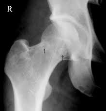
<p>Dysbaric Osteonecrosis of the Hip</p>

Dysbaric Osteonecrosis of the Hip


Contributed by UHM 2005, Vol. 32, No. 3 – Dysbaric osteonecrosis by X-ray and CT scan in Chinese divers. JIANG et al

Details

Author

Hunter Hsu

Updated:

8/8/2023 12:31:10 AM

References


[1]

Bergman J, Nordström A, Nordström P. Epidemiology of osteonecrosis among older adults in Sweden. Osteoporosis international : a journal established as result of cooperation between the European Foundation for Osteoporosis and the National Osteoporosis Foundation of the USA. 2019 May:30(5):965-973. doi: 10.1007/s00198-018-04826-2. Epub 2019 Jan 9     [PubMed PMID: 30627759]


[2]

Pascarella R, Fantasia R, Sangiovanni P, Maresca A, Massetti D, Politano R, Cerbasi S. Traumatic hip fracture-dislocation: A middle-term follow up study and a proposal of new classification system of hip joint associated injury. Injury. 2019 Aug:50 Suppl 4():S11-S20. doi: 10.1016/j.injury.2019.01.011. Epub 2019 Jan 17     [PubMed PMID: 30683569]


[3]

Belmahi N, Boujraf S, Larwanou MM, El Ouahabi H. Avascular necrosis of the femoral head: An exceptional complication of cushing's disease. Annals of African medicine. 2018 Oct-Dec:17(4):225-227. doi: 10.4103/aam.aam_75_17. Epub     [PubMed PMID: 30588938]


[4]

Duan Y, Xi Y, Cun X, Yang Y, Li B, Zhou J, Tang X, Li Z. [Treatment of avascular necrosis of femoral head in patients with human immunodeficiency virus infection by cementless total hip arthroplasty]. Zhongguo xiu fu chong jian wai ke za zhi = Zhongguo xiufu chongjian waike zazhi = Chinese journal of reparative and reconstructive surgery. 2018 Dec 15:32(12):1507-1511. doi: 10.7507/1002-1892.201806126. Epub     [PubMed PMID: 30569674]


[5]

Suksathien Y, Sueajui J. Mid-term results of short stem total hip arthroplasty in patients with osteonecrosis of the femoral head. Hip international : the journal of clinical and experimental research on hip pathology and therapy. 2019 Nov:29(6):603-608. doi: 10.1177/1120700018816011. Epub 2018 Dec 11     [PubMed PMID: 30526072]


[6]

Goyal T. Early results of surgery for femoroacetabular impingement in patients with osteonecrosis of femoral head. SICOT-J. 2018:4():47. doi: 10.1051/sicotj/2018038. Epub 2018 Nov 6     [PubMed PMID: 30398961]


[7]

Wang C, Meng H, Wang Y, Zhao B, Zhao C, Sun W, Zhu Y, Han B, Yuan X, Liu R, Wang X, Wang A, Guo Q, Peng J, Lu S. Analysis of early stage osteonecrosis of the human femoral head and the mechanism of femoral head collapse. International journal of biological sciences. 2018:14(2):156-164. doi: 10.7150/ijbs.18334. Epub 2018 Jan 14     [PubMed PMID: 29483834]


[8]

Kawano K, Motomura G, Ikemura S, Kubo Y, Fukushi J, Hamai S, Fujii M, Nakashima Y. Long-term hip survival and factors influencing patient-reported outcomes after transtrochanteric anterior rotational osteotomy for osteonecrosis of the femoral head: A minimum 10-year follow-up case series. Modern rheumatology. 2020 Jan:30(1):184-190. doi: 10.1080/14397595.2018.1558917. Epub 2019 Feb 18     [PubMed PMID: 30556788]

Level 2 (mid-level) evidence

[9]

Zhao P, Hao J. Analysis of the long-term efficacy of core decompression with synthetic calcium-sulfate bone grafting on non-traumatic osteonecrosis of the femoral head. Medecine sciences : M/S. 2018 Oct:34 Focus issue F1():43-46. doi: 10.1051/medsci/201834f108. Epub 2018 Nov 7     [PubMed PMID: 30403174]


[10]

Houdek MT, Wyles CC, Collins MS, Howe BM, Terzic A, Behfar A, Sierra RJ. Stem Cells Combined With Platelet-rich Plasma Effectively Treat Corticosteroid-induced Osteonecrosis of the Hip: A Prospective Study. Clinical orthopaedics and related research. 2018 Feb:476(2):388-397. doi: 10.1007/s11999.0000000000000033. Epub     [PubMed PMID: 29529674]


[11]

Ünal MB, Cansu E, Parmaksızoğlu F, Cift H, Gürcan S. Treatment of osteonecrosis of the femoral head with free vascularized fibular grafting: Results of 7.6-year follow-up. Acta orthopaedica et traumatologica turcica. 2016 Oct:50(5):501-506. doi: 10.1016/j.aott.2016.01.001. Epub 2016 Nov 16     [PubMed PMID: 27865611]


[12]

Sultan AA, Mohamed N, Samuel LT, Chughtai M, Sodhi N, Krebs VE, Stearns KL, Molloy RM, Mont MA. Classification systems of hip osteonecrosis: an updated review. International orthopaedics. 2019 May:43(5):1089-1095. doi: 10.1007/s00264-018-4018-4. Epub 2018 Jun 18     [PubMed PMID: 29916002]


[13]

Takashima K, Sakai T, Hamada H, Takao M, Sugano N. Which Classification System Is Most Useful for Classifying Osteonecrosis of the Femoral Head? Clinical orthopaedics and related research. 2018 Jun:476(6):1240-1249. doi: 10.1007/s11999.0000000000000245. Epub     [PubMed PMID: 29547501]


[14]

Steinberg ME, Oh SC, Khoury V, Udupa JK, Steinberg DR. Lesion size measurement in femoral head necrosis. International orthopaedics. 2018 Jul:42(7):1585-1591. doi: 10.1007/s00264-018-3912-0. Epub 2018 Apr 25     [PubMed PMID: 29691613]


[15]

Lerch TD, Vuilleumier S, Schmaranzer F, Ziebarth K, Steppacher SD, Tannast M, Siebenrock KA. Patients with severe slipped capital femoral epiphysis treated by the modified Dunn procedure have low rates of avascular necrosis, good outcomes, and little osteoarthritis at long-term follow-up. The bone & joint journal. 2019 Apr:101-B(4):403-414. doi: 10.1302/0301-620X.101B4.BJJ-2018-1303.R1. Epub     [PubMed PMID: 30929481]


[16]

Mallet C, Abitan A, Vidal C, Holvoet L, Mazda K, Simon AL, Ilharreborde B. Management of osteonecrosis of the femoral head in children with sickle cell disease: results of conservative and operative treatments at skeletal maturity. Journal of children's orthopaedics. 2018 Feb 1:12(1):47-54. doi: 10.1302/1863-2548.12.170141. Epub     [PubMed PMID: 29456754]