Continuing Education Activity
Esotropia, commonly referred to as an inward deviation of eyes, is a common clinical entity seen in the outpatient department. Esotropia may be congenital/acquired, constant/intermittent, unilateral/alternating. Clinical examination, pathophysiology, and predisposing factors are essential to understand and diagnose esotropia subtype and plan appropriate management. Congenital esotropia is the most common type of esodeviation seen in early childhood and commonly presents within the first six months of life with a large angle of esodeviation. Common associations include inferior oblique overaction, dissociated vertical deviations, or latent nystagmus. This activity highlights the role of the interprofessional team in the evaluation of refractive error and its management to help maintain a binocular single vision for the patient.
Objectives:
- Describe the etiology and types of esotropia.
- Summarize the evaluation of patients with esotropia.
- Outline the management of esotropia.
- Review the differential diagnosis and complications associated with esotropia and surgical management by the interprofessional team.
Introduction
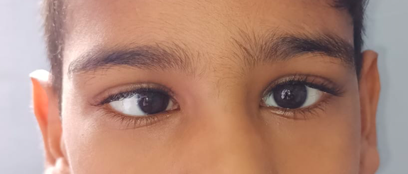
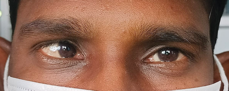
Esotropia is defined as misalignment of eyes, in which one eye deviates towards the nose.[1] The term esotropia is derived from ancient Greek, where "Eso" means "within" and "Tropia" means "a turn." It can be congenital or acquired, intermittent or constant, and may change patterns from intermittent to constant over time. Further, the deviation might remain constant or may vary in different gaze directions depending on which it is labeled as concomitant or incomitant. Squinting might appear in one or alternate eyes. The concomitant esotropias can be further classified as Infantile (congenital) esotropia, accommodative esotropia, acquired non-accommodative esotropia, sensory esotropia, and consecutive esotropia.[2][3]
Incomitant esotropias can be further divided based on underlying etiology into paralytic or non-paralytic. The main aim of this review will be to describe the concomitant esotropia and its subtypes in detail.
Etiology
The exact etiology depends on the type of esotropia. We have tried to describe the underlying etiology for each type briefly
Infantile Esotropia
The underlying factors predisposing to infantile esotropia are:
Innervational Imbalances
It is characterized by a disturbance between the tonic convergence and divergence are considered responsible for deviations.
Accommodation
The role of accommodation is considered partially or fully in congenital esotropia patients. In a few cases, the accommodative esotropia might be superadded at 2 to 3 years, over the primary deviation.[4]
Refractive Accommodative Esotropia
The most important etiology for this type is underlying uncorrected hypermetropia. Additional factors to be considered in this type include fusional divergence amplitude, AC/A ratio, and patient personality.[5]
Non-Refractive Accommodative Esotropia
An underlying high AC/A ratio is expected to predispose to this type of esotropias. The near point of accommodation is within the normal range for age in these patients.[6]
Partially Accommodative Esotropia
These cases have an uncorrected hypermetropia, with a normal AC/A ratio.[7]
Sensory Esotropia
Sensory esotropia occurs because of poor vision in one eye in early childhood. The eye with poor vision often deviates, and the other eye with good vision becomes the dominant eye.[8]
Consecutive esotropia
This often occurs due to surgical overcorrection in a previously exotropia eye.[9]
Epidemiology
As reported in a meta-analysis by Hashemi et al., the estimated prevalence of esotropia was reported as 0.77%.[10] In another study done among school-going children, it was found that strabismus was present in 3.11% of the total children screened, and the esotropia: exotropia was diagnosed in a ratio of 1:9.75. Further, it was observed that no significant differences existed related to age across 6-8 years or gender among the study population.[11]
Further, the prevalence of strabismus and its types varies among different races. In the Baltimore Pediatric Eye Disease Study, the prevalence of manifest deviations was 3.3% among whites and 2.1% among children of African American descent.[12]
The esotropias and exotropias were almost equal in both groups. A retrospective cohort-based study done among patients diagnosed with Esotropia in Minnesota reported that 36% had fully accommodative esotropia, another 17% had non-accommodative esotropia, 10% had partially accommodative esotropia. In contrast, 8% were congenital, 6.5% were paralytic, and the rest had an undetermined underlying cause of esotropia.[13]
Pathophysiology
Congenital Esotropia
The main factor responsible for congenital esotropia is the innervational imbalances between fusional convergence and divergence mechanisms. Since the fusional convergence mechanisms are more robust of the amplitude of >30prism diopters, compared to fusional divergence mechanisms of the amplitude of 4 to 6 prism diopters, the esodeviations, even though intermittent in onset, tend to become constant quickly with poor stereo acuity.
Accommodative Esotropia
The underlying pathophysiology is blurred retinal image secondary to uncorrected hypermetropia. This retinal blurring leads to extra accommodative efforts leading to excessive accommodative convergence. Further, the development of esotropia depends on the fusional divergence capacity and AC/A ratio. Those with insufficient fusional divergence develop refractive accommodative esotropia or esophoria, while those with low AC/A ratio usually remain orthotropic.[14]
Non-Accommodative Esotropia
The main etiopathogenesis is an excessive accommodative convergence associated with the normal amplitude of accommodation (3 to 5 prism dioptres). This deviation is usually small and intermittent at onset and becomes constant over time. Also, the near deviation is more with no or minimal deviation for distance.[15]
Partially Accommodative Esotropia
The underlying cause is an uncorrected high hypermetropia, with a normal AC/A ratio and the deviation that is partially corrected with the help of spectacles.[7]
History and Physical
A detailed history and meticulous examination are needed for all cases of esotropia presenting to the outpatient department. Complete history taking should include essential points like age of the patient, onset of deviation, nature of deviation – constant/intermittent, unilateral/alternating. Other critical points to be recorded are abnormal head posture, complaints of double vision, headache, asthenopia, history of closing one eye in bright sunlight. Any predisposing factors of viral illness, head trauma, and excessive near-vision work should be actively enquired. Any family history of squinting or any significant peri-natal history should also be noted down. Previous management, either optical, occlusion therapy, or surgical treatment, is also essential.[16]
Patient evaluation should include general physical examination, build, nutritional status assessment. Gross inspection of head posture, lid fissures, intercanthal distance, interpupillary distance, any mongoloid or antimongoloid slant, epicanthal folds, associated exophthalmos, or enophthalmos should be noted. An ocular examination should consist of a detailed analysis of the anterior and posterior segments. Anterior segment examination should include lid position, ptosis or pseudoptosis, any lid fissure changes associated with eye movements, Marcus Gun Jaw winking, media opacities including cornea or lenticular changes, and pupillary reactions, which give a gross indication of optic nerve head and retinal status.
Posterior segment examination should include a dilated fundus examination to rule out any pathology like macular scarring, optic nerve hypoplasia, retinoblastoma, along with an objective assessment of macular torsion. Other necessary evaluations should include assessing visual acuity using age-appropriate methods, angle of deviation measured with Hirschberg tests, prism bar cover test for near and distance, fixation pattern assessment – alternate fixation, cross-fixation, and dominance of either eye.
Accommodation Convergence/Accommodation ratio (AC/A), fusional divergence amplitudes, binocular function, stereopsis, ocular movements, and refraction with atropine cycloplegia assessments are also essential in all cases of strabismus. These tests will help identify the type of esotropia and thus help plan further patient management. Any associated clinical features like inferior oblique overaction, dissociated vertical deviation, latent nystagmus should also be examined.[17]
Evaluation
Evaluation of a patient with esotropia usually does not require specific laboratory tests. A detailed history and clinical examination are generally sufficient to reach a provisional diagnosis and help in planning further management courses. Imaging might be needed if any neurological signs or associations are expected on evaluation or cases with acute onset of esotropias.[18]
The clinical features specific to the most common types of esotropia seen in the pediatric age group are described briefly here.
Infantile Esotropia
Deviation of eyes manifests within six months from birth. These children typically have a large angle of deviation, usually more than 30 degrees, which is present for distance and near. An alternate fixation is present in the primary gaze, and cross-fixation in lateral end gazes. Dolls' head maneuver might help elicit abduction movements, which helps in distinguishing from bilateral sixth nerve palsy. Visual acuity or retinoscopy values are usually within normal limits in these children because of cross fixation, and no significant underlying refractive errors are expected.[19]
Refractive Accommodative Esotropia
These cases usually manifest around 2 to 3 years. The deviation in these cases is typically intermittent, to begin with, noticed by parents, particularly for near. This may progress from stages of esophoria to intermittent esotropia to constant esotropia. The deviation for distance and near is usually within ten prism diopters. AC/A ratio is generally normal in these cases. Atropine refraction usually reveals a high underlying hypermetropia in the range of 2 to 6 diopters.[20]
Non-Refractive Accommodative Esotropia
The esotropia in these cases is related to an underlying high AC/A ratio, with no significant underlying hypermetropia revealed on atropine refraction. This progresses from esophoria for near, to esotropia for near with esophoria for distance, to an esotropia both for distance and near. This type of esotropia also begins around 2 to 3 years. The near deviation in these cases is typically much more significant than the distance deviation.[6]
Partially Accommodative Esotropia
These cases present with a large angle esotropia, high hypermetropia, and a normal AC/A ratio. The esotropia might be more for near distance and gets partially corrected with the refractive correction alone.[21]
Sensory Esotropia
This is often characterized by monocular vision loss, amblyopia, and concomitant sensory esotropia with the dominance of the contralateral eye.[8]
Treatment / Management
Non-surgical treatment aims to treat underlying refractive errors, treatment of amblyopia, and orthoptic exercises. Surgical treatment aims to maintain binocularity, prevent amblyopia with early alignment, and promote peripheral fusion.
Non-Surgical Treatment
- Refractive error correction – It is advisable to give full hypermetropic refractive correction, i.e., without drug correction applied, as examined after cycloplegic refraction for the patient. Any hypermetropia more than +1.5D should be prescribed to the patient. Ocular deviation and AC/A ratio can be reassessed after six weeks of spectacle usage. Children with a high AC/A ratio or eyes aligned for distance and a residual esotropia for near should be considered for bifocals.[20]
- Further, it has been suggested that the higher the delay between onset of esotropia and start of refractive correction, the higher the chances of the child ending with partially accommodative esotropia.[22]
- Miotics have been tried as an alternative to glasses in uncooperative patients or those not compliant with glass usage.[23]
- Amblyopia therapy is indicated when a patient is brought late with pre-set amblyopia and fixation preference for the other eye. Most often, alternate eye patching might be advised to prevent fixation preference and thus prevent amblyopia in these cases.
- Orthoptic treatment is useful in patients with refractive or non-refractive accommodative esotropias. These exercises aim at overcoming the suppression and improving the negative fusional convergence.[24]
- Botulinum Toxin – This has been studied and proven helpful in infantile esotropia and new-onset acquired esotropia.[25] Botulinum has also been studied for residual esotropia following bilateral medial rectus recessions and lateral rectus resections.[26]
Surgical Treatment
The timing of surgery and surgical procedures may vary with the type of esotropia, amount of deviation, presence of associated clinical features, amblyopia, and compliance with non-conservative management.
We will cover the most common procedure of choice and considerations for timing for surgery for each type of esotropia separately.
Infantile/Congenital Esotropia
The ideal time for surgical intervention in these patients is between 6 months to 2 years of age. The final decision should be taken based on the child's co-operation to measure squint and understanding of the parents and the examiner. Choice of surgery also varies depending upon the surgeons' preference. Bimedial recessions are usually preferred over unilateral recession/resect procedures. If significant inferior oblique overactions are associated, combining bimedial recession/ unilateral recession-resection with bilateral inferior oblique muscles weakening is preferable. The surgery for dissociated vertical deviation should be deferred as a primary procedure and can be combined later as a staged procedure if needed.
Refractive Accommodative Esotropia
Surgery for refractive accommodative esotropia should always be avoided. Any associated vertical deviation or A/V pattern might need surgical correction.[14]
Non-Refractive Accommodative Esotropia
In this conservative management in the form of bifocals, orthoptic exercises should be preferred. Surgery might be indicated in patients with a large angle of residual deviation, which conservative methods cannot correct. Bilateral medial rectus recession with or without bilateral Faden procedure to medial rectus can be planned based on the angle of deviation for near.[27]
Partially Accommodative Esotropia
These cases require a surgical correction for the residual deviation following spectacle correction. Few studies showed improvement with occlusion therapy in these cases.[28] Favorable outcomes in stereopsis have also been reported with timely surgical intervention for the nonaccommodative component.[29]
Sensory Esotropia
Surgical intervention might be indicated for cosmetic correction in these cases. In children, any associated aphakia or traumatic cataracts should be corrected, along with a trial of occlusion therapy. Eventually, these can be planned for squint correction once the maximum best-corrected visual acuity is achieved. Surgical squint correction using unilateral recession-resection procedure should be preferred in the amblyopic eye or the eye with poor visual acuity.
Cyclic Esotropia
Botulinum toxin or surgical correction by bimedial rectus recession or recession resection procedure for medial and lateral rectus respectively might be needed. The dose of surgical correction should be based on the deviation on the day of esotropia.[30][31]
Differential Diagnosis
It is crucial to distinguish esotropia from other close differentials, as discussed below:
- Pseudoesotropia can be distinguished from true esotropia by the presence of prominent epicanthal folds, small interpupillary distance, negative angle kappa, or an excessive broad nasal bridge.
- Ciancia syndrome – This is characterized by the presence of large-angle esodeviation (>60 prism diopters). Both eyes appear to be stuck towards the nose, limited bilateral abduction with good abduction saccades, jerk endpoint nystagmus on attempted abduction, with no nystagmus on adduction, face turn towards fixing eye, and a tight medial rectus muscle on forced duction test.[32]
- Congenital fibrosis syndrome – This is also called strabismus fixus. The patient usually presents with large-angle esotropia with severe limitation of abduction of one or both eyes. Often, family history is present in these cases and shows an autosomal dominant inheritance pattern.[33]
- Congenital sixth nerve palsy – These can present as unilateral or bilateral cases. Doll's head maneuver reveals limited abduction based on laterality.
- Nystagmus blockage syndrome refers to a condition in which esotropia occurs in a child with congenital nystagmus. It is proposed that esotropia occurs secondary to an adduction or excessive convergence to reduce nystagmus. Few authors have proposed the two components of esotropia in these cases – a static angle of infantile esotropia and a superadded dynamic angle of esotropia due to convergence to dampen the nystagmus.[34]
- Cyclic Esotropia – Alternate repeated strabismic and non-strabismic phases characterize this form. The cycles may vary from 24 hours of strabismic and non-strabismic phases or 48 hours or 72 hours cycles each. This form of strabismus can be acquired at any age but is most commonly seen between 2 to 6 years.[30]
- Microtropia – This is characterized by a slight angle of heterotopia in the range of 1 to 5 degrees. Other clinical features include a relative scotoma on the fovea, abnormal retinal correspondence, mild amblyopia, defective stereo acuity, and a normal or near-normal peripheral fusion with amplitudes.
Prognosis
Overall, the prognosis of esotropia is good. Well-planned management strategy, close follow-up, and compliance with treatment are the main factors on which prognosis depends. Though most concomitant esotropia can be managed conservatively, congenital esotropia requires surgical intervention. There are two different schools of thought for early versus late surgical intervention for congenital esotropia.
It is now considered that early ocular alignment before the age of 2 years results in better visual acuity and preservation of binocular fusion to some extent, often involving peripheral fusion. Studies have shown that better long-term stability, better stereopsis, and sensorimotor development have been associated with the early alignment of eyes. It is important to note that the reoperation rates in congenital esotropia have been reported as high as 50%.[35]
Complications
The complications associated with the management of esotropia are related to loss of binocularity, chances of amblyopia, issues with compliance with treatment, and long-term follow-ups. Apart from these, these children are highly likely to lose confidence because of cosmetic concerns, developing antisocial behaviors, and underperforming in schools.
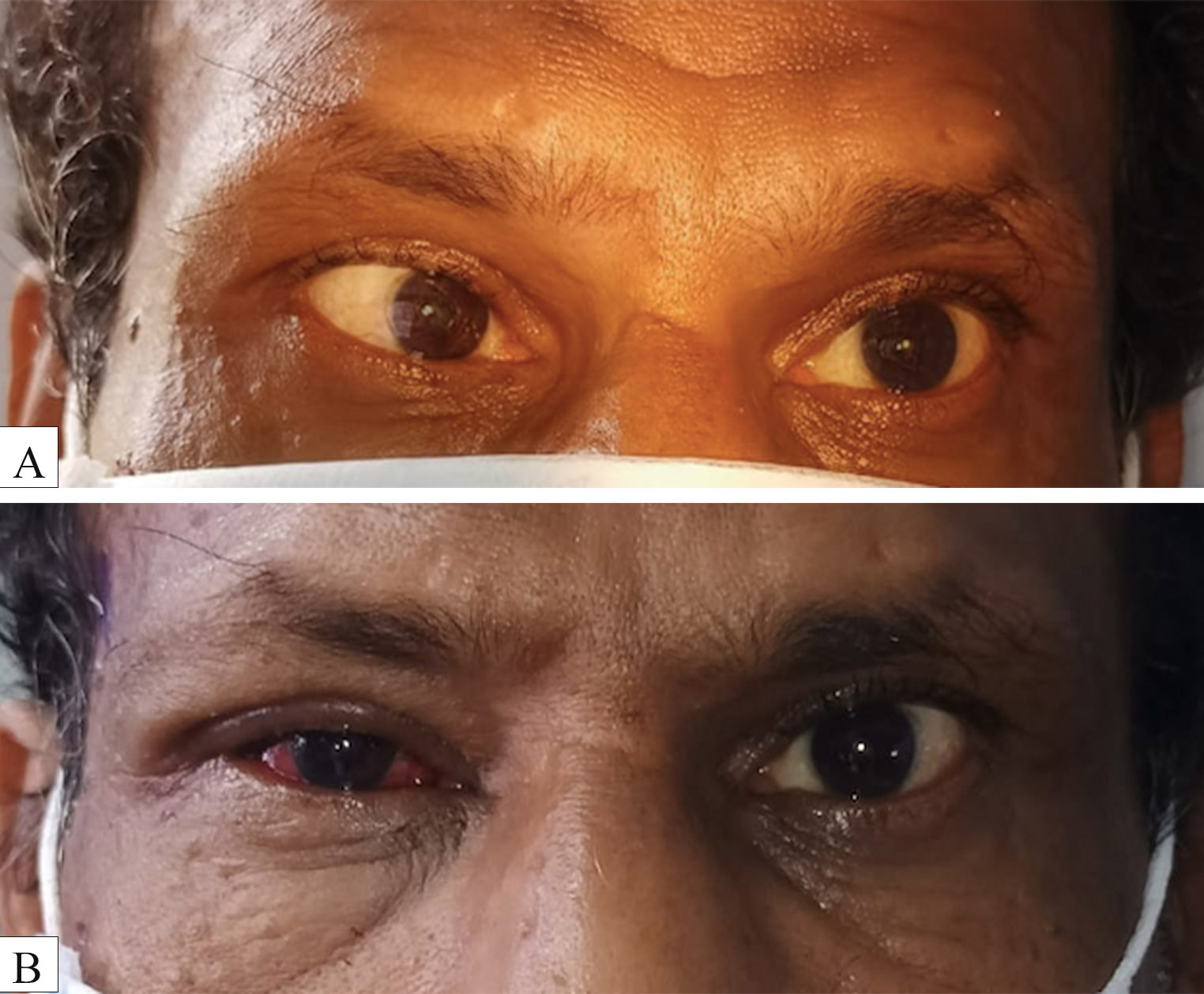
Complications associated with surgical intervention include residual esotropia, consecutive exotropia, infection, suture-related granuloma, anterior segment ischemia, slipped or lost muscle, or an oculocardiac reflex. Apart from these, the risk of general anesthesia-related complications needs to be borne in mind before planning any surgical intervention in these cases.[36]
Postoperative and Rehabilitation Care
Postoperative care involves close collaboration between the parents, treating ophthalmologists, optometrists for regular refraction, and the counselor. The nurses and counselor play an essential role in making parents understand how to use postoperative eye drops, maintain hygiene, cleaning of eyelashes and lid margins to prevent infection. The involvement of parents in the immediate postoperative period and during follow-ups is important to ensure treatment compliance. These patients need long-term close follow-ups, keeping in mind the need for correcting refractive errors, cycloplegic refraction at least once every six months, maintenance of amblyopia therapy, orthoptics exercise, repeat six-monthly squint evaluation for the development of under or overcorrection.
Deterrence and Patient Education
It is imperative to educate parents about the nature of the disease and the role of timely intervention. Congenital esotropia outcomes primarily depend on treatment compliance in the form of patching to prevent amblyopia till a surgical intervention is planned. The usual modes of presentation, risk factors, and management strategies need to be discussed with parents to ensure good outcomes.[4]
Pearls and Other Issues
It is essential to distinguish the esotropia check for refractive errors, accommodation abnormalities, and neurological issues. Grading the patients under the correct subtype is important to plan appropriate management for each case. Moreover, it is crucial to involve caregivers as the management is not a single-point treatment but requires constant planning to ensure binocularity and preserve good vision in each eye.[37]
Enhancing Healthcare Team Outcomes
Diagnosing and managing patients with esotropia is not a one-time affair. Detailed repeated examinations, with close observation for fixation patterns for each eye, retinoscopy, orthoptics evaluation, development of any eye dominance, refractive error correction, amblyopia therapy, orthoptics exercises, and planning a surgical intervention if needed at the correct time need efforts from a well-coordinated team to ensure good outcomes.
The physicians and other clinicians, health care professionals, orthoptists, and optometrists need to be well versed with the diagnosis and provide timely referrals for management of these patients to the ophthalmologists. Pediatric neurologists have an equally vital role in ruling out any underlying associations. The psychological issues, if associated, should not be ignored and need to be addressed well in time to ensure the overall sound quality of life for these patients.[38]