Posterior Polar Cataract

Earn CME/CE in your profession:


Continuing Education Activity

Posterior polar cataract (PPC) is a unique form of congenital cataract, which results in defective distance and near vision and affects the patient's daily activities. The inheritance is autosomal dominant, but a sporadic form has also been reported. Various genes have been implicated in the pathogenesis of PPC. PPC presents as an elevated plaque-like onion ring-shaped central discoid lens opacity densely adherent to the posterior capsule. It is a surgical challenge for all ophthalmologists due to the dense adherence to the posterior capsule or pre-existing posterior capsular dehiscence. Clinically Daljit Singh sign and anterior segment optical coherence tomography imaging can predict the posterior capsular dehiscence preoperatively and prevent dreaded complications like posterior capsular rupture, nucleus drop, aphakia, and vitreous prolapse. Various surgical techniques with variable success have been proposed for the management of PPC. This activity deals with etiology, epidemiology, clinical features, differential diagnosis, complications, and prognosis of posterior polar cataracts and evaluation and management by an interprofessional team.

Objectives:

  • Review the etiology of posterior polar cataracts.
  • Describe the epidemiology of posterior polar cataracts.
  • Summarize the management of posterior polar cataracts.
  • Outline the complications of posterior polar cataracts.

Introduction

Posterior polar cataract (PPC) is a unique subset of congenital cataract which forms a surgical challenge for all ophthalmologists.[1]] This challenge is due to the high incidence of intraoperative complications in the form of posterior capsular rupture and aphakia.[2] It is well known that patients with PPC have a pre-existing thin posterior capsule, and also the PPC is densely adherent to the posterior capsule, making it weak.[3] 

PPC is inherited in an autosomal dominant fashion, although sporadic occurrence has also been reported. Five genes have been linked with PPC.[3] A PPC presents as a round central discoid plaque-like onion ring-shaped opacity formed of malformed lens fibers. PPC can manifest from the end of a hyaloid artery remnant or a benign form of Mittendorf dot.[4] The incidence of posterior capsular rupture is very high during surgery, but the complication rate is less in the expert hands of a skilled cataract surgeon.[2] 

The primary aim during surgery is to remove the PPC without a posterior capsular rent. This is achievable by alleviating the stress on the posterior capsule while performing the surgery. Various surgical techniques have been proposed for managing this unique entity, but a technique giving successful results without any complication remains a challenge for most surgeons.[5]

Etiology

It has been thought that the PPC results from hyaloid artery persistence or mesoblastic tissue invasion of the lenticular substance. It manifests as PPC forms during early embryonic life and results in glare, photophobia, and defective vision during the third to fifth decade. An autosomal dominant (AD) pattern has been documented for PPC, but occasionally it can be sporadic.[6]

Positive family history has been reported in about 40 to 50%.[7] Various molecular studies focussing on genetic pathways have demonstrated an AD PPC to be a heterogeneous disease.[8] The exact cause of lens fiber malformations during lenticular development is not well understood.[9] 

PPC Linked to Five Genes (CTTP)

  • CTTP1 (OMIM 116600) on chromosome 1p36[8] 
  • CTTP2 on chromosome 11q22-q22.3
  • CTPP3 (OMIM 605387) on chromosome 20p12-q12
  • CTPP4 (OMIM 610623) on chromosome 10q25
  • CTPP5 (OMIM 610634) on chromosome 14q22-q23[10]

Additionally, the CRYAB gene, 450delA, and Pro20Ser are linked with CTTP2. The CHMP4Bgene is associated with CTTP3.  PITX3 gene, mutation of 38G > A, and 650delG are linked with CTTP4.[11][12]

Epidemiology

The incidence of PPC range from 3 to 5 in 1000.[13] It is bilateral in 65 to 80% of cases with an equal male and female ratio.[14] Among all the cases of congenital cataracts, PPC accounts for nearly 7% of eyes requiring cataract surgery.[15] In another study, researchers noted 111 cases of PPC compared to 37,000 controls without ocular defects. Vasavada et al. 9951671 reported an incidence of 36% of PCR in their analysis, whereas Osher et al. documented an incidence of 26%.[7]

Another study reported an incidence of 7 and 6%.[16] A study done in 2015 reported the incidence as low as 4%.[17] The various associations of PPC reported in the literature are retinitis pigmentosa, Wilms tumor and aniridia, anterior polar cataract, microcornea, and microphthalmia. PPC has also been reported with various dermal conditions like scleroderma, ectodermal dysplasia, Rothmund syndrome, and dyskeratosis congenita.[18][19]

Pathophysiology

The central position of PPC opacity is governed by the lenticular anatomy and the time and type of embryonal insult. The nutrition of the growing lens is dependent on tunica vasculosa lentis (TVL), a vascular network supplied by the hyaloid artery. It has been thought that PPC is a product of hyaloid artery persistence or lenticular invasion by mesoblastic tissue. Still, the exact mechanism for PPC remains a mystery. One school of thought is that it is due to gene mutation. It has been suggested that when the lens fibers fail to develop normally, it results in the formation of an opacity closely adherent to the posterior pole of the capsule.

PPC is comprised of dysplastic lenticular fibers, and these fibers migrate towards the posterior pole of the capsule from the equator, resulting in central lens opacity. These degenerative changes along with extracellular material result in the formation of onion ring-like central plaque-shaped discoid posterior pole opacity called the PPC. The posterior capsule is already thin, and when a PPC adheres to it, it results in extreme thinning and a fragile posterior capsule that is prone to dehiscence or rupture during intraocular maneuvers.

PPC has been associated with posterior capsule congenital defects in approximately 20%. Hence, based on the pathophysiologic mechanism, it can be inferred that posterior capsular rent in these cases is probably due to two main reasons. First is dense adherence of the discoid opacity centrally to the thin posterior capsule, and second, the posterior capsule is already very fragile that it gives way even with minimal surgical manipulation.[1]

History and Physical

Patients with PPC usually present with defective near and distant vision, glare, halos, difficulty reading small words, problems driving a vehicle during the night, photophobia, and reduced contrast sensitivity. The problem increases with age due to senile miosis and increased density of the PPC. The symptoms probably result from vacuole-like changes in the surrounding area of discoid opacity. If present during childhood, it can result in defective vision, amblyopia, and deviation of the eyeball manifesting as exotropia. As the age increases, the anatomical (in the bag placement of IOL without posterior capsular breach) and the patients' functional (visual) expectation also increases.[14]

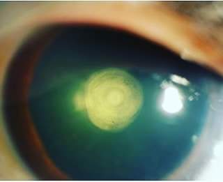
The diagnosis of PPC is clinical and can be easily made with a meticulous slit-lamp examination. Retro illumination reveals a thick white circular discoid plaque-like central opacity in the form of onion rings densely adherent to the posterior capsule. This typical appearance has also been described as a bull's eye appearance. The thickness and the breath of the opacity are a fair indicator of visual acuity also. Larger and thicker opacities affect the visual acuity more compared to the smaller ones. They are rarely picked up in infancy compared to anterior polar cataracts, which are quite evident during birth. While examining the anterior vitreous phase, it may appear like oil droplets suggesting a pre-existing posterior capsular defect. In a study, the mean PPC lens thickness was comparatively lower than that of senile sclerotic cataracts. Another study demonstrated that the mean age of patients in PPC was lesser than that of senile cataracts.[16]

PPC can also present with posterior subcapsular cataracts, cortical cataracts, and nuclear sclerotic cataracts. Sometimes dense white spots are noticed adjacent to the PPC, suggestive of the thin or absent posterior capsule. This is known as the Daljit Singh sign. When the PPC plaque is examined by a 60-degree slit, if there is a bend of rays at the center, this also signifies posterior capsule dehiscence.[13]

Classification Systems for Posterior Polar Cataract

Duke-Elder Classification System

Stationary PPC - This is the more common type seen in 65% of cases; it is a round, well-defined, localized opacity located on the posterior capsule. The onion ring appearance of PPC gives it the shape of a bull's eye. Very rarely, the opacity is masked by nuclear sclerotic cataracts. Occasionally satellite lesions are observed surrounding the opacity. The progression may be noted at any time.

Progressive PPC- The lenticular changes are present in the form of whitish opacity at the posterior cortex in the shape riders. The edges are feathery and don't involve the nucleus. It may also become symptomatic. The opacity affects the visual acuity, usually by the age of 30 to 50 years.[3]

Daljit Singh's Classification[20]

Type

Description

1

Presence of posterior polar cataract along with posterior subcapsular cataract

2

Onion ring-shaped posterior polar cataract with presence or absence of white spot at the edge

3

Discrete round or oval opacity at the center with surrounding white spots at the edge. This is usually associated with thin or absent posterior capsule

4

Combination of any of the above three along with nuclear sclerosis

Singh, in his analysis, proposed that type 2 can convert to type 3, and it is not a wise option to delay surgery in PPC cases.

Schroeder's Classification

He classified PPC in pediatric patients based on the obstruction in the red reflex through the pupil[21]

Grade

Description

Management

1

Very small PPC without any effect on the visual acuity through the clear part of the lens

Patching

2

PPC obstructing two-third part of the pupil

Patching with mydriasis and bifocal glasses

3

PPC with the surrounding area of optical distortion. A clear red reflex is appreciated only through a dilated pupil

Patching and early surgery

4

Large PPC, no red reflex is observed even through a dilated pupil

Patching and early surgery

Schroeder advised patching before surgery in PPC patients as it will be helpful to diagnose and treat amblyopia and post-surgical management. He proposed that if patching works well, surgery can be delayed in these cases. The above classification will also be helpful for planning surgery in these patients. The larger and denser the opacity, and earlier the area of pupillary obstruction surgery should be done.

Vasavada's Classification of Posterior Polar Cataract[14]

Vasavada divided PPC into three distinct varieties

  1. PPC with impending posterior capsular dehiscence
  2. Pre-existing capsular dehiscence with PPC
  3. Spontaneous dislocation of PPC

Evaluation

Visual Acuity and Refraction

Snellen’s visual acuity documentation of uncorrected and best-corrected visual, near vision, and pinhole vision is very important in these cases. The corrected distant visual acuity is an indirect indicator of the size and density of PPC.

Undilated and Dilated Fundus Evaluation

To rule out any incidental fundal pathologies and this also helps in deciding the prognosis of the case. Postoperatively, cases resulting in posterior capsular rent must undergo a retinal evaluation to rule out nucleus or cortex drop or signs of retinal detachment.

A-Scan Ultrasonography

To calculate the axial length, keratometry readings, and intraocular lens power for implantation.

B Scan Ultrasonography

In the case of dense opacities where media is hazy and the fundus is not visible, a B scan must be done to rule out retinal pathologies. In case of any retinal or optic disc pathology on B scan, the patient should be counseled in detail regarding visual prognosis. This also helps in planning future treatment of the patient with the help of a retina specialist. Guo et al. utilized 25 MHz B scan USG for delineating the posterior capsular status in PPC and proposed that this can be a valuable tool in guiding the management of these cases.[22]

Anterior Segment Optical Coherence Tomography (ASOCT)

ASOCT is a beneficial tool to assess the status of the posterior capsule preoperatively. All PPC patients must undergo preoperative ASOCT evaluation to rule out pre-existing PC dehiscence; this helps the ophthalmic surgeon plan case management and be prepared for any possible intraoperative mishap. ASOCT is a valuable tool for counseling the patients regarding visual prognosis and the outcome of the surgery.[23]

Intraoperative Anterior Segment Optical Coherence Tomography

The diagnosis and management of ASOCT have taken a leap forward, with intraoperative OCT guiding the surgical steps of PPC. Titiyal et al. described the posterior polar cortical disc defect (PPCDD) sign in PPC cases while performing phacoemulsification. They proposed that the presence of this sign is an indicator of an intact posterior capsule and the risk of PCR is comparatively lower in these patients compared to patients without PPCCD signs.[24]

Modified Posterior Optical Coherence Tomography

Pujari et al. defined conical sign with +20 dioptre and modified posterior optical coherence tomography (m-OCT). They showed that the presence of a conical defect is an indicator of pre-existing posterior capsular dehiscence and guides the intraoperative and postoperative management of these cases.[25]

Other Tests

Complete blood count, kidney function test, pulmonary function testing by a specialist, electrocardiogram, and physician evaluation are necessary when indicated before surgery. The patient should be systemically stable to undergo the surgical intervention.

Treatment / Management

Medical Treatment

Medical treatment has a limited role in PPC management. Only syndromic patients with systemic involvement may need treatment with the help of a physician or other clinician. Pediatric patients presenting with PPC should be thoroughly evaluated for rare systemic syndromes. Kronenberg et al. reported PPC in patients with neurocutaneous syndrome. Similarly, Duke-Elder reported PPC in patients with ectodermal dysplasia, ichthyosis, scleroderma, dyskeratosis congenita, and Rothmund syndrome.[1][6]

Surgical Management

Indications for Surgery

Excessive glare and photophobia, difficulty in reading fine prints, and difficulty in carrying out routine activities are all indications for surgery. Another indication is early type 1 and 2 PPC when the nucleus is soft, and the risk of complication is less. Pediatric PPC should be operated on as soon as possible to prevent the risk of blindness.[20]

Surgery of Choice

Phacoemulsification and manual small incision cataract surgery are the surgical techniques commonly employed for PPC management based on patient affordability, surgeons' expertise, and indication for surgery.[14] Extracapsular cataract extraction (ECCE) was previously used for PPC management, but recently it's being replaced MSICS due to high astigmatism and the need for multiple sutures.[26] Osher et al., in their analysis, showed that the incidence of PCR was nearly the same with phacoemulsification and ECCE.[7]

In contrast, Das et al. showed that the rate of complications was more with ECCE. Hence phacoemulsification is the surgery of choice.[26] Vasavada et al. found that PCR was most common during epinucleus removal in PPC while performing phacoemulsification.[14] In the west, phacoemulsification is more commonly performed compared to MSICS in India. In India, patients with poor socioeconomic strata usually undergo MSICS, being a commonly performed surgery. Hayashi et al., in their analysis, proposed phacoemulsification for soft PPC <4 mm and pars plana approach for PPC > 4 mm. For dense PPC >4 mm, they suggested ICCE. In a nutshell, the most commonly employed technique is phacoemulsification.[16]

Surgical Anesthesia

In today's era, topical anesthesia is the anesthesia of choice for phacoemulsification. However, some surgeons prefer peribulbar or retrobulbar anesthesia depending on the density of PPC, patients' age, systemic comorbidities, patients' cooperation on the table, other eye conditions, confidence, and surgical expertise of the surgeon while operating PPC. Sometimes during topical anesthesia, the patient squeezes the lids, which contributes to positive vitreous pressure. Excessive vitreous pressure acts as an added risk factor for intraoperative PCR. A gentle massage post peribulbar or retrobulbar block is suggested to reduce vitreous pressure.[14]

Anterior Approach

Corneal Incision Placement

The main corneal incision can be temporal or superior. The preferred incision is a temporal clear corneal 2.8 mm incision with two side port incisions. Consideration is given to straight clear corneal incision due to the ease of conversion to SICS or ECCE during posterior PCR. A straight clear corneal incision is easy to extend without causing trauma to the posterior cornea. The sclerocorneal incision helps reduce astigmatism and endothelial trauma.[27]

Ophthalmic Viscosurgical Devices ( Viscoelastic)

The viscoelastic of choice is a dispersive one; it aids in corneal endothelial protection, tamponade the vitreous during vitrectomy, and viscodissection of the nucleus.[28]

Capsulorhexis

Rhexis size should be about 5.5 mm to 6 mm for good sulcus support for IOL placement in the case of PCR. A small rhexis invites difficulty in delineation and phacoemulsification and also postoperative capsular phimosis, whereas a large rhexis helps in easy delineation and optic capture.

Hydro Procedures

As a general rule, hydrodissection is contraindicated in the case of PPC. The recommendation is for hydrodelineation by the inside-out technique. Some surgeons have also described multiple partial jets of hydrodissection while removing PPC, but it should be avoided ideally. Some surgeons prefer viscodissection for the separation of the nucleus from the epinucleus and cortex.[16]

Phacoemulsification and Dynamics

A good interplay of appropriate pharmacodynamics is important for excellent outcomes in case of PPC cases. The ideal scenario is wound with a minimum leak, adequate vitreous pressure, and perfect phaco machine parameters. For soft PPC, lower energy settings are recommended with the torsional mode in recent newer phaco machines. Nucleus rotation is contraindicated in PPC to prevent PCR and zonular dialysis. A low vacuum with a lower aspiration flow rate is recommended. The nucleus should be emulsified before manipulating the PPC component, which prevents nucleus or cortex drop in the vitreous cavity in case of PCR.

Various techniques have been described, like pre-chopping and sculpt and chop to avoid stress and manipulation of PPC. The nuclear material removal is followed by epinucleus and cortical material. In the case of soft PPC, the nuclear material should be fed to the phaco probe with the help of sinskey or the chopper.[29]

Doing this reduces tension on the central PPC component and also reduces the need to strip the cortical material under stress. Vacuum should be employed judiciously, as it can result in PCR. Dense nucleus material can be cracked into multiple small fragments and removed with minimal or no stress on the central component keeping the probe in the center. The epinucleus and cortical matter can be removed or freed leaflet by leaflet from the periphery to paracentral area avoiding stripping of the central PPC fragment. In most cases, the PPC comes in toto without compromising the integrity of the posterior capsule.

In case of a PCR, the dispersive viscoelastic and anterior vitrectomy should be ready to manage the vitreous prolapse. The other school of thought is removing the central PPC component with the help of a vitrectomy probe or by performing a posterior capsulotomy. After a thorough anterior vitrectomy. the IOL can be placed in the sulcus.[30] The morphology of PPC and status of the posterior capsule can be judged preoperatively with the help of ASOCT. Pujari et al. categorized PPC into three types conical, ectatic and moth-eaten, based on ASOCT appearance.[31]

Phacoemulsification Parameters

The ideal parameters described for management for PPC are 60% power, a bottle height of 50-70 cm, the aspiration flow rate of 15 to 25 mL/min, and a minimal vacuum of 30 to 100 mm. These parameters prevent stress on the posterior capsule and help in a very stable anterior chamber.[7]

Nucleotomy Techniques

Phacoaspiration

This process is recommended for soft PPC. While dealing with such a scenario, it is assumed that the posterior capsule is absent below the PPC.

Phaco Chop

This process is recommended for harder PPC plaques. Phaco chop help to reduce stress on the posterior capsule. It is important to avoid anterior chamber collapse as it can result in spontaneous posterior capsule rupture. Before withdrawing the phaco probe, the chamber should be filled with dispersive viscoelastic through the side port to prevent spontaneous rupture. Moreover, aggressive nuclear rotation, vigorous cracking maneuvers, and wider and deeper separation of fragments should be avoided to prevent PCR.[1]

Lambda Technique

Lee et al. described the lambda technique where the nucleus is sculpted in a λ shape. This is followed by a crack along the arms and quadrant removal of the central piece. This technique prevents stretching and rupture of the capsule.[13]

Inverse Horseshoe Technique

Salahuddin proposed this technique in which the distal portion of the nucleus is divided after sculpting. This is followed by OVD injection to elevate the two portions of the nucleus, thus forming a Visco shell. This technique helps in the hemi-dissection of the nucleus without the stretch of the posterior capsule. Later the individual nuclear fragments can be chopped and emulsified as usual.[32]

Epinucleus Pre-chop Technique

Lim and Goh described a technique of pre-chopping the epinucleus without reaching the depth of the posterior capsule and epinuclear plate. This is helpful for hard PPCs where the nucleus can be quickly mobilized by separating it from the anterior epinucleus shell.[29]

Peripheral Crack Technique

Chee described this technique for hard PPCs by partially cracking the nucleus in the periphery and further chopping the nuclear pieces in multiple quadrants without rotation. In this way, the posterior polar opacity is not manipulated, and the nucleus is separated from the outer nuclear shell with the help of the chopper.[33]

Reverse Flower Bloom Technique

In this technique, first, the nuclear material is removed without disturbing the integrity of PPC.  In the second step, the cortical matter is peeled by the outside-in technique (mimicking a reverse blooming flower), leaving the PPC intact. In the last step, the poster polar attachment is separated gently from the capsule.

Predivision Technique

Kamoi and Mochizuki described this technique where a pre-chopper creates two pre-chops on both sides of the central part. This is followed by central fragment removal, thus creating space in the capsular bag to remove further fragments easily.[34]

Posterior Approach

Pars plana lensectomy with vitrectomy has been described to prevent unwanted complications of PCR and nucleus drop. Few authors have also described the posterior approach through the pars plana route with variable outcomes. In a study of 11 eyes of 8 patients with this approach, three eyes developed posterior segment complications.[35] Another study used the pars plana approach for 2 out of  28 eyes in large PPC having a soft nucleus.[16] As a general consensus, this technique can be avoided due to the high risk of complications associated with vitrectomy

Epinucleus Removal

Vajpayee et al. described the layer by layer technique for removing epinuclear plate in cases of the pre-existing posterior capsular breach, which can be picked clinically or using ASOCT. While performing layer by layer removal of epinucleus, care should be taken to leave the central area untouched without undue stress on the posterior capsule. The PPC plaque can be easily viscodissected in the end.[33]

In another technique by Fine et al., epinucleus can be removed by pure viscodissection without unnecessary pressure on the posterior capsule.[36] Allen and Wood used the similar principle of viscodissection. They proposed that nucleus rotation should be avoided, and the epinucleus can be easily peeled off by injecting viscoelastic between the cortex and capsule.[37] In a landmark study by Taskapili et al., they compared outcomes of PPC with and without cortical viscodissection. They reported that the rate of posterior capsular rupture was higher in the without viscodissection group.[28]

Nagappa et al. described loosening of the epinucleus by phacoaspiration in the quadrant opposite the wound. This is followed by the removal of the sub incisional cortex by loosening it with hydrodissection. They reasoned that the hydrodissection wave doesn't cause posterior capsular rupture because the fluid escapes the path of least resistance in the epinuclear free area. They also proposed that this technique would be contraindicated in cases of pre-existing PCR detected clinically or by ASOCT.[38] 

Pseudohole Sign

Sometimes after removing posterior polar opacity, the impression of the onion ring is seen on the posterior capsule. This impression gives the appearance of a hole, but since the posterior capsule is intact, it has been labeled as a "pseudohole sign." Nagappa et al. were the first to describe the "fish mouth sign" as the sign of posterior capsular rupture when vitreous prolapse is seen through the PPC defect.[38]

Cortex Aspiration

The cortex removal should be performed with a bottle height of around 15 to 25 cc/min. The cortex should be pulled tangentially rather than the usual pulling centrally to avoid stress on zonules and prevent rupture of the posterior capsule. Vajpayee et al. proposed cortical removal by the "layer by layer "technique. In this technique, they described the removal of the cortex in a wedge-shaped manner and then separated with the help of a chopper; this prevents stress on the central plaque of PPC and prevents PCR. Another technique is the dry aspiration of the cortex with the help of Simcoe cannula (5 cc syringe) by filling the capsular bag with viscoelastic. It has also been proposed that capsular polishing should be avoided in these cases.[39]

Removal of the Posterior Polar Plaque

The general rule is PPC plaque should be left untouched till the end of the surgery (cortical aspiration). At the last step, the plaque should be separated by viscodissection. The separated plaque then should be lifted and can be aspirated with the help of a phaco tip or by bimanual irrigation and aspiration. Another proposed technique is filling the capsular bag with viscoelastic and then dislodge the plaque with a sinskey hook. The separated plaque can then be easily removed with the help of forceps. This will avoid the drop of PPC plaque in the vitreous cavity. Osher described the technique of "minimal residual aspiration."[28] In this technique, the PC is cleaned with the minimal touch of the PC with the help of irrigation and aspiration tips. If the PPC plaque is densely attached to the posterior capsule and the surgeon cannot dissect it by viscodissection, the plaque can be left in situ and later removed by Nd-YAG capsulotomy.[16]

Managing Posterior Capsular Tear

Once the posterior capsular tear is noticed, viscoelastic must be injected before removing the phaco probe to form the anterior chamber and prevent disruption of the anterior vitreous face. Dispersive viscoelastic must be injected to tamponade the vitreous and seal the posterior capsular rent. If possible, the rent can be fashioned into a posterior capsulorhexis, and then the intraocular lens can be easily placed in the bag. In case of an irregular tear, the IOL can be placed in the sulcus after a thorough anterior vitrectomy. 

Anterior Vitrectomy

Bimanual vitrectomy is recommended for cutting the anterior vitreous. High cut rate and low vacuum vitrectomy can be easily performed close to the rent. The vitrector is held in the dominant hand, and the infusion cannula should be held in the non-dominant hand directed towards the angle away from the rent. This helps to prevent vitreous hydration, vitreous prolapse, and enlargement of the tear.

Intraocular Lens Implantation

If the PCR is small, it can be converted to a round posterior capsulorhexis, and a single piece of IOL can be easily implanted in the capsular bag. If the PCR is large and irregular, a piece of IOL can be implanted in the ciliary sulcus, or an optic capture can be done. Optic capture helps in IOL stabilization and prevents iritis due to iridolenticular touch. Mackool, in his analysis, proposed that PMMA IOL is a better option than foldable IOL, as haptics of foldable IOL can stretch the PC and may enlarge the rent. In case of lack of adequate support for IOL, an ACIOL can be implanted or an iris-claw lens of a scleral fixated IOL.

Viscoelastic Removal, Wound Hydration, and Anterior Chamber Formation

Once the IOL has been implanted and is stable, viscoelastic should be thoroughly washed by irrigation and aspiration, and the incision should be hydrated. The anterior chamber should be formed with the self-sealing corneal incision. In case of PCR and vitreous prolapse and micro leaks, the main and side port incision should be sutured to prevent vitreous prolapse and endophthalmitis.

Differential Diagnosis

  • Posterior subcapsular cataract[40]
  • Posterior lenticonus[41][42]
  • Focal traumatic cataract[43]
  • Congenital cataract[44]
  • Lens abscess[45]
  • Lenticular intraocular foreign body

Prognosis

The prognosis of PPC depends on the stage of PPC, surgical decision-making, surgical expertise, timing, and meticulous clinical evaluation. In type 3 and 4 PPC and cases with pre-existing posterior capsular thinning or dehiscence, the prognosis might be guarded due to posterior capsular breach, vitreous prolapse in the anterior chamber, nucleus drop, and failure to implant IOL due to lack of capsular support. Cases with spontaneous rupture of PC may also have a guarded prognosis. PPC cases in skilled expert surgical hands usually have excellent visual outcomes.

Complications

Preoperatively

  • Amblyopia[44] 
  • Posterior capsular dehiscence 

Intraoperatively

  • Posterior capsular rupture
  • Zonular dialysis
  • Nucleus drop
  • Aphakia
  • Vitreous prolapse
  • Cortex drop
  • Corneal edema
  • Iridodialysis
  • Hyphema
  • Descemet membrane detachment
  • Fish mouthing of the main incision
  • Corneal burn 

Postoperatively

  • IOL decentration[46]
  • IOL drop
  • Retained cortex
  • Fibrinous uveitis
  • Hyphema
  • Secondary glaucoma[47]
  • Vitreous touch syndrome
  • Vitreous wick syndrome
  • Wound leak
  • Descemet membrane detachment[48]
  • Macular edema

Postoperative and Rehabilitation Care

In cases with no intraocular complications, the routine postoperative cataract topical drug regimen should be followed using steroids and antibiotics. Topical prednisolone 1% or dexamethasone 0.1% or steroid and antibiotics combination like 0.3% moxifloxacin with 1% prednisolone or 0.3% gatifloxacin with 0.1% dexamethasone should be started 6/5/4/3/2/1 one week each in tapering doses.

In cases with plain steroids formulation, a separate antibiotic can be prescribed in the form of 0.3% ofloxacin, 0.3% moxifloxacin, or 0.3% gatifloxacin four times per day for 20 days to safeguard the patient from secondary bacterial infection. Some ophthalmologists also prescribe 5% homatropine two times per day for 15 days to reduce postoperative iritis. In cases landing up with intraoperative posterior capsular rent and vitreous prolapse, besides routine postoperative drugs as mentioned above, oral antibiotic tablet ciprofloxacin 750 mg two times per day, oral anti-inflammatory tablet 50 mg diclofenac and 10 mg serratiopeptidase two times per day, and oral antacid tablet pantoprazole 40 mg 2 times per day for five days must be prescribed to prevent the risk of endophthalmitis.

A vitreoretinal surgeon should evaluate cases with an intraoperative nucleus, or cortical matter drop postoperatively to decide whether a secondary surgical procedure is necessary or conservative management is warranted. PPC cases with intraoperative aphakia should be planned for secondary IOL implantation, whether scleral fixated or sulcus fixated based on the capsular support. The ideal time of implantation is 1 to 2 months postoperatively when the inflammation has been controlled. The patients should be meticulously followed up and counseled for an excellent postoperative outcome.

Consultations

Any case of PPC presenting to the routine ophthalmic outpatient department should be meticulously evaluated and requires referral to an experienced cataract surgeon skilled in managing these challenging cases. The cataract surgeon routinely performing these complex anterior segment cases can best safeguard vision in these cases.

Deterrence and Patient Education

The patient must be educated regarding the type of congenital cataract they are having and all the associated complication with it. The patient should be educated regarding the prognosis of the condition in expert hands and the management options available. The patient should also be counseled regarding the postoperative drug regimen and the importance of treatment compliance and regular follow-up. Besides this, the patient should be alerted regarding the screening of family members for PPC.

Pearls and Other Issues

To sum up, PPC is an autosomal dominant or rarely sporadic congenital form of cataract that affects the patients' daily activities. It presents as a round onion ring-like discoid opacity dense adherent to the central posterior capsule. The PPC plaque is densely adherent to the posterior capsule and can result in posterior capsular rupture during cataract surgery. In expert hands, the surgical outcome is usually excellent. Cases landing up with intraoperative PCR can be managed by sulcus fixated, iris claw, iris sutured, scleral fixated, or anterior chamber IOL. Dreaded complications like nucleus drop, cortical matter in vitreous or IOL drop can be managed in conjunction with a vitreoretinal surgeon.

Enhancing Healthcare Team Outcomes

Any case of PPC presenting to the outpatient department should be thoroughly evaluated. These cases should be managed by a skilled cataract surgeon who is thorough with PPC management techniques. The nursing team has a significant role to play in preoperative counseling of the patient regarding the intraoperative complications and postoperative outcome. They also play a crucial role in obtaining consent from the patient or the next of kin.

A physician or an internist should evaluate PPC patients having systemic complaints to rule out any occult systemic pathologies. The pharmacist also plays an important role in drug chart preparation and procurement of drugs for the patient. Cases with intraoperative complications can be easily managed by the cataract surgeon himself, and cases requiring vitreoretinal intervention need expert inputs by the vitreoretinal team for lensectomy and pars plana vitrectomy. The prognosis is usually good in expert hands and has excellent long-term anatomical and functional outcomes.


(Click Image to Enlarge)
Classical slit lamp image of the patient depicting onion ring shaped discoid plaque like opacity densely adherent to the cent
Classical slit lamp image of the patient depicting onion ring shaped discoid plaque like opacity densely adherent to the central posterior capsule suggestive of posterior polar cataract
Contributed by Dr. Bharat Gurnani, MBBS, DNB, FCRS, FICO, MRCS Ed, MNAMS

(Click Image to Enlarge)
Higher magnification slit lamp image of the patient depicting onion ring shaped plaque like discoid opacity occupying the central capsule
Higher magnification slit lamp image of the patient depicting onion ring shaped plaque like discoid opacity occupying the central capsule. Careful look reveals no bend of central rays, a clinical indication of intact posterior capsule (Daljit Singh Sign)
Contributed by Dr. Bharat Gurnani, MBBS, DNB, FCRS, FICO, MRCS Ed, MNAMS
Details

Editor:

Kirandeep Kaur

Updated:

6/11/2023 6:25:34 AM

References


[1]

Kalantan H. Posterior polar cataract: A review. Saudi journal of ophthalmology : official journal of the Saudi Ophthalmological Society. 2012 Jan:26(1):41-9. doi: 10.1016/j.sjopt.2011.05.001. Epub 2011 May 7     [PubMed PMID: 23960967]


[2]

Chakrabarti A, Nazm N. Posterior capsular rent: Prevention and management. Indian journal of ophthalmology. 2017 Dec:65(12):1359-1369. doi: 10.4103/ijo.IJO_1057_17. Epub     [PubMed PMID: 29208816]


[3]

Vasavada AR, Raj SM, Vasavada V, Shrivastav S. Surgical approaches to posterior polar cataract: a review. Eye (London, England). 2012 Jun:26(6):761-70. doi: 10.1038/eye.2012.33. Epub 2012 Mar 23     [PubMed PMID: 22441026]


[4]

JONES HE. HYALOID REMNANTS IN THE EYES OF PREMATURE BABIES. The British journal of ophthalmology. 1963 Jan:47(1):39-44     [PubMed PMID: 14186858]


[5]

Hua X, Dong Y, Du J, Yang J, Yuan X. Phacoemulsification with hydrodelineation and OVD-assisted hydrodissection in posterior polar cataract. BMC ophthalmology. 2018 Jul 9:18(1):165. doi: 10.1186/s12886-018-0845-8. Epub 2018 Jul 9     [PubMed PMID: 29986674]


[6]

Yamada K, Tomita HA, Kanazawa S, Mera A, Amemiya T, Niikawa N. Genetically distinct autosomal dominant posterior polar cataract in a four-generation Japanese family. American journal of ophthalmology. 2000 Feb:129(2):159-65     [PubMed PMID: 10682967]


[7]

Osher RH, Yu BC, Koch DD. Posterior polar cataracts: a predisposition to intraoperative posterior capsular rupture. Journal of cataract and refractive surgery. 1990 Mar:16(2):157-62     [PubMed PMID: 2329471]


[8]

Ionides AC, Berry V, Mackay DS, Moore AT, Bhattacharya SS, Shiels A. A locus for autosomal dominant posterior polar cataract on chromosome 1p. Human molecular genetics. 1997 Jan:6(1):47-51     [PubMed PMID: 9002669]


[9]

Bidinost C, Matsumoto M, Chung D, Salem N, Zhang K, Stockton DW, Khoury A, Megarbane A, Bejjani BA, Traboulsi EI. Heterozygous and homozygous mutations in PITX3 in a large Lebanese family with posterior polar cataracts and neurodevelopmental abnormalities. Investigative ophthalmology & visual science. 2006 Apr:47(4):1274-80     [PubMed PMID: 16565358]


[10]

Pras E, Mahler O, Kumar V, Frydman M, Gefen N, Pras E, Hejtmancik JF. A new locus for autosomal dominant posterior polar cataract in Moroccan Jews maps to chromosome 14q22-23. Journal of medical genetics. 2006 Oct:43(10):e50     [PubMed PMID: 17047090]


[11]

Semina EV, Ferrell RE, Mintz-Hittner HA, Bitoun P, Alward WL, Reiter RS, Funkhauser C, Daack-Hirsch S, Murray JC. A novel homeobox gene PITX3 is mutated in families with autosomal-dominant cataracts and ASMD. Nature genetics. 1998 Jun:19(2):167-70     [PubMed PMID: 9620774]


[12]

Feltoon L. Eleven excuses for not having a will--what's yours? The Pennsylvania nurse. 1992 Sep:47(9):32     [PubMed PMID: 1528616]


[13]

Lee MW, Lee YC. Phacoemulsification of posterior polar cataracts--a surgical challenge. The British journal of ophthalmology. 2003 Nov:87(11):1426-7     [PubMed PMID: 14609852]


[14]

Vasavada A, Singh R. Phacoemulsification in eyes with posterior polar cataract. Journal of cataract and refractive surgery. 1999 Feb:25(2):238-45     [PubMed PMID: 9951671]


[15]

Mistr SK, Trivedi RH, Wilson ME. Preoperative considerations and outcomes of primary intraocular lens implantation in children with posterior polar and posterior lentiglobus cataract. Journal of AAPOS : the official publication of the American Association for Pediatric Ophthalmology and Strabismus. 2008 Feb:12(1):58-61     [PubMed PMID: 18029213]


[16]

Hayashi K, Hayashi H, Nakao F, Hayashi F. Outcomes of surgery for posterior polar cataract. Journal of cataract and refractive surgery. 2003 Jan:29(1):45-9     [PubMed PMID: 12551666]


[17]

Vasavada AR, Vasavada V, Vasavada S, Srivastava S, Vasavada V, Raj S. Femtodelineation to enhance safety in posterior polar cataracts. Journal of cataract and refractive surgery. 2015 Apr:41(4):702-7. doi: 10.1016/j.jcrs.2015.02.021. Epub     [PubMed PMID: 25840293]


[18]

Siatiri H, Moghimi S. Posterior polar cataract: minimizing risk of posterior capsule rupture. Eye (London, England). 2006 Jul:20(7):814-6     [PubMed PMID: 16254595]


[19]

Nada M, Rattan KN, Magu S, Parshad S. Aniridia and Wilm's tumor. Indian journal of pediatrics. 2003 Oct:70(10):837-8     [PubMed PMID: 14649484]


[20]

. Consultation section. Cataract surgical problem. Journal of cataract and refractive surgery. 1997 Jul-Aug:23(6):819-24     [PubMed PMID: 9292661]


[21]

Schroeder HW. The management of posterior polar cataract: the role of patching and grading. Strabismus. 2005 Dec:13(4):153-6     [PubMed PMID: 16361185]


[22]

Guo Y, Lu C, Wu B, Gao J, Li J, Yuan X, Tang X. Application of 25 MHz B-Scan Ultrasonography to Determine the Integrity of the Posterior Capsule in Posterior Polar Cataract. Journal of ophthalmology. 2018:2018():9635289. doi: 10.1155/2018/9635289. Epub 2018 Mar 26     [PubMed PMID: 29785304]


[23]

Kymionis GD, Diakonis VF, Liakopoulos DA, Tsoulnaras KI, Klados NE, Pallikaris IG. Anterior segment optical coherence tomography for demonstrating posterior capsular rent in posterior polar cataract. Clinical ophthalmology (Auckland, N.Z.). 2014:8():215-7. doi: 10.2147/OPTH.S55763. Epub 2014 Jan 10     [PubMed PMID: 24453477]


[24]

Titiyal JS, Nair S, Kaur M, Rawat J, Mazumdar SA. Intraoperative posterior polar cortical disc defect: sign of intact posterior capsule. Journal of cataract and refractive surgery. 2021 Aug 1:47(8):1039-1043. doi: 10.1097/j.jcrs.0000000000000578. Epub     [PubMed PMID: 33577272]


[25]

Pujari A, Selvan H, Yadav S, Urkude J, Singh R, Mukhija R, Makwana T, Sharma N. Preoperative assessment of posterior capsular integrity using a posterior segment OCT with a +20 D lens: The 'conical sign' to suggest capsular deficiency in posterior polar cataracts. Journal of cataract and refractive surgery. 2020 Jun:46(6):844-848. doi: 10.1097/j.jcrs.0000000000000197. Epub     [PubMed PMID: 32304483]


[26]

Das S, Khanna R, Mohiuddin SM, Ramamurthy B. Surgical and visual outcomes for posterior polar cataract. The British journal of ophthalmology. 2008 Nov:92(11):1476-8. doi: 10.1136/bjo.2007.129403. Epub 2008 Aug 26     [PubMed PMID: 18728051]


[27]

Pong JC, Lai JS. Managing the hard posterior polar cataract. Journal of cataract and refractive surgery. 2008 Apr:34(4):530; author reply 530-1. doi: 10.1016/j.jcrs.2007.12.027. Epub     [PubMed PMID: 18361960]


[28]

Taskapili M, Gulkilik G, Kocabora MS, Ozsutcu M. Phacoemulsification with viscodissection in posterior polar cataract: minimizing risk of posterior capsule tear. Annals of ophthalmology (Skokie, Ill.). 2007 Spring:39(2):145-9     [PubMed PMID: 17984504]


[29]

Lim Z, Goh J. Modified epinucleus pre-chop for the dense posterior polar cataract. Ophthalmic surgery, lasers & imaging : the official journal of the International Society for Imaging in the Eye. 2008 Mar-Apr:39(2):171-3     [PubMed PMID: 18435348]


[30]

Vasavada AR, Raj SM. Inside-out delineation. Journal of cataract and refractive surgery. 2004 Jun:30(6):1167-9     [PubMed PMID: 15177589]


[31]

Pujari A, Yadav S, Sharma N, Khokhar S, Sinha R, Agarwal T, Titiyal JS, Sharma P. Study 1: Evaluation of the signs of deficient posterior capsule in posterior polar cataracts using anterior segment optical coherence tomography. Journal of cataract and refractive surgery. 2020 Sep:46(9):1260-1265. doi: 10.1097/j.jcrs.0000000000000246. Epub     [PubMed PMID: 32618831]


[32]

Salahuddin A. Inverse horse-shoe technique for the phacoemulsification of posterior polar cataract. Canadian journal of ophthalmology. Journal canadien d'ophtalmologie. 2010 Apr:45(2):154-6. doi: 10.3129/i09-231. Epub     [PubMed PMID: 20379301]


[33]

Chee SP. Management of the hard posterior polar cataract. Journal of cataract and refractive surgery. 2007 Sep:33(9):1509-14     [PubMed PMID: 17720063]


[34]

Kamoi K, Mochizuki M. Pre-surround division technique: precise cracks surrounding the posterior opacity prior to phacoemulsification in posterior polar cataract surgery. Journal of cataract and refractive surgery. 2014 Nov:40(11):1764-7. doi: 10.1016/j.jcrs.2014.09.002. Epub 2014 Oct 23     [PubMed PMID: 25442881]


[35]

Ghosh YK, Kirkby GR. Posterior polar cataract surgery - a posterior segment approach. Eye (London, England). 2008 Jun:22(6):844-8     [PubMed PMID: 17332769]


[36]

Fine IH, Packer M, Hoffman RS. Management of posterior polar cataract. Journal of cataract and refractive surgery. 2003 Jan:29(1):16-9     [PubMed PMID: 12551661]


[37]

Allen D, Wood C. Minimizing risk to the capsule during surgery for posterior polar cataract. Journal of cataract and refractive surgery. 2002 May:28(5):742-4     [PubMed PMID: 11978449]


[38]

Nagappa S, Das S, Kurian M, Braganza A, Shetty R, Shetty B. Modified technique for epinucleus removal in posterior polar cataract. Ophthalmic surgery, lasers & imaging : the official journal of the International Society for Imaging in the Eye. 2011 Jan-Feb:42(1):78-80. doi: 10.3928/15428877-20101025-01. Epub 2010 Oct 28     [PubMed PMID: 21053813]


[39]

Vajpayee RB, Sinha R, Singhvi A, Sharma N, Titiyal JS, Tandon R. 'Layer by layer' phacoemulsification in posterior polar cataract with pre-existing posterior capsular rent. Eye (London, England). 2008 Aug:22(8):1008-10     [PubMed PMID: 17417622]


[40]

Gurnani B, Kaur K, Gireesh P. A rare presentation of anterior dislocation of calcified capsular bag in a spontaneously absorbed cataractous eye. Oman journal of ophthalmology. 2021 May-Aug:14(2):120-121. doi: 10.4103/ojo.OJO_65_2019. Epub 2021 Jun 28     [PubMed PMID: 34345149]


[41]

Kaur K,Gurnani B,Jezeela K,Veena K, Ruptured unilateral anterior lenticonus with congenital hypertrophy of retinal pigment epithelium: A rare association. Indian journal of ophthalmology. 2020 Nov;     [PubMed PMID: 33120654]


[42]

Kaur K, Gurnani B, Gireesh P. Hearing Loss and Decreasing Visual Acuity. Journal of pediatric ophthalmology and strabismus. 2019 Jul 1:56(4):212. doi: 10.3928/01913913-20190520-01. Epub     [PubMed PMID: 31322708]


[43]

Gurnani B, Kaur K, Sekaran S. First case of coloboma, lens neovascularization, traumatic cataract, and retinal detachment in a young Asian female. Clinical case reports. 2021 Sep:9(9):e04743. doi: 10.1002/ccr3.4743. Epub 2021 Aug 30     [PubMed PMID: 34484773]

Level 3 (low-level) evidence

[44]

Kaur K, Kannusamy V, Mouttapa F, Gurnani B, Venkatesh R, Khadia A. To assess the accuracy of Plusoptix S12-C photoscreener in detecting amblyogenic risk factors in children aged 6 months to 6 years in remote areas of South India. Indian journal of ophthalmology. 2020 Oct:68(10):2186-2189. doi: 10.4103/ijo.IJO_2046_19. Epub     [PubMed PMID: 32971637]


[45]

Balamurugan S, Gurnani B, Kaur K, Gireesh P, Narayana S. Traumatic intralenticular abscess-What is so different? The Indian journal of radiology & imaging. 2020 Jan-Mar:30(1):92-94. doi: 10.4103/ijri.IJRI_369_19. Epub 2020 Mar 30     [PubMed PMID: 32476758]


[46]

Gurnani B, Kaur K. Rare traumatic anterior dislocation of capsular tension ring-intraocular lens complex in-toto. Indian journal of ophthalmology. 2020 Nov:68(11):2529. doi: 10.4103/ijo.IJO_807_20. Epub     [PubMed PMID: 33120674]


[47]

Christy J, Jain N, Gurnani B, Kaur K. Twinkling Eye -A Rare Presentation in Neovascular Glaucoma. Journal of glaucoma. 2019 May 23:():. doi: 10.1097/IJG.0000000000001287. Epub 2019 May 23     [PubMed PMID: 31135586]


[48]

Gurnani B, Kaur K. Comment on: Two-year clinical outcome after Descemet membrane endothelial keratoplasty using a standardized protocol. Indian journal of ophthalmology. 2021 Jun:69(6):1637-1638. doi: 10.4103/ijo.IJO_3442_20. Epub     [PubMed PMID: 34011764]

Level 2 (mid-level) evidence