Minimally Invasive Glaucoma Surgery

Earn CME/CE in your profession:


Continuing Education Activity

Glaucoma is a chronic progressive ocular pathology that affects the optic nerve and results in irreversible vision loss. The condition is also called the silent thief of sight, as it slowly damages the optic nerve before any noticeable central vision loss can occur. The damage is due to persistently raised intraocular pressure (IOP), which damages the optic nerve and causes visual field defects. The medical management of glaucoma is by either topical antiglaucoma medications, laser peripheral iridotomy, or laser trabeculoplasty to lower the IOP. The surgical management is in the form of trabeculoplasty or glaucoma drainage devices. The surgical management options are associated with many intraoperative and postoperative complications, require long-term follow-up, and may have prolonged recovery time. The surgical management options are indicated for severe diseases when medical management fails. Over the years, there have been limited options for mild-to-moderate illness patients with uncontrolled IOP. To fill this gap, novel microinvasive or minimally invasive glaucoma surgery (MIGS) has been introduced. Patients with mild-to-moderate glaucoma who are noncompliant or intolerant to topical drugs are ideal cases for MIGS. The MIGS implant can be implanted during phacoemulsification surgery, reducing dependence on topical medications. This activity highlights the indications, contraindications, classification, technique, complications, and clinical significance of performing MIGS by an interprofessional team.

Objectives:

  • Describe the technique of minimally invasive glaucoma surgery.
  • Review the indications of minimally invasive glaucoma surgery.
  • Summarize the contraindications of minimally invasive glaucoma surgery.
  • Explain the clinical significance of minimally invasive glaucoma surgery and the role of interprofessional collaboration in improving outcomes.

Introduction

Glaucoma is a chronic progressive optic neuropathy characterized by visual field changes and optic nerve cupping. It is the second most common cause of irreversible blindness across the globe.[1] Managing glaucoma in developed and developing countries poses a substantial economic and social burden. Glaucoma accounts for a total of 8% percent of blindness worldwide.[2] The modifiable risk factor for glaucoma is intraocular pressure (IOP) which is the main line of treatment.[3]

The conventional treatment modalities for mild to moderate glaucoma are antiglaucoma medications and laser procedures in the form of laser iridotomy and laser trabeculoplasty. Advanced cases or cases with non-resolving intraocular pressure require a target IOP to safeguard vision and field. The treatment for advanced stages can be trabeculectomy and glaucoma drainage devices (GDD).[4]

Scientists and Ophthalmic researchers have always searched for alternative surgical treatment modalities for rapid and effective reduction of IOP, maintaining a safety profile, using ab-interno procedures with minimal tissue trauma and faster recovery.[5] These procedures have been labeled microinvasive or minimally invasive glaucoma surgery (MIGS).[6]

MIGS work either by bypassing the trabecular meshwork (Hydrus stent, iStent, Trabectome, Kahook dual blade), increased aqueous outflow through Schlemm's canal (canaloplasty), increased uveoscleral outflow (iStent supra), aqueous shunt through subconjunctival space (XEN implant) or by ciliary body ablation (endocyclophotocoagulation).[7]

Although long-term results are awaited, MIGS has revolutionized glaucoma management with minimal tissue trauma, good post-operative recovery, and improved patient satisfaction. The MIGS options have opened a whole new bunch of options for all the glaucoma specialists with promising results. Another advantage is that MIGS can be combined with phacoemulsification reducing surgical time. This activity deals with various available MIGS options, their design, efficacy, the technique of implantation, and safety profile for better patient management.[8]

MIGS Characteristics

  • High safety- reduced risk of complications like choroidal detachment, hypotony, hemorrhage, and effusion
  • Minimal alteration of normal angle anatomy- MIGS improves the physiological aqueous outflow by minimal disruption to angle structures
  • Ab interno procedure- MIGS is performed through the clear corneal incision using an ab interno approach with direct visualization of the anatomical target
  • MIGS is an excellent alternative to traditional angle surgery in lowering the IOP - the amount of IOP reduction is lesser than trabeculectomy but should be at least 20%.
  • Good post-operative recovery with minimal bedtime for patients - MIGS offers ease to patients as well as surgeons[8]

Minimally Invasive Glaucoma Surgery Classification

There are four main approaches for IOP reduction by MIGS

1. Increasing Aqueous Outflow from Trabecular Meshwork and Schlemm Canal

Stents

  • iStent
  • iStent Inject
  • Hydrus

Tissue Removal

  • Gonioscopy-assisted transluminal trabeculotomy (GATT)
  • Kahook dual blade goniotomy
  • Trabectome
  • TRAB 360 system
  • Excimer laser trabeculostomy

Through Schlemm Canal

  • VISCO360 device
  • Ab Interno canaloplasty (ABiC) using the iTrack microcatheter system[9]

2. Increased Uveoscleral Outflow through Suprachoroidal Space

  • CyPass micro-stent[10]

3. Aqueous Shunt through Subconjunctival Space

4. Ciliary Process Ablation Resulting in Reduced Aqueous Outflow

  • Endocyclophotocoagulation[12]

Table Depicting Various Types of MIGS Based On Mechanism of Action

S. No

MIGS Mechanism

Type of MIGS

Remarks

1

Increased Trabecular Outflow

iStent Micro-Bypass

Made of titanium, the stent has a heparin coating and is non-ferromagnetic. Dimensions 1 mm x 0.3 mm. Ab interno insertion into Schlemm's canal.


Gonioscopy-assisted transluminal trabeculotomy (GATT)

In this technique microcatheter or prolene/ nylon suture is passed through Schlemm's canal 360 degrees under gonioscopic assistance. This is an ab interno trabeculotomy. 


Trabectome

This is an ab interno technique which is a combination of electrocautery, irrigation, and aspiration.


TRAB 360 Trabeculotomy

This is performed with the help of a disposable, non-powdered device. A flexible nylon trabeculotome is advanced in the Schlemm canal 180 degrees and then lysed.


Kahook Dual Blade

This is a type of ab interno trabeculotomy which is done using a stainless steel blade.


Ab interno canaloplasty

This is a microcatheter used to vasodilate the Schlemm's canal.


Hydrus Microstent

This is a crescent-shaped ab interno nickel-titanium device inserted into the Schlemm canal.

2

Increase Uveoscleral / Suprachoroidal/ Supraciliary Outflow


CyPass Micro-Stent 

This stent has fenestrations made up of biocompatible polyamide material—inserted ab internally between the sclera and suprachoroidal space.


iStent Supra

This is a heparin-coated stent with a polyethersulfone and titanium sleeve inserted internally between the sclera and suprachoroidal space. 

3

Increase Subconjunctival Outflow


XEN Glaucoma  Treatment System 

This is a tube implant made up of gelatin and glutaraldehyde. Inserted ab internally into the anterior chamber, then through the sclera into subconjunctival space, forming a bleb.


InnFocus MicroShunt (PRESERFLO MicroShunt)

It is a flexible micro shunt composed of SIBS (poly(styrene-block-isobutylene-block-styrene)) inserted ab externally into the anterior chamber through subconjunctival space, forming a bleb.


4

Decrease Aqueous Production


Endocyclophotocoagulation

Cyclodestruction of the ciliary body using continuous mode energy


Anatomy and Physiology

Minimally invasive glaucoma surgery lowers IOP by altering various aspects of aqueous humor dynamics. MIGS can bypass the trabecular meshwork. The resistance to aqueous outflow is bypassed by using a stent. The stent allows the aqueous to flow directly from the anterior chamber to Schlemm's canal and, in turn, reduces IOP.[13] 

The second approach to bypass the trabecular meshwork resistance is goniotomy or trabeculectomy. The surgical alteration of tissue by excision or incision allows improved aqueous drainage through Schlemm's canal. Another approach is by dilating the Schlemm canal using viscoelastic, which enhances the normal physiological aqueous outflow.[14]

The next approach of MIGS is to increase uveoscleral outflow. A microstent can be placed in the suprachoroidal space. If this is not possible, then aqueous can be bypassed into the subconjunctival space by a small incisional ab interno approach.[15] 

The last approach reduces the aqueous production through ciliary processes by ciliary body ablation. This is done by endocyclophotocoagulation, in which an endoscopic laser is inserted through a clear corneal incision, and the ciliary body is ablated.[16]

Indications

Minimally invasive glaucoma surgery may be considered in the following:

  • Mild to moderate glaucoma cases
  • Primary open angle glaucoma (POAG), pigmentary glaucoma and pseudoexfoliation glaucoma (PXFG)
  • Intraocular pressure is not reduced even after maximal antiglaucoma therapy and laser trabeculoplasty
  • Noncompliance to treatment
  • Adverse drug reaction[17]

Contraindications

MIGS is contraindicated in the following situations:

  • Active neovascular glaucoma (NVG)
  • Primary angle-closure glaucoma (PACG) or secondary angle-closure glaucoma (SACG)
  • Corneal opacity
  • Angle dysgenesis
  • Elevated episcleral venous pressure, e.g., Sturge Weber syndrome, thyroid eye disease, retrobulbar tumors[18]

Equipment

The following equipment will be required:

  • iStent
  • iStent Inject
  • Hydrus implant
  • Gonioscope
  • Trabectome
  • Kahook dual blade
  • Excimer laser
  • Viscoelastic
  • iTrack 360 microcatheter
  • Xen implant
  • Cypass implant
  • Cyclodiode

Personnel

Ophthalmologists, particularly glaucoma specialists, are involved in providing the surgical options of MIGS to their patients. Operation theater technicians, optometrists, nurses, pharmacists, and anesthetists also form a crucial part of the management team to provide optimal outcomes.

Technique or Treatment

Trabecular Meshwork Bypass

The juxtacanalicular trabecular meshwork is the site of greatest resistance to aqueous outflow. Through the technique of MIGS, the tissue can be ablated or excised, or trabecular meshwork can be bypassed through stents, thus allowing direct bypass of aqueous humor into the Schlemm's canal.[19] 

This is indicated in POAG, PXFG, pigmentary glaucoma, and ocular hypertension with a target IOP of 15 to 16 mm Hg. TM bypass is contraindicated in PACG, NVG, angle dysgenesis, corneal opacity, and elevated episcleral venous pressure cases like Sturge Weber syndrome.[20] The complications associated with TM bypass include hyphema, stent displacement, malposition, obstruction, peripheral anterior synechiae, and transient IOP elevation.[21]

Stents

iStent

iStent is the first FDA-approved MIGS that came on the market in 2012. This is a very small stent coated with heparin and made of titanium non-ferromagnetic material. This is a ridge shaped like a snorkel.[22]

The stent measures 1 mm x 0.3 mm, and the snorkel measures 0.25 mm in height with a central lumen of 120 um, which will project in the anterior chamber. The device is designed to have three arches for retention that ensure its proper placement in the Schlemm's canal at the nasal angle. iStent is implanted with the help of an ab interno approach using a pre-loaded inserter through a clear corneal incision under gonioscopic guidance.

iStent implantation results in an IOP reduction of approximately 25% in nearly 70% of the patients, significantly reducing the medication burden. Single iStent placement may result in an IOP reduction of 23 to 27%, whereas multiple iStent may cause a 40 to 44% reduction in IOP in approximately 60% of patients by one year, as per previous studies.[23]

iStent Inject 

This device was FDA approved in 2018 and is a second-generation TM bypass stent. This is considered to be a small medical implant in the human body. Like iStent, this is heparin-coated and non-ferromagnetic, made up of titanium. The stent dimensions are the height of 360 um and 230 um in diameter with a central lumen of 80 microns.[24]

The stent has a head with the tapering end that sits in the Schlemm's canal, and the thorax enters the TM and anterior chamber. iStent inject is inserted by ab interno approach through gonioscopic visualization.[22]

The device is available as a pre-loaded injector (G2-M-IS injector system) with two stents placed nasally in the TM and Schlemm canal 30 to 60 degrees apart. Fea et al., in their analysis, documented a reduction in IOP of 35 to 39% with a decrease in medication load, and 65 to 75% of patients were free of medications at one year. One of the previous studies showed that the IOP reduction by implantation of two stents is equivalent to IOP reduction with two antiglaucoma drugs after one year.[8]

Hydrus Stent

This stent is a small crescent-shaped stent of 8 mm in length and 290 um in diameter. This has a small inlet and three window-shaped openings which sit in the anterior chamber. This stent is made up of nitinol material, which has good shaping memory. The device can be implanted in the nasal or inferior temporal quadrant through a pre-loaded injection using a clear corneal incision.[25]

The device has small intracanalicular scaffolds which occupy nearly three clock hours of Schlemm's canal and spare the collector channel ostia in the posterior portion of the canal. This stent works on the unique trimodal mechanism. It bypasses the trabecular meshwork and allows aqueous flow. It dilates the Schlemm's canal 4 to 5 times, making outflow through collector channels easier. The HORIZON study concluded that there was more than a 20% reduction in IOP in 80% of the cases, and 73% of eyes were free of antiglaucoma medications at 24 months.[26]

Another study concluded a 20% IOP reduction in 77.3% of eyes at 24 months post-surgery compared to antiglaucoma medications. The COMPARE study compared Hydrus vs. iStent and concluded that 39.7% of eyes showed more than 20% IOP reduction than 13.3% of eyes in the iStent group. Approximately 46.6% were medication-free in the Hydrus group compared to 24% in the iStent group.[27]

Tissue Excision (Trabeculotomy)- Bypassing the TM

Kahook Dual Blade (KDB) Goniotomy

This is a stainless steel disposable dual-side blade having a sharp tip. The ends are tapered for a smooth entry through the trabecular meshwork. The blade's heel snugs easily within the Schlemm's canal for smooth access, and the front part of the blade generates stretch. The double blade makes parallel incisions in the TM. This helps in the total removal of the TM with minimal tissue damage and less fibrosis over time, giving a long-term outcome.[28]

The blade is used by the ab interno approach under gonioscopic visualization. Previous studies have documented approximately 20% reduction in IOP in 70% of eyes with at least one medication with KDB at one-year follow-up. Good results are seen in patients with pseudoexfoliation glaucoma, pigmentary glaucoma, and secondary open-angle glaucoma, as it helps remove pigments from the TM. KDB has also been shown to be beneficial in congenital glaucoma; and advanced and refractory glaucoma cases.[29]

Trabectome

The trabectome was invented by Baerveld, Chuck, and Irvine and got FDA approval in 2004 to manage adult and pediatric cataracts. This comprises a handpiece of 19.5 gauzes, automated irrigation and aspiration, electronic cautery, and a foot pedal to control the irrigation and aspiration.[30]

The handpiece's footplate stretches the TM and protects the surrounding tissue from the thermal burn. The handpiece tip also has a bipolar cautery which generates plasma at 550 kHz, which ablated the TM by creating ionizing molecules. This creates an opening from AC into the Schlemm canal and collector channels. Trabectome is documented to reduce IOP by approximately 40%. It has also been proven effective in narrow-angle glaucoma, failed trabeculectomy, shunts, and refractory glaucoma cases.[31]

Laser Trabeculostomy

This was first described by Berlin in 1987 using an excimer laser and used by Vogel and Lauritzen in 1997. This is performed using a fiber optic delivery system and a short pulse of 80ns, 308 nm xenon chloride laser to the TM. The energy pulse delivered is 1.2 mJ, and the pulse duration is 10 to 60 ns with a frequency of 20 Hz. This procedure helps remove the tissue obstructing the aqueous outflow with significantly less damage to the surrounding tissue. A total of 10 microchannels over 90 degrees are created, 500 um apart.

This process is also labeled pneumatic canaloplasty as it dilates the Schlemm canal and collector channels. LT is also an ab interno procedure performed under direct visualization of gonioscope. This process has a moderate reduction of IOP between 20 to 40%.[32]

Gonioscopy Assisted Transluminal Trabeculotomy (GATT)

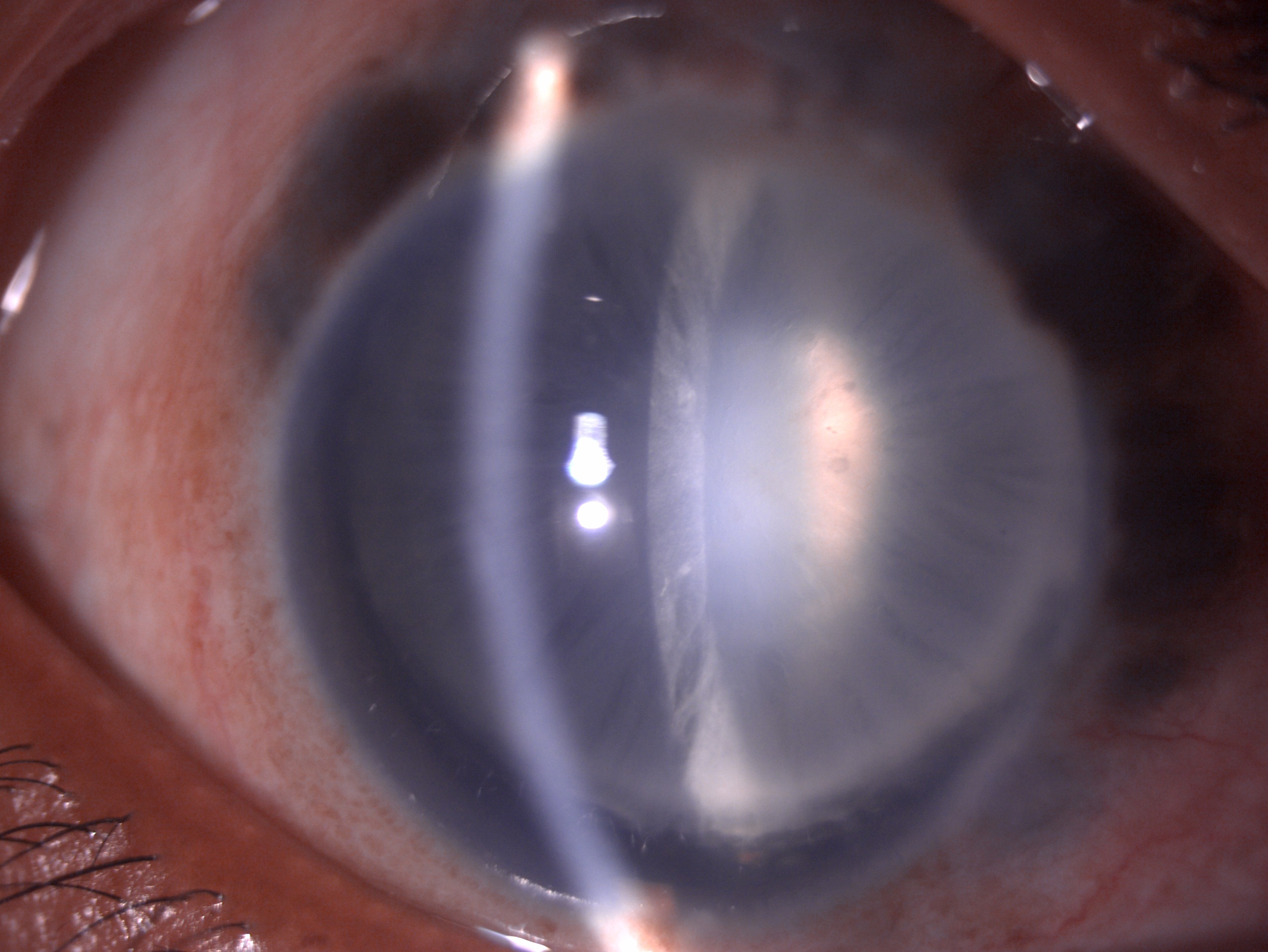
This was first described by Grover et al. in 2014. This consists of a microsurgical forceps and an iTrack microcatheter with a 200 um diameter shaft having a lubricating coating and a distal tip with illumination to monitor the catheter location. In this ab interno approach, the catheter or suture is passed through the nasal quadrant under gonioscopic visualization into the Schlemm canal and then advanced to 360 degrees with the help of micro-forceps.[33] Then traction is applied to break the TM, thus resulting in 360-degree trabeculectomy. GATT has shown to be effective in approximately 70 to 90% of cases compared to the ab externo approach with an IOP reduction of 30 to 40%. GATT was primarily used for congenital glaucoma and juvenile open-angle glaucoma. This is an essential option for refractory glaucoma and posts trabeculectomy cases. GATT is contraindicated in patients who require anticoagulants or have bleeding diatheses.[34]

Ab Interno Canaloplasty (AbiC)

This is performed with the help of the iTrack microcatheter. The catheter is 200 um thick with a bulbous 250u tip and has a lubricating coating for smooth passage, viscoinjector, and fiber optic enabled illumination. The microcatheter is passed through Schlemm's canal. Then it i's removed as the Visco surgical device is inserted to Visco dilate all the sites of resistance outflows, such as the Schlemm canal and collector channels. Visco-dilatation helps to create perforations within the TM, which increase aqueous outflow. This technique results in 30 to 40% IOP reduction at 12 months with less than two antiglaucoma drugs.[35]

Suprachoroidal Shunts

These shunts help direct the aqueous outflow to the suprachoroidal space in a controlled manner and thus increase the uveoscleral outflow. This acts as a permanent reservoir between the anterior chamber and supraciliary space.[36]
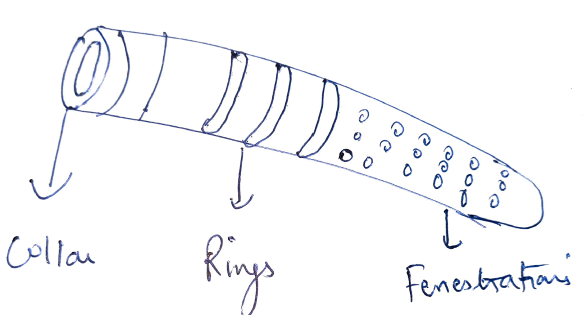
Cypass Stent

They were first used in 2016 and FDA approved for glaucoma management. The stent is a polyamide stent, flexible, fenestrated, micro-sized of 6.35 mm x 510 um, having a lumen of 300 um. This is available as a pre-loaded device with a guidewire confirming the shape of the sclera to help dissection and insertion between the anterior chamber/ sclera and suprachoroidal space. The device results in a higher rate of endothelial cell loss; hence was withdrawn from the market in 2018.[37]

iStent Supra

This is a curved heparin-coated tube with a lumen of 0.16 mm. It is made up of biocompatible polyether sulfone consisting of a titanium sleeve and retention ridge to hold it in place. The stent is mounted with a guide so that it can be easily inserted in the anterior chamber and suprachoroidal space by ab interno approach under gonioscopic guidance. iStent supra, along with Travoprost, is shown to reduce IOP by 20% in patients with advanced glaucoma.[38]

Subconjunctival Filtration Stents

Xen Implant (Xen Gel Stent)

This subconjunctival implant allows the aqueous flow to the subconjunctival space. This is a kind of bleb-forming procedure like trabeculectomy. Xen implant is implicated for open-angle glaucoma in moderate to an advanced stage, refractory glaucoma; post failed trabeculectomy, phakic or pseudophakic glaucoma. The device is contraindicated in angle-closure glaucoma, conjunctival scarring, neovascularization, inflammation, and silicone oil or vitreous in the anterior chamber.[38]

This hydrophilic tube is made up of porcine collagen-derived gelatin, which is crosslinked with glutaraldehyde. The device is in a rigid state and straight when dry and mouldable when soft and hydrated. This is 6 mm in length and has three types XEN 140, XEN 63, and XEN 45. XEN 45 is FDA-approved with an inner lumen of 45 um and an outer diameter of 150 um. The device has the advantage that it prevents the risk of hypotony. This is available as a pre-loaded device with a disposable 27G injector inserted in the anterior chamber through an ab interno approach.[39]

The device is placed in the superonasal or superior quadrant and is passed through the sclera to be placed in the subconjunctival space. There is a bleb formation at the end of the procedure, and mitomycin C can be used to reduce fibrosis. The Xen gel implant can migrate into the anterior chamber or gape through the conjunctiva, or there may be fragmentation of the stent. Hypotony, hyphema, and stent obstruction can also occur. Xen gel implant causes a 29 to 41% reduction in IOP in nearly 45% of patients without antiglaucoma drugs.[40]

InnFocus MicroShunt (PRESERFLO MicroShunt)

This is an 8.5 mm long micro shunt with a 1 mm fin that divides the shunt into a long proximal (4.5 mm) and short distal (3 mm) part. The material is poly (styrene-block-isobutylene-block-styrene) or SIBS. This material has been used for coronary stents and is biologically inert. The external lumen is 350 um, and the internal lumen is 75 um. The placement of the device requires fornix-based peritomy, dissection of attachments of the tenon, and application of mitomycin-c (MMC) soaked sponges to the scleral bed. After washing with saline, a point 3 mm behind the limbus is marked through which the device is entered intra-sclerally to open inside the anterior chamber parallel to the iris. The tenon and conjunctiva are sutured after confirming the patency of the device.

Batlle and colleagues showed that the IOP was controlled within low teens in most patients three years after receiving the shunt with MMC.[41] Most complications, including transient hypotony and transient choroidal effusion, resolved spontaneously.[41] In a retrospective study on 164 eyes, complete success and qualified success were achieved at one year in 76.9% and 92.5% of eyes, respectively.[42] 

Endocytophotocoagulation (ECP)

This results in decreased aqueous production by ciliary body ablation and, hence, a reduction in IOP. The mechanism is localized ciliary process shrinkage with temporary occlusive vasculopathy. If the ciliary processes are visualized directly, it results in nominal collateral damage. This is indicated in mild to moderate glaucoma cases along with cataract extraction with sparing of the conjunctiva, refractory open-angle glaucoma cases, pediatric glaucoma, neovascular glaucoma, and contraindicated in active inflammation. Rarely ECP may result in zonular damage or cataract progression.[43]

The device is a probe with a laser unit having a diode laser. The wave energy is emitted at a wavelength of 810 nm with 175 W xenon light, helium-neon laser, and a video camera imaging system. The probe consists of a fiber optic system that transmits the signals. The probe can be inserted through a clear corneal incision and can be straight or curved. The video monitor allows the endoscopic view; thus, the surgeon can control the foot pedal to fire the laser. The laser power is between 0.2 to 0.25 watts, and the power is titrated to see blanching. At one moment, a 200 to 300-degree angle is treated.

In refractory glaucoma cases, 1 to 2 mm of pars plana is also coagulated to reduce the IOP aggressively; this is labeled as ECP plus. This is done through the pars plana approach and is preceded by pars plana vitrectomy. The adverse effects reported are secondary glaucoma, hyphema, post-operative inflammation, retinal detachment, choroidal detachment, cystoid macular edema, IOL (intraocular lens) dislocation, and capsular opacification. It results in approximately 35% IOP reduction in patients with one medication. Combined with phacoemulsification, ECP reduces IOP by 20 % in about 60% of patients, and a success rate of 43% is noted in pediatric patients.[44]

Complications

  • Hyphema
  • Hypotony
  • Stent displacement
  • Stent malposition
  • Stent obstruction
  • Stent migration
  • Stent fragmentation
  • Conjunctival gaping
  • Peripheral anterior synechiae
  • Transient IOP elevation
  • Post-operative inflammation
  • Retinal detachment
  • Choroidal detachment
  • Cystoid macular edema
  • IOL dislocation
  • IOL subluxation
  • Posterior capsular opacification[45]

Clinical Significance

Minimally invasive glaucoma surgery has revolutionized glaucoma management over the past few years. It is a relatively new modality of glaucoma management that is gaining popularity as large numbers of surgeons are opting for it. The postoperative outcomes and patient satisfaction rate are good with these procedures.

The safety profile and efficacy are also good. MIGS has opened a new door for mild to moderate glaucoma management, reducing the medication burden on the patients. Recent studies have provided highly encouraging results, and more large-scale studies with long-term follow-up will be needed to give better insight and understanding of MIGS.[46]

Enhancing Healthcare Team Outcomes

Any patient presenting to the outpatient department or with a family history of glaucoma should be thoroughly screened for glaucoma. Ophthalmologists play a crucial role in diagnosing and deciding the type of intervention for glaucoma severity. The ophthalmologist chooses the type of MIGS needed to reduce the IOP. The optometrists, nursing staff, operation theater assistants, and counselors play a vital role in the mild to moderate glaucoma management with MIGS.[47]

Nursing, Allied Health, and Interprofessional Team Interventions

The nursing, allied health, and interprofessional team help manage patients undergoing MIGS by assisting in OPD (outpatient department), OR (operating room), and counseling regarding the procedure and complications associated with it.

Nursing, Allied Health, and Interprofessional Team Monitoring

The nursing, allied health, and interprofessional team also help monitor these patients with regular IOP monitoring, whenever needed with OCT (optical coherence tomography) and HFA (Humphrey field analyzer), and monitoring the regular follow-up of these patients.


(Click Image to Enlarge)
Digital image of the patient depicting mild congestion, scleral thinning, uveal show, deep anterior chamber, zonular dialysis
Digital image of the patient depicting mild congestion, scleral thinning, uveal show, deep anterior chamber, zonular dialysis and subluxated cataract who has been planned for MIGS
Contributed by Dr. Bharat Gurnani, MBBS, DNB, FCRS, FICO, MRCS Ed, MNAMS

(Click Image to Enlarge)
iStent micro inject
iStent micro inject
Contributed by Dr. Bharat Gurnani, MBBS, DNB, FCRS, FICO, MRCS Ed, MNAMS

(Click Image to Enlarge)
Cypass MIGS stent
Cypass MIGS stent
Contributed by Dr. Bharat Gurnani, MBBS, DNB, FCRS, FICO, MRCS Ed, MNAMS
Details

Updated:

8/25/2023 3:04:40 AM

References


[1]

GBD 2019 Blindness and Vision Impairment Collaborators, Vision Loss Expert Group of the Global Burden of Disease Study. Causes of blindness and vision impairment in 2020 and trends over 30 years, and prevalence of avoidable blindness in relation to VISION 2020: the Right to Sight: an analysis for the Global Burden of Disease Study. The Lancet. Global health. 2021 Feb:9(2):e144-e160. doi: 10.1016/S2214-109X(20)30489-7. Epub 2020 Dec 1     [PubMed PMID: 33275949]


[2]

Delgado MF, Abdelrahman AM, Terahi M, Miro Quesada Woll JJ, Gil-Carrasco F, Cook C, Benharbit M, Boisseau S, Chung E, Hadjiat Y, Gomes JA. Management Of Glaucoma In Developing Countries: Challenges And Opportunities For Improvement. ClinicoEconomics and outcomes research : CEOR. 2019:11():591-604. doi: 10.2147/CEOR.S218277. Epub 2019 Sep 27     [PubMed PMID: 31632107]


[3]

Weinreb RN, Aung T, Medeiros FA. The pathophysiology and treatment of glaucoma: a review. JAMA. 2014 May 14:311(18):1901-11. doi: 10.1001/jama.2014.3192. Epub     [PubMed PMID: 24825645]


[4]

Muñoz-Negrete FJ, Moreno-Montañés J, Hernández-Martínez P, Rebolleda G. Current Approach in the Diagnosis and Management of Uveitic Glaucoma. BioMed research international. 2015:2015():742792. doi: 10.1155/2015/742792. Epub 2015 Oct 19     [PubMed PMID: 26558280]


[5]

Chen DZ, Sng CCA. Safety and Efficacy of Microinvasive Glaucoma Surgery. Journal of ophthalmology. 2017:2017():3182935. doi: 10.1155/2017/3182935. Epub 2017 Apr 23     [PubMed PMID: 28512578]


[6]

Costagliola C, Sbordone M, Gandolfi S, Cesari L, Furneri G, Fea AM. Minimally Invasive Surgery in Mild-to-Moderate Glaucoma Patients in Italy: Is It Time to Change? Clinical ophthalmology (Auckland, N.Z.). 2020:14():2639-2655. doi: 10.2147/OPTH.S264839. Epub 2020 Sep 14     [PubMed PMID: 32982157]


[7]

Gillmann K, Mansouri K. Minimally Invasive Glaucoma Surgery: Where Is the Evidence? Asia-Pacific journal of ophthalmology (Philadelphia, Pa.). 2020 May-Jun:9(3):203-214. doi: 10.1097/APO.0000000000000294. Epub     [PubMed PMID: 32501895]


[8]

Pillunat LE, Erb C, Jünemann AG, Kimmich F. Micro-invasive glaucoma surgery (MIGS): a review of surgical procedures using stents. Clinical ophthalmology (Auckland, N.Z.). 2017:11():1583-1600. doi: 10.2147/OPTH.S135316. Epub 2017 Aug 29     [PubMed PMID: 28919702]


[9]

Xin C, Song S, Wang N, Wang R, Johnstone M. Effects of Schlemm's Canal Expansion: Biomechanics and MIGS Implications. Life (Basel, Switzerland). 2021 Feb 23:11(2):. doi: 10.3390/life11020176. Epub 2021 Feb 23     [PubMed PMID: 33672433]


[10]

Schachtschabel U, Lindsey JD, Weinreb RN. The mechanism of action of prostaglandins on uveoscleral outflow. Current opinion in ophthalmology. 2000 Apr:11(2):112-5     [PubMed PMID: 10848216]

Level 3 (low-level) evidence

[11]

Wang J, Barton K. Aqueous shunt implantation in glaucoma. Taiwan journal of ophthalmology. 2017 Jul-Sep:7(3):130-137. doi: 10.4103/tjo.tjo_35_17. Epub     [PubMed PMID: 29034151]


[12]

Hou B, Wang F, Ye Z, Jin X, Fu Y, Li Z. Study of minimally invasive radiofrequency ablation of the ciliary body for the treatment of glaucoma in rabbits. Molecular medicine reports. 2020 Mar:21(3):1071-1076. doi: 10.3892/mmr.2019.10906. Epub 2019 Dec 31     [PubMed PMID: 31894284]


[13]

Huang AS, Francis BA, Weinreb RN. Structural and functional imaging of aqueous humour outflow: a review. Clinical & experimental ophthalmology. 2018 Mar:46(2):158-168. doi: 10.1111/ceo.13064. Epub 2017 Oct 12     [PubMed PMID: 28898516]


[14]

Jordan JF, Wecker T, van Oterendorp C, Anton A, Reinhard T, Boehringer D, Neuburger M. Trabectome surgery for primary and secondary open angle glaucomas. Graefe's archive for clinical and experimental ophthalmology = Albrecht von Graefes Archiv fur klinische und experimentelle Ophthalmologie. 2013 Dec:251(12):2753-60. doi: 10.1007/s00417-013-2500-7. Epub 2013 Oct 26     [PubMed PMID: 24158374]


[15]

Shah M. Micro-invasive glaucoma surgery - an interventional glaucoma revolution. Eye and vision (London, England). 2019:6():29. doi: 10.1186/s40662-019-0154-1. Epub 2019 Sep 29     [PubMed PMID: 31583261]


[16]

Dastiridou AI, Katsanos A, Denis P, Francis BA, Mikropoulos DG, Teus MA, Konstas AG. Cyclodestructive Procedures in Glaucoma: A Review of Current and Emerging Options. Advances in therapy. 2018 Dec:35(12):2103-2127. doi: 10.1007/s12325-018-0837-3. Epub 2018 Nov 17     [PubMed PMID: 30448885]

Level 3 (low-level) evidence

[17]

Mahabadi N, Foris LA, Tripathy K. Open Angle Glaucoma. StatPearls. 2024 Jan:():     [PubMed PMID: 28722917]


[18]

Dhingra D, Bhartiya S. Evaluating glaucoma surgeries in the MIGS context. Romanian journal of ophthalmology. 2020 Apr-Jun:64(2):85-95     [PubMed PMID: 32685772]


[19]

Carreon T, van der Merwe E, Fellman RL, Johnstone M, Bhattacharya SK. Aqueous outflow - A continuum from trabecular meshwork to episcleral veins. Progress in retinal and eye research. 2017 Mar:57():108-133. doi: 10.1016/j.preteyeres.2016.12.004. Epub 2016 Dec 24     [PubMed PMID: 28028002]


[20]

Junttila TL, Alberto N, Winkels M, Greenwood MD. Successful Reduction of Intraocular Pressure in a Patient with Glaucoma Secondary to Sturge-Weber Syndrome Using a Suprachoroidal Shunt. Journal of current glaucoma practice. 2020 Jan-Apr:14(1):43-46. doi: 10.5005/jp-journals-10078-1266. Epub     [PubMed PMID: 32581468]


[21]

Brandão LM, Grieshaber MC. Update on Minimally Invasive Glaucoma Surgery (MIGS) and New Implants. Journal of ophthalmology. 2013:2013():705915. doi: 10.1155/2013/705915. Epub 2013 Nov 27     [PubMed PMID: 24369494]


[22]

Le K, Saheb H. iStent trabecular micro-bypass stent for open-angle glaucoma. Clinical ophthalmology (Auckland, N.Z.). 2014:8():1937-45. doi: 10.2147/OPTH.S45920. Epub 2014 Sep 23     [PubMed PMID: 25284980]


[23]

Spiegel D, Wetzel W, Haffner DS, Hill RA. Initial clinical experience with the trabecular micro-bypass stent in patients with glaucoma. Advances in therapy. 2007 Jan-Feb:24(1):161-70     [PubMed PMID: 17526473]

Level 3 (low-level) evidence

[24]

Voskanyan L, García-Feijoó J, Belda JI, Fea A, Jünemann A, Baudouin C, Synergy Study Group. Prospective, unmasked evaluation of the iStent® inject system for open-angle glaucoma: synergy trial. Advances in therapy. 2014 Feb:31(2):189-201. doi: 10.1007/s12325-014-0095-y. Epub 2014 Jan 23     [PubMed PMID: 24452726]

Level 3 (low-level) evidence

[25]

Stoeckel D, Pelton A, Duerig T. Self-expanding nitinol stents: material and design considerations. European radiology. 2004 Feb:14(2):292-301     [PubMed PMID: 12955452]


[26]

Samuelson TW, Chang DF, Marquis R, Flowers B, Lim KS, Ahmed IIK, Jampel HD, Aung T, Crandall AS, Singh K, HORIZON Investigators. A Schlemm Canal Microstent for Intraocular Pressure Reduction in Primary Open-Angle Glaucoma and Cataract: The HORIZON Study. Ophthalmology. 2019 Jan:126(1):29-37. doi: 10.1016/j.ophtha.2018.05.012. Epub 2018 Jun 23     [PubMed PMID: 29945799]


[27]

Ahmed IIK, Fea A, Au L, Ang RE, Harasymowycz P, Jampel HD, Samuelson TW, Chang DF, Rhee DJ, COMPARE Investigators. A Prospective Randomized Trial Comparing Hydrus and iStent Microinvasive Glaucoma Surgery Implants for Standalone Treatment of Open-Angle Glaucoma: The COMPARE Study. Ophthalmology. 2020 Jan:127(1):52-61. doi: 10.1016/j.ophtha.2019.04.034. Epub 2019 Apr 26     [PubMed PMID: 31034856]

Level 1 (high-level) evidence

[28]

Dorairaj SK, Seibold LK, Radcliffe NM, Aref AA, Jimenez-Román J, Lazcano-Gomez GS, Darlington JK, Mansouri K, Berdahl JP. 12-Month Outcomes of Goniotomy Performed Using the Kahook Dual Blade Combined with Cataract Surgery in Eyes with Medically Treated Glaucoma. Advances in therapy. 2018 Sep:35(9):1460-1469. doi: 10.1007/s12325-018-0755-4. Epub 2018 Aug 4     [PubMed PMID: 30078175]

Level 3 (low-level) evidence

[29]

Sieck EG, Epstein RS, Kennedy JB, SooHoo JR, Pantcheva MB, Patnaik JL, Wagner BD, Lynch AM, Kahook MY, Seibold LK. Outcomes of Kahook Dual Blade Goniotomy with and without Phacoemulsification Cataract Extraction. Ophthalmology. Glaucoma. 2018 Jul-Aug:1(1):75-81. doi: 10.1016/j.ogla.2018.06.006. Epub 2018 Jul 6     [PubMed PMID: 32672636]


[30]

Kaplowitz K, Schuman JS, Loewen NA. Techniques and outcomes of minimally invasive trabecular ablation and bypass surgery. The British journal of ophthalmology. 2014 May:98(5):579-85. doi: 10.1136/bjophthalmol-2013-304256. Epub 2013 Dec 12     [PubMed PMID: 24338085]


[31]

Minckler D, Baerveldt G, Ramirez MA, Mosaed S, Wilson R, Shaarawy T, Zack B, Dustin L, Francis B. Clinical results with the Trabectome, a novel surgical device for treatment of open-angle glaucoma. Transactions of the American Ophthalmological Society. 2006:104():40-50     [PubMed PMID: 17471324]


[32]

Durr GM, Töteberg-Harms M, Lewis R, Fea A, Marolo P, Ahmed IIK. Current review of Excimer laser Trabeculostomy. Eye and vision (London, England). 2020:7():24. doi: 10.1186/s40662-020-00190-7. Epub 2020 May 5     [PubMed PMID: 32391398]


[33]

Rahmatnejad K, Pruzan NL, Amanullah S, Shaukat BA, Resende AF, Waisbourd M, Zhan T, Moster MR. Surgical Outcomes of Gonioscopy-assisted Transluminal Trabeculotomy (GATT) in Patients With Open-angle Glaucoma. Journal of glaucoma. 2017 Dec:26(12):1137-1143. doi: 10.1097/IJG.0000000000000802. Epub     [PubMed PMID: 29035912]


[34]

Guo CY, Qi XH, Qi JM. Systematic review and Meta-analysis of treating open angle glaucoma with gonioscopy-assisted transluminal trabeculotomy. International journal of ophthalmology. 2020:13(2):317-324. doi: 10.18240/ijo.2020.02.17. Epub 2020 Feb 18     [PubMed PMID: 32090043]

Level 2 (mid-level) evidence

[35]

Gallardo MJ, Supnet RA, Ahmed IIK. Viscodilation of Schlemm's canal for the reduction of IOP via an ab-interno approach. Clinical ophthalmology (Auckland, N.Z.). 2018:12():2149-2155. doi: 10.2147/OPTH.S177597. Epub 2018 Oct 23     [PubMed PMID: 30425450]


[36]

Hoeh H, Ahmed II, Grisanti S, Grisanti S, Grabner G, Nguyen QH, Rau M, Yoo S, Ianchulev T. Early postoperative safety and surgical outcomes after implantation of a suprachoroidal micro-stent for the treatment of open-angle glaucoma concomitant with cataract surgery. Journal of cataract and refractive surgery. 2013 Mar:39(3):431-7. doi: 10.1016/j.jcrs.2012.10.040. Epub     [PubMed PMID: 23506920]


[37]

Reiss G, Clifford B, Vold S, He J, Hamilton C, Dickerson J, Lane S. Safety and Effectiveness of CyPass Supraciliary Micro-Stent in Primary Open-Angle Glaucoma: 5-Year Results from the COMPASS XT Study. American journal of ophthalmology. 2019 Dec:208():219-225. doi: 10.1016/j.ajo.2019.07.015. Epub 2019 Aug 1     [PubMed PMID: 31377287]


[38]

Lenzhofer M, Hohensinn M, Strohmaier C, Reitsamer HA. [Subconjunctival minimally invasive glaucoma surgery : Methods and clinical results]. Der Ophthalmologe : Zeitschrift der Deutschen Ophthalmologischen Gesellschaft. 2018 May:115(5):381-387. doi: 10.1007/s00347-018-0669-1. Epub     [PubMed PMID: 29500714]


[39]

Vera V, Sheybani A, Wustenberg W, Romoda L, Camejo L, Liu X, Lewis R. Compatibility and durability of the gel stent material. Expert review of medical devices. 2022 May:19(5):385-391. doi: 10.1080/17434440.2022.2081073. Epub 2022 May 31     [PubMed PMID: 35615918]


[40]

Vera V, Gagne S, Myers JS, Ahmed IIK. Surgical Approaches for Implanting Xen Gel Stent without Conjunctival Dissection. Clinical ophthalmology (Auckland, N.Z.). 2020:14():2361-2371. doi: 10.2147/OPTH.S265695. Epub 2020 Aug 17     [PubMed PMID: 32903875]


[41]

Batlle JF, Fantes F, Riss I, Pinchuk L, Alburquerque R, Kato YP, Arrieta E, Peralta AC, Palmberg P, Parrish RK 2nd, Weber BA, Parel JM. Three-Year Follow-up of a Novel Aqueous Humor MicroShunt. Journal of glaucoma. 2016 Feb:25(2):e58-65. doi: 10.1097/IJG.0000000000000368. Epub     [PubMed PMID: 26766400]


[42]

Schlenker MB, Durr GM, Michaelov E, Ahmed IIK. Intermediate Outcomes of a Novel Standalone Ab Externo SIBS Microshunt With Mitomycin C. American journal of ophthalmology. 2020 Jul:215():141-153. doi: 10.1016/j.ajo.2020.02.020. Epub 2020 Mar 13     [PubMed PMID: 32173344]


[43]

Falkenberry SM, Siegfried CJ. Endocyclophotocoagulation. Middle East African journal of ophthalmology. 2009 Jul:16(3):130-3. doi: 10.4103/0974-9233.56225. Epub     [PubMed PMID: 20142978]


[44]

Chen J, Cohn RA, Lin SC, Cortes AE, Alvarado JA. Endoscopic photocoagulation of the ciliary body for treatment of refractory glaucomas. American journal of ophthalmology. 1997 Dec:124(6):787-96     [PubMed PMID: 9402825]


[45]

Yook E, Vinod K, Panarelli JF. Complications of micro-invasive glaucoma surgery. Current opinion in ophthalmology. 2018 Mar:29(2):147-154. doi: 10.1097/ICU.0000000000000457. Epub     [PubMed PMID: 29256897]

Level 3 (low-level) evidence

[46]

Fingeret M, Dickerson JE Jr. The Role of Minimally Invasive Glaucoma Surgery Devices in the Management of Glaucoma. Optometry and vision science : official publication of the American Academy of Optometry. 2018 Feb:95(2):155-162. doi: 10.1097/OPX.0000000000001173. Epub     [PubMed PMID: 29370021]


[47]

Azuara-Blanco A, Burr J, Thomas R, Maclennan G, McPherson S. The accuracy of accredited glaucoma optometrists in the diagnosis and treatment recommendation for glaucoma. The British journal of ophthalmology. 2007 Dec:91(12):1639-43     [PubMed PMID: 17537783]