Posterior Cortical Atrophy

Earn CME/CE in your profession:


Continuing Education Activity

This activity describes and reviews the role of physicians and nursing providers in evaluating and managing patients with posterior cortical atrophy. The article highlights the difference in the presentation of posterior cortical atrophy compared to typical Alzheimer's disease. It describes the common underlying etiologies for this condition. It reviews the most common symptoms upon presentation, course of the illness, co-morbid psychiatric symptoms and describes neuropsychological and radiological findings. This activity examines the possible early interventions, including safety measures required to be implemented, developing compensation strategies, psychosocial interventions, and patient education with prompt recognition and diagnosis of this neurodegenerative disorder. This activity emphasizes the interprofessional team's approach involving neurologists, psychiatrists, occupational therapists, physical therapists, and social workers.

Objectives:

  • Describe the etiology of posterior cortical atrophy.
  • Identity the presentation (both in cognitive assessment and radiological findings) of a patient with posterior cortical atrophy.
  • Review the diagnostic criteria via cognitive assessment findings and radiological imaging findings associated with posterior cortical atrophy.
  • Explain the treatment strategies that will improve outcomes in patients with posterior cortical atrophy.

Introduction

Posterior cortical atrophy (PCA) is a rare heterogeneous, progressive neurodegenerative condition that primarily affects the occipital and parietal cortex, leading to visuoperceptual, visuospatial, praxis skills, and literacy impairment.[1] Typically, the age of onset is between 50 to 65 years. Benson and colleagues originally introduced the term posterior cortical atrophy in 1988 while describing the deficits in higher-order visual processing that are similar to Gerstmann and Balint syndromes with relatively preserved episodic memory.[2] 

Though considered a visual variant of Alzheimer disease, PCA is a distinct clinical syndrome. Patients with posterior cortical atrophy have a good memory, language, insight, and a greater degree of posterior atrophy on Brain MRI than Alzheimer's disease controls.[3]

Etiology

Alzheimer disease (AD) is the commonest underlying pathology, and hence it is frequently labeled as a "visual variant" of AD.[4] Lewy body disease, corticobasal degeneration, and the Heidenham variant of Creutzfeldt-Jakob disease (CJD) have also been described as less common alternative pathologies to posterior cortical atrophy.[5] 

A new consensus criterion for the diagnosis and classification of PCA was published in 2017. A classification framework has also been proposed that subtypes PCA into PCA-pure, PCA-plus, and other pathologic subtypes depending on the presentation and biomarker evidence of underlying pathology.[6] This facilitates the interpretation of data from research studies across a broad range of scenarios.

Epidemiology

Posterior cortical atrophy typically presents early (< 65 years). An international study of 302 patients with posterior cortical atrophy showed a peak onset between 50 and 65 years of age with diminishing incidence with age.[7] The mean age of onset was determined to be 58.9 years. A precise estimation of the incidence and prevalence of posterior cortical atrophy is difficult given the changing definitions and the relative rarity of the disorder.

The proportion of patients diagnosed with posterior cortical atrophy depends on the clinical context. It varies from 5% in specialty cognitive clinics to 13 % when looking at early-onset Alzheimer disease data.[8][9] Some studies have reported PCA to be more common in women, reflecting the increased prevalence of AD in women. While other studies have reported no sex differences.[10]

Pathophysiology

There is a clear difference in the distribution of senile plaques and neurofibrillary tangles in posterior cortical atrophy as compared to Alzheimer's disease. This can be seen in post-mortem findings where primary visual cortices and visual association areas are mainly involved compared to patients with typical Alzheimer disease.[11] 

There is a greater density of senile plaques and neurofibrillary tangles in the occipitoparietal area than in the frontal regions.[12] In contrast, no significant differences in amyloid burden have been reported across studies.[13] CSF biomarker levels of Aβ, T-tau, and P-tau are similar to that of Alzheimer disease.[14]

History and Physical

The course of posterior cortical atrophy is usually insidious and primarily affects the central visual deficits without peripheral visual deficits.[15] On average, there is a diagnostic delay of around four years.[13] Symptoms are often present for months or years before they present to the neurologists or psychiatrists. Initially, patients often present to optometrists/ophthalmologists, and it is not uncommon to see a history of several unsuccessful corrections to eyeglasses or even surgical procedures to correct the symptoms. Patients can often present with nonspecific anxiety very early on in the illness.

Further exploration of the functioning can reveal difficulties in judging distances and traffic speed while driving. History is also likely to show impairment in reading analog clocks and digitalized pixelated signs. It is not uncommon for patients to bump into door frames while walking or locating door knobs due to visual processing difficulties. Patients can also report anxiety while going down the escalators, especially when going down and difficulty with revolving doors. Considerable difficulty reading degraded letters and numbers compared to reading the same letters in a completed form is highly suggestive of apperceptive agnosia.[16] 

Patients can present with features of acalculia, agraphia, finger agnosia, and left-right disorientation, which are features of Gerstmann syndrome, or with Balint syndrome characterized by ocular apraxia, optic ataxia, and simultagnosia. Most of the patients present with components of these syndromes (simultanagnosia and acalculia) in the initial course of the disease. Patients with PCA often fail to see the picture as a whole and only see specific features.

There is a marked inability to describe a complex picture in its entirety, and this classic finding is referred to as simultanagnosia. These patients have defects in visual attention, thereby causing only 'local capturing and 'partonomic' errors. These cohorts show an inability to read pseudoisochromatic plates, despite intact color vision and have problems interpreting a Navon figure.[17]

Neurological examination is typically unrevealing. Mild gait abnormalities may be a consequence of poor balance due to visual processing difficulties. However, a small percentage of the patients may show myoclonus or other motor signs. Symptoms of parkinsonism may suggest underlying Lewy body disease. Asymmetric motor symptoms of dystonia, dyspraxia, myoclonus, and the alien limb may be secondary to underlying corticobasal degeneration. 

Crutch et al. described it as a clinical-radiological syndrome with the following core features: Clinically presenting as insidious onset with gradual progression and visual disturbances being prominent early in the course (with or without other posterior cognitive functions). The following cognitive features are seen in the decreasing order of frequency: Deficits in space perception, simultanagnosia, deficits in the perception of objects, constructional dyspraxia, environmental agnosia, oculomotor apraxia, dressing apraxia, optic ataxia, alexia, inability to differentiate left and right side, acalculia, limb apraxia, apperceptive prosopagnosia, agraphia, homonymous visual field defects, and finger agnosia. At least three of the above cognitive features must be present as a presenting feature or early in the course of illness with or without evidence of impairment in activities of daily living. 

The criteria emphasize the relative sparing of all of the following: anterograde memory, speech, and nonvisual language functions, executive functions, personality, and behavior. On neuroimaging (MRI/FDG-PET/SPECT), occipitoparietal or occipitotemporal atrophy/hypometabolism/hypoperfusion is predominant. Exclusion criteria include the presence of structural lesions, including brain tumor or vascular lesions, and afferent visual pathology involving optic nerve, optic chiasm, or optic tract. The presence of other causes of cognitive impairment, such as renal failure, also needs to be ruled out.

The PCA should always be considered among patients presenting with:

  1. Spatial disorientation
  2. Problem with Ishihara charts despite a normal color vision
  3. Homonymous visual field defects
  4. Omission of letters on the acuity chart[6]

Evaluation

Visual field testing: Typically shows normal acuity and fundi. But can also show hemifield impairments or constrictions and variable field deficits that may not be typical of classical cortical lesions.[18] Homonymous hemianopia or quadrantanopia is seen in almost 50% of the cases.[6]

Neuropsychological testing will demonstrate preservation of orientation, repetition, and recall, with difficulties in calculation, spelling, copying figures, and clock drawing dependent on parietal lobe functioning. Right parietal dysfunction is also evidenced through difficulties in dot counting, line bisection, or clock drawing. Neuropsychological research criteria for PCA require fulfillment of clinical criteria with an impaired performance below the 5th percentile in at least two of the following four parietal tests object perception, space perception, calculation, spelling, AND evidence of performance above the 5th percentile in recognition memory.[19]

Structural neuroimaging: The distribution of amyloid deposition as noted in amyloid PET is typically widespread and is very similar to that of patients with typical Alzheimer's disease.

Functional Neuroimaging: Fludeoxyglucose PET scan is likely to demonstrate hypometabolism in parietooccipital cortices. However, when there is a typical history of PCA, an absence of marked parietooccipital atrophy should not exclude the diagnosis.[16] Cortical blood flow as measured by arterial spin labeling MRI may also present a similar pattern reflecting low blood flow than metabolism.[20]

Genetic testing: Testing for autosomal dominant forms of the disease and APOE testing is typically not indicated. Regarding APOE ε4 testing, the presence of the APOE ε4 allele may be less likely when compared to the typical cases and hence not recommended.[7]

CSF testing: CSF amyloid-β (Aβ) is typically decreased, and both total and phosphorylated tau increase. This is reflective of the underlying AD pathophysiology. However, some studies suggest that the degree of total and phosphorylated tau elevation is not as high as in typical AD. Hence, the tau to Aβ ratios may be lower than typical AD.[21]

Recent advances:[6]

  1. Tau-PET is more specific since it is a tauopathy, thereby mirrors the regions of hypometabolism and atrophy compared to PET-amyloid scan.
  2. 11C-PBR28-PET, microglia-specific imaging, shows higher binding in the occipital cortex bilaterally.
  3. The visual evoked potential (VEP) may be a good adjunct in the diagnosis.

The proposed algorithmic steps suggested during the evaluation of PCA include:[22]

  1. Is it neurodegenerative pathology?
  2. Is it a posteriorly based cortical entity?
  3. Is it pure PCA or PCA plus Alzheimer disease (AD), Lewi body dementia (LBD), corticobasal degeneration (CBD), or prion lesion?
  4. Does it have positivity to any pertinent biomarkers such as cerebrospinal fluid (CSF) Ab 1-42, Tau, p-Tau for AD, and 14-3-3 protein for prion disease? 

Treatment / Management

Pharmacological management: Data on anticholinesterase inhibitors in PCA is lacking. However,  since the majority of the cases of PCA are secondary to underlying AD pathology, targeting the underlying pathophysiology using anticholinesterase inhibitors and memantine is an appropriate choice. There are individual case reports that describe the responsiveness of PCA to anticholinesterase inhibitors.[23] 

As the visual impairment progresses with preservation of insight and the emergence of increasing dependence, patients commonly deal with significant feelings of overwhelming anxiety and depression.[24] Patients may benefit from selective serotonin reuptake inhibitors (SSRIs) to cope with anxiety and mood changes. 

Non-pharmacological management: The mainstay of the treatment approach is non-pharmacological, with interventions geared toward providing more coping strategies, psychological support, and care.[17] The goal would be to compensate at least partially for deficits with improved daily functioning, everyday skills, self-care, and quality of life.[25] 

It is of paramount importance that an early diagnosis is made so that safety measures are implemented on time, such as ensuring safety by avoiding driving, removing unsafe furniture, and ensuring safety adaptations to the home settings. Encouraging audiobooks can help compensate for deficits in reading as the disease progresses.

Use of voice recognition, smartphone apps, labeling around doorknobs and hallways, home adaptions in the kitchen, bathroom, and staircase can help compensate for the visuospatial deficits and navigate everyday living. In addition, referring patients and families to support groups for PCA can immensely benefit. 

Differential Diagnosis

Posterior cortical atrophy needs to be differentiated from the typical Alzheimer disease. Patients with posterior cortical atrophy tend to be younger with early and marked involvement of visuospatial tasks, reading, and writing with relative preservation of memory.[15] 

History should also be directed at ruling out other alternative etiologies such as Lewy body disease, corticobasal syndrome, and Creutzfeldt-Jakob disease (CJD). Features that suggest dementia with Lewy bodies include REM sleep behavior disorder, fluctuation of mental status, visual hallucinations, parkinsonism, and neuroleptic sensitivity. Clinical features favoring corticobasal syndrome include asymmetrical parkinsonism, myoclonus, muscle stiffness/dystonia, sensory deficits, motor apraxia, eyelid opening apraxia, and a feeling that a limb does not belong to the patient ("alien" limb phenomenon). Symptoms secondary to involvement of the pyramidal tract, such as spasticity, hyperreflexia, and weakness, are likely to be present in corticobasal degeneration.

Diagnosing Creutzfeldt-Jakob disease (Heidenhain variant) may be more challenging as the presentation can be variable. The disease follows a rapid course leading to death typically within a year. Patients may present with visual impairment secondary to involvement of the visual cortex, features of Balint syndrome (optic ataxia, oculomotor apraxia, and simultagnosia), or Gerstmann syndrome (agraphia, acalculia, finger agnosia, and left-right disorientation). These may also be accompanied by ataxia, myoclonus, and atypical sensory phenomenon. Cortical blindness (Anton syndrome) is not a feature of posterior cortical atrophy and is generally seen in Creutzfeldt-Jakob disease. The most striking feature of CJD in MRI is the presence of a "cortical ribbon sign" in the parieto-occipital region.[26]

Salient Differences from the Common Differential of the Entity

  1. PCA has more significant atrophy within the visual association cortex, contrary to the hippocampal atrophy observed in amnestic AD. Patients often have a good insight into their symptoms, contrary to anosognosia observed among patients with AD.[6]

  2. Lewi body dementia has visual hallucinations and parkinsonian features. FDG-PET scan shows an extension to involve the orbitofrontal and temporal cortex. Dopamine transporter SPECT shows low uptake within the basal ganglia.

  3. Heidenhain variant of Creutzfeldt-Jakob disease characteristically has a fulminant course and presents with Anton's syndrome. The CSF analysis is positive for 14-3-3 protein.

  4. Asymmetric parkinsonism,  eyelid opening apraxia, and alien limb syndrome are hallmarks of Corticobasal degeneration.[22]

Prognosis

Patients with posterior cortical atrophy have progressive deterioration of visuospatial and visuoperceptual abilities. The cognitive symptoms invariably and inevitably worsen with patients becoming functionally blind, leading to considerable distress, especially in those who retain insight into the deficits and are aware of the increasing dependency for self-care and day-to-day activities. It can also lead to significant depression and feelings of disempowerment.[24] As the cognitive decline proceeds to later stages, PCA is indistinguishable from advanced typical Alzheimer disease.

Complications

Early recognition of the deficits and making a prompt diagnosis are essential in preventing falls and avoiding driving accidents. Making changes to the home environment and ensuring adequate supervision in challenging situations due to functional impairment from visuospatial deficits will prevent these disastrous complications.

Deterrence and Patient Education

It is of paramount importance that patient and their families are informed about the diagnosis and accompanying functional disabilities. Patients should be encouraged to reduce clutter around the house, use non-visual cues and arrange for twenty-four-hour supervision when required. Assistance with potentially dangerous and demanding tasks like cooking and formal driving evaluations by specialized therapists will help determine and ensure safety. Patients will likely benefit by working with occupational therapists and adopting compensation strategies tailored to individual deficits.

Pearls and Other Issues

 Three forms of PCA have been described:[17]

  1. A biparietal form affects the dorsal ‘where’ visual pathway and causes visuospatial deficits such as Bálint syndrome, apraxia, and neglect.
  2. An occipitotemporal form affects the ventral ‘what’ visual pathway and causes visual agnosia, prosopagnosia, alexia.
  3. The occipital form presents with visual field defects such as homonymous hemianopia or quadrantanopia.[17]

Enhancing Healthcare Team Outcomes

Although posterior cortical atrophy is a rare neurodegenerative type of dementia, it is an important condition that needs to be recognized early. Patients with this condition are often misdiagnosed or have significant delays in the diagnosis. It can present as an enigmatic tale with both the patients and the clinician 'looking but not seeing.'[17]

Awareness and a high suspicion index among general practitioners, ophthalmologists, and optometrists can lead to a prompt referral for a neuropsychiatric evaluation. Early recognition and determination of the underlying condition can ensure the initiation of safety measures, psychological support for patients and families, and other appropriate pharmacological and non-pharmacological measures.

An interprofessional and collaborative approach involving geriatricians, neurologists, nursing care, physical therapy, occupational therapy, pharmacy, and social work will likely lead to better outcomes in this challenging and progressive disorder.


(Click Image to Enlarge)
Single-participant axial images for one control participant and five patients with PCA showing cerebral blood flow (ASL), glucose metabolism (FDG-PET), atrophy (structural MRI), and amyloid deposition (florbetapir-PET)
Single-participant axial images for one control participant and five patients with PCA showing cerebral blood flow (ASL), glucose metabolism (FDG-PET), atrophy (structural MRI), and amyloid deposition (florbetapir-PET). For clinical purposes, 18F-florbetapir images should be read on a grey scale. ASL, arterial spin labelling; CBF, cerebral blood flow; FDG-PET,18 F-labelled fluorodeoxyglucose positron emission tomography; PCA, posterior cortical atrophy; SUVR, standard uptake value ratio.
Contributed from Lehmann et al., 2016, Figure 1. (CC BY 4.0 https://creativecommons.org/licenses/by/4.0/).

(Click Image to Enlarge)
MRI findings in posterior cortical atrophy
Contributed by Sunil Munakomi, MD
Details

Editor:

Sunil Munakomi

Updated:

8/13/2023 2:54:06 AM

References


[1]

Crutch SJ,Lehmann M,Schott JM,Rabinovici GD,Rossor MN,Fox NC, Posterior cortical atrophy. The Lancet. Neurology. 2012 Feb;     [PubMed PMID: 22265212]


[2]

Benson DF,Davis RJ,Snyder BD, Posterior cortical atrophy. Archives of neurology. 1988 Jul;     [PubMed PMID: 3390033]


[3]

Mendez MF,Ghajarania M,Perryman KM, Posterior cortical atrophy: clinical characteristics and differences compared to Alzheimer's disease. Dementia and geriatric cognitive disorders. 2002;     [PubMed PMID: 12053130]


[4]

Galton CJ,Patterson K,Xuereb JH,Hodges JR, Atypical and typical presentations of Alzheimer's disease: a clinical, neuropsychological, neuroimaging and pathological study of 13 cases. Brain : a journal of neurology. 2000 Mar;     [PubMed PMID: 10686172]

Level 3 (low-level) evidence

[5]

Yerstein O,Parand L,Liang LJ,Isaac A,Mendez MF, Benson's Disease or Posterior Cortical Atrophy, Revisited. Journal of Alzheimer's disease : JAD. 2021;     [PubMed PMID: 34057092]


[6]

Crutch SJ,Schott JM,Rabinovici GD,Murray M,Snowden JS,van der Flier WM,Dickerson BC,Vandenberghe R,Ahmed S,Bak TH,Boeve BF,Butler C,Cappa SF,Ceccaldi M,de Souza LC,Dubois B,Felician O,Galasko D,Graff-Radford J,Graff-Radford NR,Hof PR,Krolak-Salmon P,Lehmann M,Magnin E,Mendez MF,Nestor PJ,Onyike CU,Pelak VS,Pijnenburg Y,Primativo S,Rossor MN,Ryan NS,Scheltens P,Shakespeare TJ,Suárez González A,Tang-Wai DF,Yong KXX,Carrillo M,Fox NC,Alzheimer's Association ISTAART Atypical Alzheimer's Disease and Associated Syndromes Professional Interest Area., Consensus classification of posterior cortical atrophy. Alzheimer's     [PubMed PMID: 28259709]

Level 3 (low-level) evidence

[7]

Schott JM,Crutch SJ,Carrasquillo MM,Uphill J,Shakespeare TJ,Ryan NS,Yong KX,Lehmann M,Ertekin-Taner N,Graff-Radford NR,Boeve BF,Murray ME,Khan QU,Petersen RC,Dickson DW,Knopman DS,Rabinovici GD,Miller BL,González AS,Gil-Néciga E,Snowden JS,Harris J,Pickering-Brown SM,Louwersheimer E,van der Flier WM,Scheltens P,Pijnenburg YA,Galasko D,Sarazin M,Dubois B,Magnin E,Galimberti D,Scarpini E,Cappa SF,Hodges JR,Halliday GM,Bartley L,Carrillo MC,Bras JT,Hardy J,Rossor MN,Collinge J,Fox NC,Mead S, Genetic risk factors for the posterior cortical atrophy variant of Alzheimer's disease. Alzheimer's     [PubMed PMID: 26993346]


[8]

Snowden JS,Stopford CL,Julien CL,Thompson JC,Davidson Y,Gibbons L,Pritchard A,Lendon CL,Richardson AM,Varma A,Neary D,Mann D, Cognitive phenotypes in Alzheimer's disease and genetic risk. Cortex; a journal devoted to the study of the nervous system and behavior. 2007 Oct;     [PubMed PMID: 17941342]


[9]

Koedam EL,Lauffer V,van der Vlies AE,van der Flier WM,Scheltens P,Pijnenburg YA, Early-versus late-onset Alzheimer's disease: more than age alone. Journal of Alzheimer's disease : JAD. 2010;     [PubMed PMID: 20061618]


[10]

Renner JA,Burns JM,Hou CE,McKeel DW Jr,Storandt M,Morris JC, Progressive posterior cortical dysfunction: a clinicopathologic series. Neurology. 2004 Oct 12;     [PubMed PMID: 15477534]


[11]

Hof PR,Vogt BA,Bouras C,Morrison JH, Atypical form of Alzheimer's disease with prominent posterior cortical atrophy: a review of lesion distribution and circuit disconnection in cortical visual pathways. Vision research. 1997 Dec;     [PubMed PMID: 9425534]


[12]

Levine DN,Lee JM,Fisher CM, The visual variant of Alzheimer's disease: a clinicopathologic case study. Neurology. 1993 Feb;     [PubMed PMID: 8437694]

Level 3 (low-level) evidence

[13]

Tang-Wai DF,Graff-Radford NR,Boeve BF,Dickson DW,Parisi JE,Crook R,Caselli RJ,Knopman DS,Petersen RC, Clinical, genetic, and neuropathologic characteristics of posterior cortical atrophy. Neurology. 2004 Oct 12;     [PubMed PMID: 15477533]

Level 3 (low-level) evidence

[14]

Seguin J,Formaglio M,Perret-Liaudet A,Quadrio I,Tholance Y,Rouaud O,Thomas-Anterion C,Croisile B,Mollion H,Moreaud O,Salzmann M,Dorey A,Bataillard M,Coste MH,Vighetto A,Krolak-Salmon P, CSF biomarkers in posterior cortical atrophy. Neurology. 2011 May 24;     [PubMed PMID: 21525425]


[15]

McMonagle P,Deering F,Berliner Y,Kertesz A, The cognitive profile of posterior cortical atrophy. Neurology. 2006 Feb 14;     [PubMed PMID: 16476930]


[16]

Schott JM,Crutch SJ, Posterior Cortical Atrophy. Continuum (Minneapolis, Minn.). 2019 Feb;     [PubMed PMID: 30707187]


[17]

Beh SC,Muthusamy B,Calabresi P,Hart J,Zee D,Patel V,Frohman E, Hiding in plain sight: a closer look at posterior cortical atrophy. Practical neurology. 2015 Feb     [PubMed PMID: 25216669]


[18]

Pelak VS,Smyth SF,Boyer PJ,Filley CM, Computerized visual field defects in posterior cortical atrophy. Neurology. 2011 Dec 13;     [PubMed PMID: 22131540]


[19]

Lehmann M,Crutch SJ,Ridgway GR,Ridha BH,Barnes J,Warrington EK,Rossor MN,Fox NC, Cortical thickness and voxel-based morphometry in posterior cortical atrophy and typical Alzheimer's disease. Neurobiology of aging. 2011 Aug;     [PubMed PMID: 19781814]


[20]

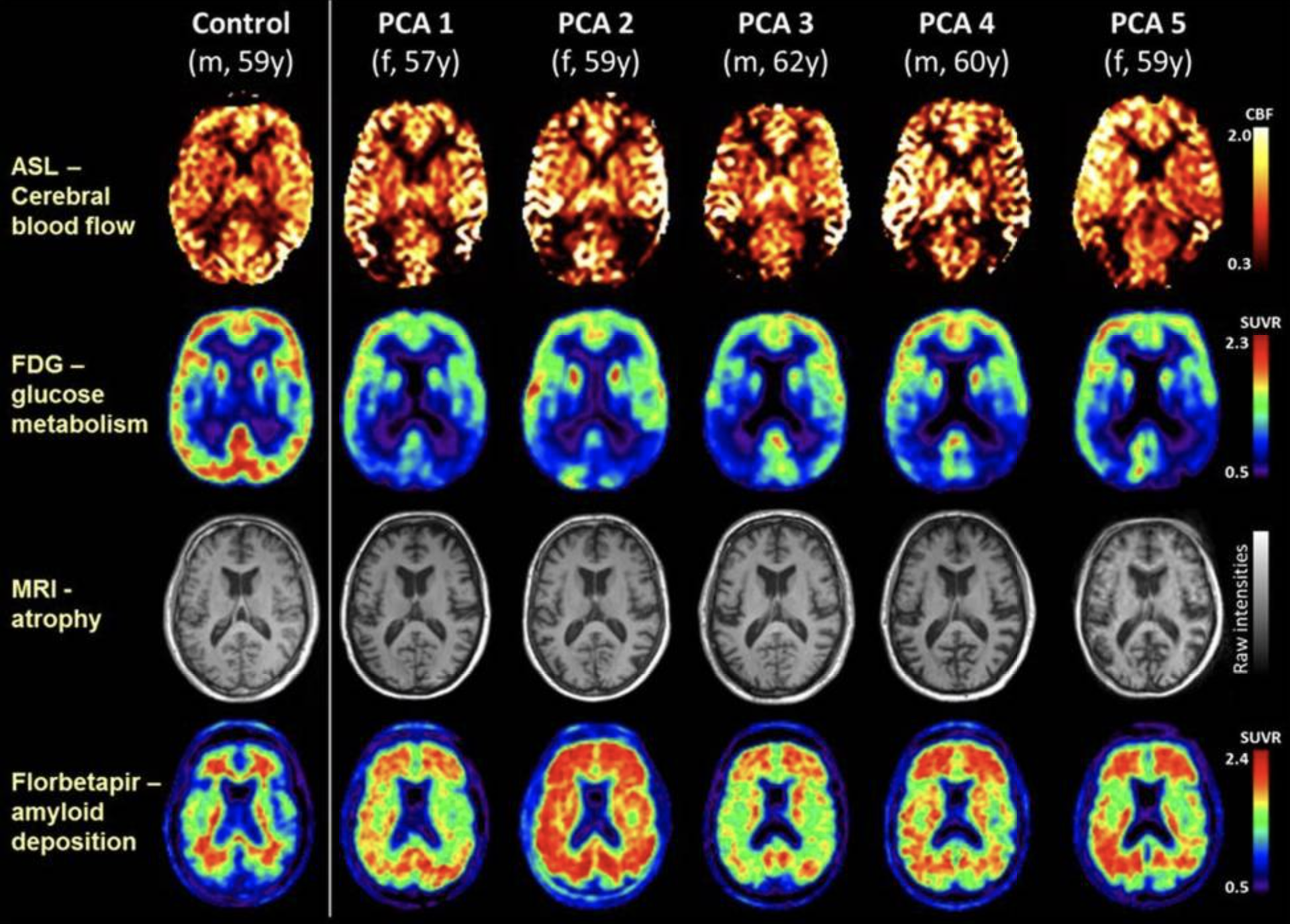
Lehmann M,Melbourne A,Dickson JC,Ahmed RM,Modat M,Cardoso MJ,Thomas DL,De Vita E,Crutch SJ,Warren JD,Mahoney CJ,Bomanji J,Hutton BF,Fox NC,Golay X,Ourselin S,Schott JM, A novel use of arterial spin labelling MRI to demonstrate focal hypoperfusion in individuals with posterior cortical atrophy: a multimodal imaging study. Journal of neurology, neurosurgery, and psychiatry. 2016 Sep;     [PubMed PMID: 26733599]


[21]

Paterson RW,Toombs J,Slattery CF,Nicholas JM,Andreasson U,Magdalinou NK,Blennow K,Warren JD,Mummery CJ,Rossor MN,Lunn MP,Crutch SJ,Fox NC,Zetterberg H,Schott JM, Dissecting IWG-2 typical and atypical Alzheimer's disease: insights from cerebrospinal fluid analysis. Journal of neurology. 2015 Dec;     [PubMed PMID: 26410752]


[22]

Maia da Silva MN,Millington RS,Bridge H,James-Galton M,Plant GT, Visual Dysfunction in Posterior Cortical Atrophy. Frontiers in neurology. 2017     [PubMed PMID: 28861031]


[23]

Kim E,Lee Y,Lee J,Han SH, A case with cholinesterase inhibitor responsive asymmetric posterior cortical atrophy. Clinical neurology and neurosurgery. 2005 Dec;     [PubMed PMID: 16311158]

Level 3 (low-level) evidence

[24]

Suárez-González A,Henley SM,Walton J,Crutch SJ, Posterior cortical atrophy: an atypical variant of Alzheimer disease. The Psychiatric clinics of North America. 2015 Jun;     [PubMed PMID: 25998111]


[25]

Weill-Chounlamountry A,Alves J,Pradat-Diehl P, Non-pharmacological intervention for posterior cortical atrophy. World journal of clinical cases. 2016 Aug 16;     [PubMed PMID: 27574605]

Level 3 (low-level) evidence

[26]

Townley RA,Dawson ET,Drubach DA, Heterozygous genotype at codon 129 correlates with prolonged disease course in Heidenhain variant sporadic CJD: case report. Neurocase. 2018 Feb;     [PubMed PMID: 29436943]

Level 3 (low-level) evidence