Fluoroscopic Percutaneous Coronary Interventions, Assessment, Protocols, and Interpretation

Earn CME/CE in your profession:


Continuing Education Activity

Fluoroscopic guided percutaneous coronary intervention is a widely performed procedure to treat coronary artery disease. This activity reviews the importance of fluoroscopic guided percutaneous coronary intervention and highlights the interprofessional team's role in performing this procedure.

Objectives:

  • Identify the indications for fluoroscopic guided percutaneous coronary intervention.
  • Explain in detail the technique for fluoroscopic guided percutaneous coronary intervention.
  • Review the clinical significance of the fluoroscopic guided percutaneous coronary intervention.
  • Summarize how employing an interprofessional team approach can improve patient outcomes during fluoroscopic guided percutaneous coronary intervention.

Introduction

Ischemic heart disease is the leading cause of death globally. Percutaneous coronary intervention (PCI) has played a vital role in managing obstructive coronary artery disease.[1] Nearly all percutaneous coronary intervention was carried out under fluoroscopic guidance by utilizing ionizing radiation. It is essential to take appropriate fluoroscopic views according to vessel anatomy during percutaneous coronary intervention so that the diseased segment should not be missed.[2][3][4] 

The knowledge of appropriate views for percutaneous coronary intervention helps cover the diseased segment of the artery and reduces radiation exposure to both patients and healthcare workers by decreasing fluoroscopic time and radiation dose. In this chapter, we will discuss percutaneous coronary intervention under fluoroscopic guidance in detail.

Anatomy and Physiology

The heart is Supplied by Three Major Coronary Arteries

  1. Left anterior descending with Septal and diagonal branches
  2. Left circumflex artery
  3. The right coronary artery bifurcates into the right posterior descending artery and right posterior left ventricular branch.

However, in a patient with a previous history of coronary artery bypass graft, grafts are placed distal to the narrowed segment of the coronary artery. The most commonly used vessels for grafts are saphenous vein graft, left internal mammary artery, right internal mammary artery, radial artery, gastroepiploic artery, and inferior epigastric artery. It is essential to know the site and number of grafts before the graft study.

Indications

2011, ACCF/AHA/SCAI Guideline for Percutaneous Coronary Intervention

Indications of percutaneous coronary intervention to improve mortality:[5]

  • In unprotected left main artery with ST-elevation myocardial infarction: If distal flow to vessel less than TIMI III and percutaneous coronary intervention can be done much quicker and safer than CABG (Class II-a)
  • In unprotected left main artery with non-ST-elevation myocardial infarction or Unstable angina: If CABG can not be performed (Class II-a)
  • In unprotected left main artery with stable ischemic heart disease: If two of the following present 1) LOW-risk PCI (Syntax<22) and ostial or trunk involvement 2) Increase risk of surgical adverse events ( STS score < 5 (Class II-a)
  • In triple vessel coronary artery disease with or without Proximal LAD: (Class II-a)
  • In two-vessel disease with proximal LAD: (Class II-b)
  • In two-vessel disease without proximal LAD: (Class II-b)
  • In one vessel disease with proximal LAD: (Class II-b)
  • For patients with the survivor of sudden cardiac death with presumed ischemia-mediated ventricular tachycardia (Class I)

Indications of percutaneous coronary intervention to improve symptoms:

  • Percutaneous coronary intervention in patients with persistent angina despite optimal medical management to improve symptoms with single or more significant (>70% diameter) coronary artery disease and amenable to revascularization. (Class I)
  • Percutaneous coronary intervention in patients with persistent angina to improve symptoms with single or more significant (>70% diameter) coronary artery disease and medical management can not be given because of medication contraindication, adverse effects, or patient preferences. (Class II-a)
  • Percutaneous coronary intervention in patients with persistent angina despite optimal medical management to improve symptoms with single or more significant (>70% diameter) coronary artery disease and associated with ischemia. (Class II-a)

Contraindications

The following are the contraindications of percutaneous coronary intervention 

Absolute Contraindication

  1. Intolerance to dual antiplatelets
  2. Severe illness which reducing the lifespan to less than one year

Relative Contraindication

  1. Coronary artery vessel diameter < 2.5 mm
  2. Anatomy not feasible for coronary intervention
  3. Diffusely disease saphenous vein graft

Equipment

For fluoroscopic percutaneous coronary intervention, the following equipment is essential:

  • Fluoroscopic machine
  • Contrast agent
  • Guide catheters
  • Guide wires
  • Coronary balloon
  • Coronary stent

To prevent radiation exposure during the fluoroscopic guided percutaneous coronary intervention, each medical staff require protective measure during the procedure which includes:

  • Lead apron
  • Thyroid collar
  • Radiation protective glasses
  • Radiation monitoring badges

Personnel

  • Intervention cardiologist
  • Radiation technician
  • Cath lab technician
  • Staff nurse

Preparation

Informed consent should be taken before the procedure. It is essential to take a complete history and perform a physical examination pre-procedure to determine the indication of the procedure. The patient should be premedicated with antiplatelets.

Technique or Treatment

It is essential to know in which view a specific part of the artery will be visible while performing coronary angioplasty under fluoroscopic guidance.[6][7]

Vessels

Fluoroscopic views

Additional views

Left Main Coronary Artery (LM)

Ostial part of LM

LAO (left anterior oblique) caudal 

AP (Anteroposterior) Caudal

Mid part of LM

RAO (Right anterior oblique)cranial

RAO (Right anterior oblique)caudal

AP (Anteroposterior) Caudal

Distal part of LM

LAO(left anterior oblique) caudal

RAO (Right anterior oblique) caudal 

LAO(left anterior oblique) cranial

Left Anterior Descending Artery (LAD)

Ostial part of LAD

LAO(left anterior oblique) caudal  

AP(Anteroposterior) Caudal

RAO(Right anterior oblique) caudal

Mid part of LAD

RAO (Right anterior oblique) cranial

LAO(left anterior oblique) cranial

RAO (Right anterior oblique) caudal

AP(Anteroposterior) Cranial

Distal  part of LAD

RAO (Right anterior oblique)  cranial 

LAO(left anterior oblique) cranial

RAO (Right anterior oblique) caudal 

Left Circumflex Artery (LCX)

Ostial part of LCX

LAO(left anterior oblique) caudal  

AP(Anteroposterior) Caudal

Mid part of LCX

RAO (Right anterior oblique)

LAO(left anterior oblique) 

LAO(left anterior oblique) cranial

AP(Anteroposterior) Caudal

Distal  part of LCX

RAO (Right anterior oblique)

LAO(left anterior oblique) 

LAO(left anterior oblique) cranial 

Right Coronary Artery (RCA)

Osteoproximal part of RCA

LAO(left anterior oblique)   

AP(Anteroposterior) Caudal

LAO(left anterior oblique) Caudal

Mid part of RCA

RAO (Right anterior oblique)

LAO(left anterior oblique)  

The distal part of RCA at the level of bifurcation to RPDA(Right posterior descending artery) and RPLV (Right posterior left ventricular branch)

LAO(left anterior oblique) Cranial


AP (Anteroposterior) Cranial

RPDA

RAO (Right anterior oblique)


AP (Anteroposterior) Cranial

RPLV

AP(Anteroposterior) Caudal


Saphenous Vein-Graft to Coronary Artery

Saphenous vein graft(SVG) to LAD

AP (Anteroposterior) Cranial

RAO (Right anterior oblique)cranial


Lateral

Saphenous vein graft(SVG) to Left circumflex or Obtuse marginal artery

RAO (Right anterior oblique)

AP(Anteroposterior) Caudal

Saphenous vein graft(SVG) to Right coronary artery

LAO(left anterior oblique)

LAO(left anterior oblique) Cranial


Left Internal Mammary Artery to LAD

Osteoproximal part for Left internal mammary artery to LAD

LAO(left anterior oblique) Cranial


Mid Part of LIMA

RAO (Right anterior oblique)  cranial 


The insertion site of LIMA to LAD

Left Lateral (90 degrees)


Images are shown before and after the percutaneous coronary intervention of RCA.

Complications

Following are the complications associated with the percutaneous coronary intervention.

  1. Coronary artery dissection
  2. Coronary artery perforation
  3. Distal embolization of clot
  4. Side branch occlusion
  5. Stent thrombosis
  6. Access site bleeding
  7. Access site hematoma
  8. Acute kidney injury
  9. Stroke
  10. Allergic reaction to contrast

Following are the complications associated with fluoroscopic radiation exposure to patients or staff.[8]

Tissue Reaction

  • Skin Injury: It is the most common tissue reaction at the beam entry site following fluoroscopic guided percutaneous coronary intervention and varies according to time of exposure:
  1. Few hours  to days of post-exposure: Mild skin erythema
  2. A week to several weeks of post-exposure: Significant erythema
  3. After 4 to 8 weeks of post-exposure: Severe skin injury including ulceration in rare cases
  • Bone Injury: In rare cases, radiation can cause necrosis of superficial bones such as ribs.
  • Eye Injury: The single-dose threshold of radiation that can cause a cataract to believe to be 500 mGy with a minimum latency period of one year.

Stochastic Effects: Radiation-Induced Cancer

It is the most important and catastrophic result of radiation exposure. It is vital to provide knowledge of the scholastic risk of radiation exposure to both patients and healthcare workers. These steps are necessary to reduce the risk, which includes the use of radiation protection instruments.

Clinical Significance

Fluoroscopic percutaneous coronary intervention is a widely performed procedure for the last several years. In comparison to thrombolysis, percutaneous coronary intervention reduces morbidity and mortality significantly in patients with myocardial infarction. In chronic coronary syndrome patients, it has proven to improve the quality of life. Compared to CABG, it is associated with a shorter hospital stay, no scar on the chest, and early mobilization after the procedure. Because of advancement in percutaneous coronary intervention over time, survival improvement in patients with coronary artery disease was noted.

Enhancing Healthcare Team Outcomes

Fluoroscopic percutaneous coronary intervention requires an interprofessional team including an intervention cardiologist, cath lab technician, radiation technician, and staff nurse. To improve outcomes, good collaboration among team members is essential. It is vital that every team member should be aware of their roles to avoid complications.

Nursing, Allied Health, and Interprofessional Team Interventions

The nursing staff gives medications and monitors vitals during the procedure. The radiographic technician set views on the fluoroscopy machine for the procedure and supports performing procedures such as intravascular imaging and invasive physiological assessment. The cath lab technician prepares equipment such as a coronary balloon, coronary stent and guides catheters for the procedure.


(Click Image to Enlarge)
RCA lesion before angioplasty
Contributed by Ghufran Adnan

(Click Image to Enlarge)
RCA lesion after percutaneous coronary intervention
Contributed by Ghufran Adnan
Details

Author

Ghufran Adnan

Updated:

4/17/2023 4:29:45 PM

References


[1]

. Invasive compared with non-invasive treatment in unstable coronary-artery disease: FRISC II prospective randomised multicentre study. FRagmin and Fast Revascularisation during InStability in Coronary artery disease Investigators. Lancet (London, England). 1999 Aug 28:354(9180):708-15     [PubMed PMID: 10475181]

Level 1 (high-level) evidence

[2]

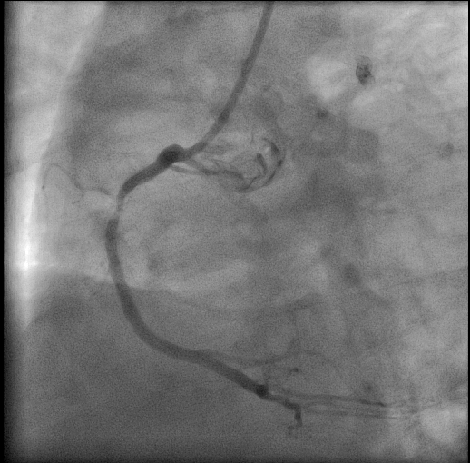
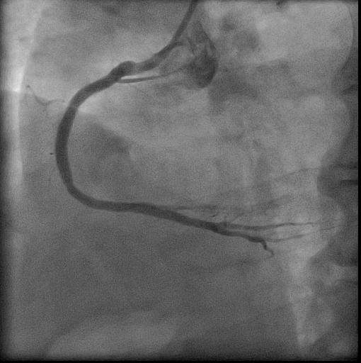
Lespérance J, Saltiel J, Petitclerc R, Bourassa MG. Angulated views in the sagittal plane for improved accuracy of cinecoronary angiography. The American journal of roentgenology, radium therapy, and nuclear medicine. 1974 Jul:121(3):565-74     [PubMed PMID: 4846573]


[3]

Sos TA, Lee JG, Levin DC, Baltaxe HA. New lordotic projection for improved visualization of the left coronary artery and its branches. The American journal of roentgenology, radium therapy, and nuclear medicine. 1974 Jul:121(3):575-82     [PubMed PMID: 4846574]


[4]

Aldridge HE, McLoughlin MJ, Taylor KW. Improved diagnosis in coronary cinearteriography with routine use of 110 degrees oblique views and cranial and caudal angulations. Comparison with standard transverse oblique views in 100 patients. The American journal of cardiology. 1975 Oct 6:36(4):468-73     [PubMed PMID: 1190051]


[5]

Levine GN, Bates ER, Blankenship JC, Bailey SR, Bittl JA, Cercek B, Chambers CE, Ellis SG, Guyton RA, Hollenberg SM, Khot UN, Lange RA, Mauri L, Mehran R, Moussa ID, Mukherjee D, Nallamothu BK, Ting HH. 2011 ACCF/AHA/SCAI Guideline for Percutaneous Coronary Intervention: a report of the American College of Cardiology Foundation/American Heart Association Task Force on Practice Guidelines and the Society for Cardiovascular Angiography and Interventions. Circulation. 2011 Dec 6:124(23):e574-651. doi: 10.1161/CIR.0b013e31823ba622. Epub 2011 Nov 7     [PubMed PMID: 22064601]

Level 1 (high-level) evidence

[6]

Elliott LP, Green CE, Rogers WJ, Mantle JA, Papapietro S, Hood WP Jr. The importance of angled right anterior oblique views in improving visualization of the coronary arteries. Part I: Caudocranial view. Radiology. 1982 Mar:142(3):631-6     [PubMed PMID: 7063677]


[7]

Green CE, Elliott LP, Rogers WJ, Mantle JA, Papapietro S, Hood WP Jr. The importance of angled right anterior oblique views in improving visualization of the coronary arteries. Part II: Craniocaudal view. Radiology. 1982 Mar:142(3):637-41     [PubMed PMID: 7063678]


[8]

Hirshfeld JW Jr, Ferrari VA, Bengel FM, Bergersen L, Chambers CE, Einstein AJ, Eisenberg MJ, Fogel MA, Gerber TC, Haines DE, Laskey WK, Limacher MC, Nichols KJ, Pryma DA, Raff GL, Rubin GD, Smith D, Stillman AE, Thomas SA, Tsai TT, Wagner LK, Wann LS. 2018 ACC/HRS/NASCI/SCAI/SCCT Expert Consensus Document on Optimal Use of Ionizing Radiation in Cardiovascular Imaging: Best Practices for Safety and Effectiveness: A Report of the American College of Cardiology Task Force on Expert Consensus Decision Pathways. Journal of the American College of Cardiology. 2018 Jun 19:71(24):e283-e351. doi: 10.1016/j.jacc.2018.02.016. Epub 2018 May 2     [PubMed PMID: 29729877]

Level 3 (low-level) evidence