Continuing Education Activity
The main goal of forearm splinting is to prevent pronation and supination of the forearm. There are a variety of indications for forearm splinting, including soft tissue strains or sprains, fractures, joint dislocations, tendon lacerations, and compression neuropathy. Forearm splints aim to decrease pain and debility, prevent further injury and facilitate the healing process. In the acute setting, forearm splints are often used for distal radius and/or ulna fractures, radial and/or ulnar shaft fractures, flexor tendon lacerations within the forearm and radial nerve palsies that manifest with wrist drop. In the chronic setting, forearm splinting is commonly used for De Quervain’s tenosynovitis, medial and lateral epicondylitis and any compressive neuropathies affecting the nerves in the forearm. This activity describes the indications, contraindications, and complications of forearm splinting and highlights the role of the interprofessional team in caring for patients requiring forearm splints.
Objectives:
- Identify the technique involved in forearm splinting.
- Describe the indications for forearm splinting.
- Review the contraindications to forearm splinting.
- Explain interprofessional team strategies for enhancing care coordination and communication to improve the utilization of proper technique for forearm splinting.
Introduction
The technique of splinting can be found throughout multiple fields of medicine including emergency medicine, orthopedics, primary care, and podiatry. It is primarily used to immobilize a joint or limb to allow for pain control, injury stabilization, and ultimately tissue healing. In the acute setting, splinting is useful as a temporizing treatment for sprains, strains, joint dislocations, fractures, and soft tissue lacerations. In the chronic setting, splinting is useful mainly for inflammatory conditions. The main goal of forearm splinting is to prevent rotation about the entire forearm. Specific conditions in which forearm splinting would be useful include injuries to any part of the radius and/or ulna or soft tissue structures located within the forearm.
Anatomy and Physiology
The anatomy of the forearm can be broken down into osteology, musculature, and neurovascular structures.
The osteology of the forearm is composed of the radius and ulna. The radius is a cylindrical long bone which has a bow within the shaft, whereas the ulna is purely a straight bone with a triangular cross-section. This bow in the radius allows for rotation of the radius around a stationary ulna bone thus allowing for pronation and supination of the forearm.[1] There 4 main articulations with regard to the radius. These include the radiocapitellar joint, proximal radial-ulnar joint, distal radial-ulnar joint, and radial-carpal joint. The ulna shares the same articulations, save for the radiocapitellar joint, and instead has an ulno-humeral articulation proximally. The radius has a tuberosity proximally where the biceps inserts and a styloid distally where the brachioradialis inserts. The ulna also has a tuberosity proximally where the brachialis inserts and a styloid distally.
The musculature of the forearm can be broken down into the anterior and posterior compartment. The anterior compartment is composed of the flexor muscles. The superficial flexors include the pronator teres, flexor carpi radialis, palmaris longus, flexor digitorum superficialis and flexor carpi ulnaris. The deep flexors include the flexor digitorum profundus, flexor pollicis longus and pronator quadratus. The posterior compartment is composed of the extensor muscles. The superficial extensors include the anconeus, extensor digitorum communis, extensor digit minimi and extensor carpi ulnaris. There is a subdivision within the superficial extensors called the "mobile wad" which is composed of the brachioradialis, extensor carpi radialis longus and extensor carpi radialis brevis. The deep extensors include supinator, abductor pollicis longus, extensor pollicis brevis, extensor pollicis longus and extensor indicis proprius.
The nervous structures located within the forearm include the median, radial, ulnar and musculocutaneous nerves. The median nerve comes from the medial and lateral cords of the brachial plexus and provides an only motor function in the forearm. The anterior interosseous nerve is a continuation of the median nerve and provides a motor function in the forearm and volar wrist capsule sensation. The radial nerve comes from the posterior cord of the brachial plexus and provides motor function and sensation to the posterior forearm via the posterior cutaneous nerve of the forearm. The posterior interosseous nerve is a continuation of the radial nerve and provides motor function and dorsal wrist capsular sensation. The ulnar nerve comes from the medial cord of the brachial plexus and provides purely motor function in the forearm. The musculocutaneous nerve comes from the lateral cord of the brachial plexus and provides the only sensation to the radial aspect of the forearm via the lateral antebrachial cutaneous nerve.
The vascular structures in the forearm include the radial and ulnar arteries, which are a continuation of the brachial artery. Both the radial and ulnar artery continue throughout the forearm and ultimately form the deep and superficial palmar arch, respectively, in the hand.
Indications
The main goal of forearm splinting is to prevent pronation and supination about the entire forearm.[2] There are a variety of reasons as to when forearm splinting would be appropriate, including soft tissue strains or sprains, fractures, joint dislocations, tendon lacerations, and compression neuropathies. In any case, the main objectives are to decrease pain and debility, prevent further injury and facilitate the healing process. Common reasons encountered which would necessitate forearm splinting in the acute setting include distal radius and/or ulna fractures, radial and/or ulnar shaft fractures, flexor tendon lacerations within the forearm and a radial nerve palsy which manifests clinically as a wrist drop. In the chronic setting, common reasons include De Quervain’s tenosynovitis, medial and lateral epicondylitis and any compressive neuropathies affecting the nerves in the forearm.
Contraindications
While there are no absolute contraindications to splinting, there needs to be appropriate clinical decision making prior to splinting, especially in the acute setting. Situations in which caution must be exercised include the presence of thermal or electrical burns, open fractures, grossly contaminated wounds and significant soft tissue swelling.[3] In general, splinting may be performed so long as any concomitant injury is addressed prior to splinting. Furthermore, splinting in the setting of significant soft tissue swelling is preferred over casting as the splint is not circumferential or constricting and may be easily loosened or removed.[4][5]
Equipment
- Stockinet
- Webril/cast padding
- Plaster or prefabricated splinting material
- Water: Cold water will maximize molding time; warm water will facilitate faster hardening
- Elastic bandages
- Scissors
Personnel
Depending on the application, forearm splinting may be done alone or may require an assistant. If the patient can follow direction, an assistant may not be necessary. If the patient is not cooperative or is sedated, an assistant will be required to hold the forearm.
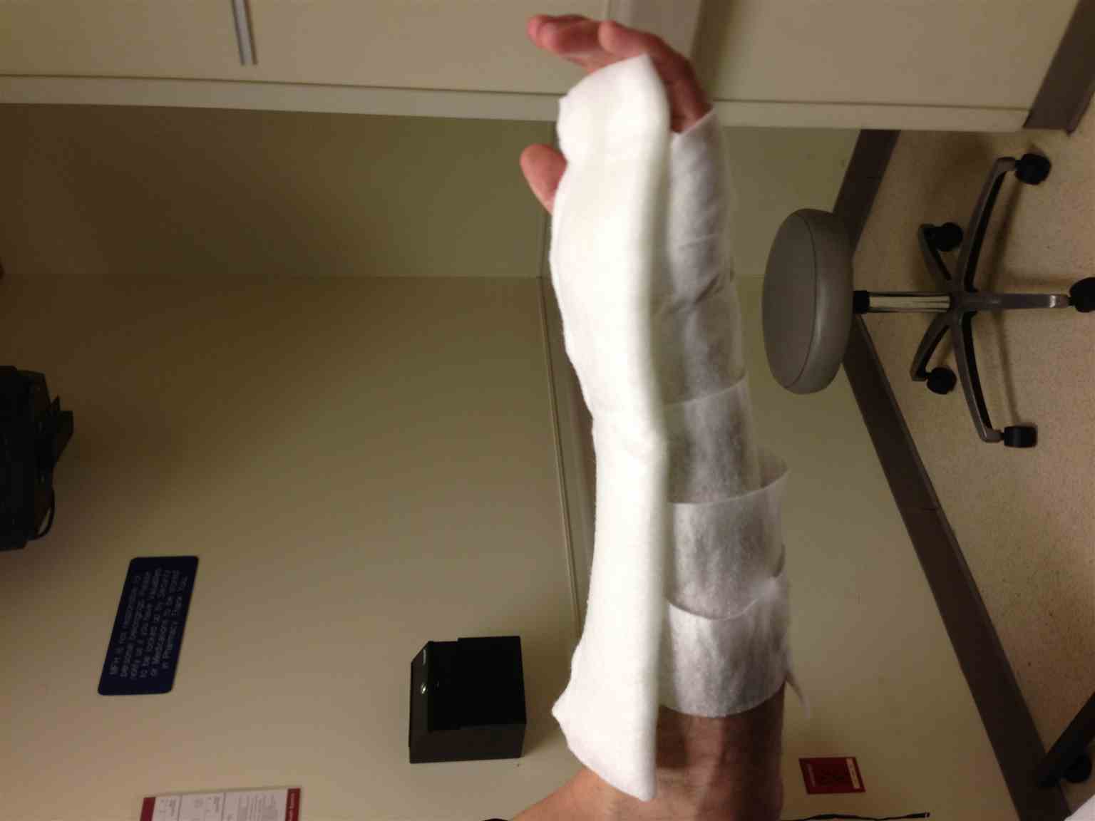
Preparation
Preparation begins first with identifying the injury being treated. Once this is identified, the type of splint may be determined. The most common forearm splint is the sugar-tong splint. Other forearm splints include a sugar tong with a posterior mold and a volar-forearm splint. It is necessary to determine the splint being used as the required length of the splinting materials differs. For the sugar-tong splint, the forearm needs to be in the neutral rotation, and the elbow flexed to 90 degrees. The splint should begin at the distal palmar crease, wrap around the flexed elbow, and stop at the dorsal metacarpal heads. If necessary, to add a posterior slab to the sugar tong, it should begin distal to the wrist on the ulnar aspect of the forearm and stop proximal to the elbow, which prevents elbow flexion and extension. The volar forearm splint also begins at the distal palmar crease and stops proximal to the antecubital fossa, thus allowing for elbow flexion and some pronation and supination. Whether using plaster or prefabricated splinting material, the required length may be measured directly on the patient using a sheet of cotton cast padding (Webril) as a proxy. The splinting material can then be cut to the length of the measured cotton cast padding. Plaster needs to be at least 8 sheets thick to provide adequate strength but no more than 12 sheets thick to avoid thermal injury. Plaster produces heat as it hardens and care must be taken to prevent thermal injury. Stockinet is cut so that there is extra on both ends. The extra stockinet will be folded down over the plaster which allows the creation of padded cuffs at the ends of the splint for patient comfort. Cotton cast padding is used to wrap the forearm directly over the stockinet. The plaster splinting material is wet, wrung and bonded and placed on the forearm. Webril may be used to wrap over the splinting material once more before overwrapping with an elastic bandage. If necessary, appropriate molding of the splint may commence.
Technique or Treatment
- Determine appropriate type of splint, in this case, a sugar tong.
- The forearm should be in neutral rotation with elbow flexed to 90 degrees throughout the process.
- Measure the appropriate length of plaster, 8 to 12 sheets thick, from the distal palmar crease, around a flexed elbow, ending at the dorsal metacarpal heads.
- The measure, cut, and place stockinet on the forearm. Be sure to cut a hole in stockinet for the thumb. There should be at least one inch extra of stockinet distal to the distal palmar crease and extend to the mid brachium.
- Wrap forearm with cotton cast padding beginning at the distal palmar crease and ending distal to the mid brachium, ensuring 50% overlap of padding with a minimum of 2 layers. Ensure that bony prominence is well padded. Attempt to avoid bunching or wrinkles in padding.
- Dip plaster into the water to thoroughly wet.
- Wring wet plaster and bond together between fingers.
- Apply plaster to cotton cast padding on the forearm, ensuring that the plaster begins at the distal palmar crease, wraps around the elbow and ends at the dorsal metacarpal heads.
- Overwrap plaster with one layer of cotton cast padding to avoid plaster sticking to an elastic bandage.
- Fold excess stockinet over plaster and cotton cast padding so that there is a cuff at each end of the splint.
- Loosely wrap entire splint with an elastic bandage.
- Apply appropriate mold while the plaster is hardening.
- Avoid placing a splint on pillows or blanket during hardening process as this has an insulating effect.
- Once hard, re-assess neurovascular status of hand and fingers.
Complications
Complications of forearm splinting include pressure necrosis, which can begin as soon as 2 hours after application, compartment syndrome if wrapped too tightly, thermal injury from plaster if too thick, and joint stiffness, specifically the metacarpophalangeal joints if the splint extends past the distal palmar crease.[6][7][8]
Clinical Significance
Forearm splinting is an excellent way to immobilize the forearm to help alleviate pain, stabilize injuries, prevent further damage to bones, muscles, nerves and/or arteries, and prevents a closed fracture from becoming an open one. Furthermore, due to the non-circumferential nature of the splint, as opposed to a cast, it can allow for soft tissue swelling and can be easily removed by the clinician to evaluate any wounds beneath.[9][10] In the acute setting, it is an excellent way to temporize forearm injuries.
After placement of the forearm splint, it is important to educate the patient on proper splint care. The splint needs to remain clean and dry. If the splint gets wet, the plaster loses its strength and the padding beneath will not dry. This can lead to maceration and breakdown of intact skin. Furthermore, if there is an open wound already beneath, it can lead to infection. In instances where the splint becomes wet, the patient should return to the place where the splint was placed. The patient should be instructed to avoid removing the splint, especially if the splint was applied for a fracture or dislocation. Removal in this instance can cause re-displacement of the fracture or re-dislocation. Most importantly, should the patient develop new-onset numbness or tingling of the hand or fingers they should first elevate the forearm and if it does not resolve, return to the place of splint application or emergency department for further evaluation.
Enhancing Healthcare Team Outcomes
Forearm splinting is done by many healthcare professionals that includes the emergency department physician, orthopedic surgeon, therapist, primary care provider, orthopedic nurse and the urgent care physician. Forearm splinting is an excellent way to immobilize the forearm to help alleviate pain, stabilize injuries, prevent further damage to bones, muscles, nerves and/or arteries, and prevents a closed fracture from becoming an open one. Furthermore, due to the non-circumferential nature of the splint, as opposed to a cast, it can allow for soft tissue swelling and can be easily removed by the clinician to evaluate any wounds beneath.[9][10] In the acute setting, it is an excellent way to temporize forearm injuries.