Continuing Education Activity
Bursitis is a swelling or inflammation of a bursa, which is a synovium-lined, sac-like structure found throughout the body near bony prominences and between bones, muscles, tendons, and ligaments. There are many causes of bursitis, including overuse injury, infectious disease, trauma, and inflammatory disorders. This activity explains the risk factors, evaluation, and management of bursitis and highlights the importance of the interprofessional team in enhancing care for affected patients.
Objectives:
- Describe the pathophysiology of patients with bursitis.
- Outline the evaluation of patients with bursitis.
- Explain the treatment strategies for patients with bursitis.
- Employ interprofessional team strategies for improving care coordination and communication to advance the evaluation, management, and rehabilitation of patients with bursitis and optimize outcomes.
Introduction
Bursitis is a swelling or inflammation of a bursa, which is a synovium-lined, sac-like structure found throughout the body near bony prominences and between bones, muscles, tendons, and ligaments. There are over 150 known bursae in the human body, and their function is to facilitate movement in the musculoskeletal system, creating a cushion between tissues that move against one another. When bursitis occurs, the bursa enlarges with fluid, and any movement against or direct pressure upon the bursa will precipitate pain for the patient. There are many causes of bursitis, including overuse injury, infectious disease, trauma, and inflammatory disorders. The name bursitis itself is often a misnomer, as not all forms of bursitis are due to a primary inflammatory process but are rather a swelling of the bursa due to a noxious stimulus.[1][2][3][4]
Etiology
There are myriad causes of bursitis of which the clinician must be knowledgeable. The most common etiology is prolonged pressure, whereby the bursa is stressed between a hard surface and bony prominence. Examples of prolonged pressure causing bursitis include students who frequently rest their elbows on their desks and people who work on their knees without adequate padding. Likewise, repetitive motions can also irritate the bursa and result in bursitis. The second most common cause of bursitis is trauma when direct pressure is applied to the bursa. Often the patient will not be able to recall the inciting incident as it may have seemed benign at the time.
Traumatic bursitis puts the patient at risk for septic bursitis, which is most often caused by direct penetration of the bursa through the skin. Septic bursitis can also be provoked through the hematogenous spread; however, due to the relatively poor blood supply to the bursa, this is rare. Staphylococcus aureus causes the majority of septic bursitis. Another important cause of bursitis is autoimmune conditions and systemic inflammatory conditions, as well as arthropathies, including rheumatoid arthritis, osteoarthritis, systemic lupus erythematosus, scleroderma, spondyloarthropathy, and gout. Lastly, bursitis can be idiopathic in origin, and septic bursitis, in particular, can be induced by invasive procedures.[5][6][7]
Epidemiology
In general, bursitis is encountered equally in the male and female populations. However, some types of bursitis have documented female predilection, specifically pes anserine and trochanteric bursitis. Furthermore, these forms of bursitis are more common in individuals who are obese. Men are more often affected by olecranon bursitis due to the increased rate of men who perform manual labor for a living (plumbers, gardeners, mechanics, and construction workers, among others). Since certain occupational stressors increase one's risk of developing bursitis, many colloquial terms for specific types of bursitis have been coined to reflect these epidemiologic connections. For example, prepatellar bursitis is also known as "housemaid's knee," while olecranon bursitis is sometimes referred to as "student's elbow."
Likewise, infrapatellar bursitis is often called "clergyman's knee," while ischial bursitis is called "weaver's bottom." Bursitis of the subcutaneous calcaneal bursa can be provoked by footwear that is too tight or ill-fitting, and this is frequently encountered in dancers and figure skaters. In each of these cases, the cause of the bursitis is usually prolonged pressure over the affected bursa. While bursitis affects people of all ages, the elderly may be at greater risk, given that many older people are afflicted by osteoarthritis and other chronic diseases, which can increase the risk of bursitis. For septic bursitis, immunocompromised patients, such as people with diabetes, those with certain rheumatologic disorders, people who suffer from alcoholism, or those with HIV, are at increased risk.
Pathophysiology
The bursa itself is a synovial lining that represents a potential space insofar as it is collapsed upon itself until a resulting trigger causes the bursa to become irritated and fill with synovial fluid. The patient experiences pain when the inflamed bursa is then compressed against bone, muscle, tendon, ligaments, or skin. Despite the moniker, not all bursitis is associated with an overt inflammatory process. That said, the examination of the subacromial bursa in patients with subacromial bursitis has revealed increased inflammatory mediators such as tumor necrosis factor-alpha, cyclooxygenases, and specific interleukins.
History and Physical
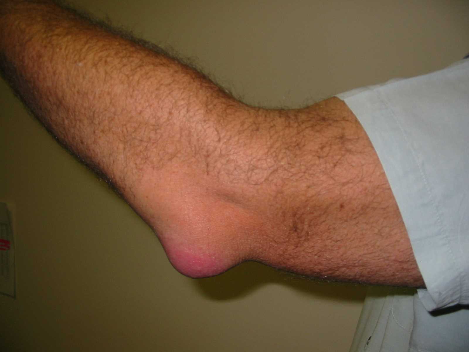
Two forms of bursitis exist, chronic and acute, and the presentations of each will manifest differently from one another. A detailed medical history, as well as an understanding of the patient's daily routine, will help the clinician differentiate the 2 types of bursitis from each other and from other diagnoses. Acute bursitis typically arises from trauma, infection, or crystalline joint disease, while chronic bursitis is more likely the result of inflammatory arthropathies and repetitive pressure/overuse, or "micro traumas." In acute bursitis, patients generally present with pain on palpation of the bursa. The range of motion of the involved joint may be decreased secondary to pain. Active motion involving the affected bursa also elicits pain; however, this is dependent on the location of the bursa and the biomechanics involved in moving the bones, muscles, and tissues around the bursa. For instance, many patients will experience pain with active motion but not with passive motion. When the surrounding muscles are not activated and, therefore, not compressing the bursa, there is little to no pain. Some acute bursitis will produce pain with flexion of the affected joint, but there will be no pain on extension (these findings are commonly seen with prepatellar and olecranon bursitis).
Contrary to the physical exam findings in acute bursitis, chronic bursitis is often painless. The bursa itself has had time to expand to accommodate the increased fluid, and the result is significant swelling and thickening of the bursa. An examination of the skin is very important in the evaluation of acute or chronic bursitis. The skin should be evaluated for trauma, erythema, and warmth. One study found that a temperature increase of just 2.2 °C between the skin overlying the affected bursa compared to that overlying the unaffected contralateral bursa was highly sensitive and specific for septic bursitis. However, deep bursitis, even when acute, may produce no tenderness with palpation of the overlying structures or any obvious skin changes. Lastly, musculoskeletal imbalances or certain anatomic variants are sometimes associated with the development of bursitis. Decreased core strength and chronic back pain can exacerbate trochanteric bursitis, which itself is often precipitated by gluteus minimus or medius tendinopathy, while mechanical factors such as pes planus and genu valgum are risk factors for the development of pes anserine bursitis.
Evaluation
The diagnosis of certain types of bursitis can be made clinically and without further studies; however, imaging plays a role in the diagnosis and management of bursitis. Imaging can be helpful to narrow down the differential diagnosis or even provide a precise answer in cases of diagnostic uncertainty. Plain film imaging of the affected joint or bursa should be considered in cases where there is a history of trauma or concern for a foreign body or fracture causing swelling or pain. MRI can be used to evaluate the deeper bursa, as can ultrasound which has the added benefit of showing real-time images within a joint or area surrounding the bursa and can be used to observe changes with active and passive movement.
Ultrasound is particularly helpful for visualizing cobblestoning of the fat overlying a bursa, which can help differentiate cellulitis from infectious bursitis. Color Doppler can likewise be used to show signs of infection, such as hyperemia of the bursa and the surrounding tissues.
Aspiration of the inflamed bursa can be helpful when there is a question of septic bursitis or bursitis secondary to crystalline disease. Aspirated fluid should be sent for cell count, Gram stain and culture, glucose, and analysis for crystals. A white blood cell count of less than 500/mm3 from the aspirated fluid is consistent with noninfectious and noncrystalline bursitis.[8][9][10]
Treatment / Management
The vast majority of bursitis will heal on its own. However, there are several modalities for improving the patient's pain and ensuring a return to complete functionality of the affected area. Conservative treatment involves the use of rest, ice, compression, and elevation for symptomatic improvement.
Patients should be educated on proper ergonomics for avoiding exacerbating movements, and certain superficial bursa can be protected with padding for those in whom prolonged pressure on the elbows or knees is an everyday occupational occurrence. A foam donut can be used for patients with ischial bursitis, and stretching as well as core strengthening exercises both play a role in improving and alleviating symptoms.
For bursitis occurring near the Achilles tendon, proper-fitting footwear that reduces pressure on the area should be encouraged. For analgesia, NSAIDs and/or acetaminophen are first-line agents. For the deeper bursa, corticosteroid injections, sometimes with a local anesthetic, can provide symptomatic relief.
Local injections of corticosteroids are not recommended for the superficial bursa, however, as this carries an increased risk of iatrogenic septic bursitis, local tendon injury, skin atrophy, or draining sinus tracts. Another danger of corticosteroid injections is that they may improve pain and, therefore, delay the diagnosis of another condition, such as a rotator cuff tear, in which there is an optimal time frame for surgical repair. In general, the evidence supporting the use of corticosteroid injections for chronic bursitis is lacking, and a recent study suggested no benefit. Physical therapy and range of motion exercises play a role in increasing the strength of the muscles that support the area around the bursa. This is particularly important in subacromial bursitis, where immobilization may result in atrophy, retraction, and a frozen shoulder.
In bursitis caused by systemic inflammatory conditions, it is important that the physician treats the underlying condition. For septic bursitis, systemic antibiotics with activity against gram-positive organisms are the first-line therapy. The majority of patients with septic bursitis can be treated as an outpatient with oral antibiotics, and admission is only required if systemic or whole-joint involvement is suspected or if the patient appears unstable. For certain recalcitrant cases, the bursa can be excised surgically, usually by endoscopic or arthroscopic procedures.
Differential Diagnosis
There are many other pathologic processes that may be confused for bursitis or even occur concomitantly in the same location. The differential for joint pain is wide and encompasses many different disorders, and many forms of bursitis can mimic osteoarthritis, rheumatoid arthritis, or other inflammatory conditions. Additionally, the differential is heavily influenced by the location of the presumed bursitis. In a patient with shoulder pain, the differential includes rotator cuff or labral tears and shoulder impingement.
Often these pathologies occur together, and one may, in fact, have precipitated bursitis. Gout can mimic bursitis as well, especially at the olecranon, prepatellar, and infrapatellar bursa, as these joints are common locations for the formation of gouty tophi or pain from pseudogout. Ischial bursitis may be confused for sciatica, as the bursa itself is near the sciatic nerve, and patients may even complain of lancinating pain. However, the pain will be more pronounced on sitting, which helps distinguish ischial bursitis from sciatica.
Ischial bursitis can also be mistaken for ankylosing spondylitis, an inflammatory enthesopathy, or other conditions causing sacroiliitis. Trochanteric bursitis must be differentiated from iliotibial band syndrome; however, tenderness in IT band syndrome will be more distal compared to the more proximal location of the trochanteric bursa. Iliopsoas bursitis may present similarly to arthritis, overuse injuries from running, synovitis, labral tears, or avascular necrosis of the femoral head. Medial collateral ligament bursitis and pes anserine bursitis may appear similar to MCL strains or tears, meniscal injuries, or even tibial plateau fractures.
Bursitis of the knee does not usually produce an effusion, so this can help the clinician differentiate bursitis from the above pathologies. Retrocalcaneal bursitis, upon initial evaluation, may appear like Achilles tendinitis, an enthesopathy, pain from bone spurs, or even plantar fasciitis. Like many other forms of bursitis, these maladies may coexist or could have precipitated one another in the first place. Lastly, a septic bursa can be confused with a septic joint or even simple cellulitis of the skin overlying the bursa. It is important that the clinician distinguishes between these infectious processes, as their management may drastically differ, and a failure to recognize an infected joint may result in significant morbidity and mortality for the patient.
Prognosis
Bursitis is not a fatal disorder, and most patients have a good outcome. The vast majority are managed as outpatients. However, patients who do not avoid the trigger or continue with the same activity tend to develop recurrences.
Enhancing Healthcare Team Outcomes
Bursitis is very common in clinical practice and managed by many clinicians. However, to have uniform treatment and outcomes, the condition is best managed by an interprofessional team that includes a nurse practitioner, a sports physician, primary care provider, an emergency department physician, a rheumatologist, and an orthopedic surgeon.
Once the diagnosis is made, the treatment, in most cases, is supportive. Most cases of non-infectious bursitis resolve on their own in a few weeks. However, if the fluid is infected, a consult should be obtained from the infectious disease specialist and orthopedic surgeon.
After treatment, to restore functionality, some patients may benefit from physical therapy. Patients should be educated to refrain from the same activities that triggered bursitis.
Patients not responding to conservative measures may require steroid injections for pain relief. However, orthopedic specialty nurses and physical therapists should assist in teaching range of motion exercises that play a role in increasing the strength of the muscles that support the area around the bursa. During follow-up, the nurse practitioner, primary care provider, and nurse must monitor the patient for improvement in symptoms.
Surgery is the last resort and is only recommended for cases that fail conservative treatment.
Outcomes
The outcomes in most patients with bursitis are good.[11][12] [Level 5]