Introduction
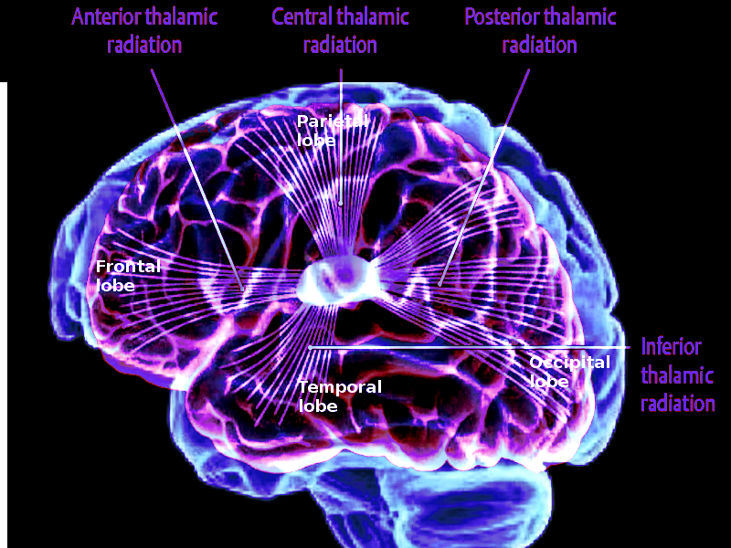
The thalamus serves as the main relay station for the brain. Motor pathways, limbic pathways, and sensory pathways besides olfaction all pass through this central structure. The thalamus can divide into approximately 60 regions called nuclei.[1] Each nucleus has unique pathways as inputs and various projections as outputs, most of which send information to the cerebral cortex.
Structure and Function
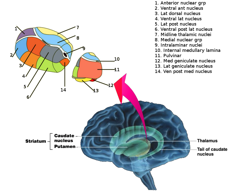
The thalamus is a paired structure located in the center of the brain. Each side can divide into three groups of thalamic nuclei: a lateral nuclear group, a medial nuclear group, and an anterior nuclear group. These three groups get split by the internal medullary lamina, a Y-shaped structure present on each side of the thalamus. There is also an area of thin, midline thalamic nuclei next to the third ventricle, and an enveloping thalamic reticular nucleus that covers each lateral thalamus.
In addition to grouping by anatomic location, the thalamic nuclei can be categorized by function as well. There are three categories[2]:
- Relay nuclei (lateral nuclear group, medial nuclear group, anterior nuclear group)
- Reticular nucleus
- Intralaminar nuclei
Relay nuclei represent the majority of the thalamus. Their projections to the cortex localize to more specific regions than the reticular and intralaminar nuclei. Relay nuclei subdivide into the three groups, as mentioned above, of lateral nuclear, medial nuclear, and anterior nuclear. The most clinically relevant nuclei all fall into the lateral nuclear group. These include the ventral posterolateral, ventral posteromedial, lateral geniculate, medial geniculate, and ventral lateral nuclei. The discussion of their relevance appears in detail in the section on Clinical Significance.
The reticular nucleus envelops each lateral thalamus. Lateral to it is the internal capsule. This nucleus is unique in that its projections do not go to the cortex. Its projections circle back to the thalamus itself, from which it received its inputs. Thus, the reticular nucleus serves to regulate the activity of the thalamus.
Intralaminar nuclei also send projections to the cortex. Their inputs, however, come from the basal ganglia.
Categorization of thalamic nuclei:
Relay nuclei
- Lateral nuclear group
- Ventral posterolateral nucleus (VPL)
- Ventral posteromedial nucleus (VPM)
- Lateral geniculate nucleus (LGN)
- Medial geniculate nucleus (MGN)
- Ventral lateral nucleus (VL)
- Ventral anterior nucleus (VA)
- Pulvinar
- Lateral dorsal nucleus
- Lateral posterior nucleus
- Ventral medial nucleus
- Medial nuclear group
- Anterior nuclear group
- Midline thalamic nuclei
- Paraventricular
- Parataenial
- Interanteromedial
- Intermediodorsal
- Rhomboid
- Medial ventral
Reticular nucleus
Intralaminar nuclei
- Rostral intralaminar nuclei
- Central medial nucleus
- Paracentral nucleus
- Central lateral nucleus
- Caudal intralaminar nuclei
- Centromedian nucleus
- Parafascicular nucleus
Embryology
Embryologically, the parts of the brain can be allotted to three origins: prosencephalon, mesencephalon, and rhombencephalon. The prosencephalon becomes the forebrain, the mesencephalon becomes the midbrain, and the rhombencephalon becomes the hindbrain. These three divisions can be further split into subdivisions so that the prosencephalon consists of the telencephalon and diencephalon, the mesencephalon does not subdivide, and the rhombencephalon is made up of the metencephalon and myelencephalon.
In week 3 of embryologic development, ectoderm forms the neural tube. The cranial part of the neural tube then becomes the prosencephalon, mesencephalon, and rhombencephalon. By week 5, the thalamus arises from the diencephalon along the sides of the third ventricle. Other structures that arise from the diencephalon include the pituitary gland, hypothalamus, epithalamus, optic vesicle, the optic cup, which forms the retina, and optic stalk.
A developmental relationship exists between the thalamus and the cortex. Both parts of the brain influence one another by affecting proliferation, connections of axons, and interneuron maturation.[3]
Categorization of the embryologic origins of the brain:
- Prosencephalon
- Telencephalon
- Diencephalon
- Mesencephalon
- Rhombencephalon
- Metencephalon
- Myelencephalon
Blood Supply and Lymphatics
Several arteries supply the thalamus with blood, all of which are branches of the posterior cerebral artery. These include the tuberothalamic, inferolateral, paramedian, and posterior choroidal arteries.[4][5]
Physiologic Variants
The artery of Percheron is an anatomic variation to the blood supply of the brain. This artery branches from the posterior cerebral artery to supply the thalamus. The prevalence of the artery of Percheron is estimated to be 4% to 12%.[6] Occlusion of this artery can result in the symmetric infarction of both sides of the thalamus.[7]
Clinical Significance
The most clinically relevant nuclei all fall into the lateral nuclear group. These include the ventral posterolateral, ventral posteromedial, lateral geniculate, medial geniculate, and ventral lateral nuclei. The ventral posterolateral nucleus gets fed by the spinothalamic tracts and dorsal columns of the spinal cord. As a result, this nucleus handles sensations of temperature and pain, along with vibration, pressure, fine touch, and proprioception. Signals then proceed to the primary somatosensory cortex within the postcentral gyrus.[8] The ventral posteromedial nucleus receives inputs from the trigeminal pathway and gustatory, or taste, pathway. Facial sensation and taste sensation get relayed here. Similar to its lateral counterpart, the ventral posteromedial nucleus sends this information to the primary somatosensory cortex within the postcentral gyrus. The third major thalamic nucleus is the lateral geniculate nucleus. Visual sensory information from the eyes is sent through the optic nerves, through the optic chiasm where some fibers cross and others remain ipsilateral, and finally through the optic tracts before entering this nucleus. From the lateral geniculate nucleus, visual information gets sent to the primary visual cortex, which is in the calcarine sulcus of the occipital lobe.[9] The medial geniculate nucleus, on the other hand, relays auditory sensory information from the superior olive and inferior colliculus of the tectum. Auditory information then travels to the auditory cortex of the temporal lobe. Finally, the ventral lateral nucleus receives input from the cerebellum and basal ganglia. Thus, it handles motor information and sends it to the precentral (motor) cortex.
The reticular nucleus receives inputs from the other thalamic nuclei, but also the reticular activating system and basal forebrain. As a result, the thalamus plays a role in controlling alertness and attention.[10]
The central medial nucleus, one of the intralaminar nuclei, is another clinically relevant thalamic nucleus. It receives inputs from the globus pallidus internus, deep cerebellar nucleus, and reticular activating system and primarily sends that information to the cerebral cortex and striatum. The central medial nucleus is located deep within the brain and handles alertness, motor information, consciousness, and awareness.[11]