Continuing Education Activity
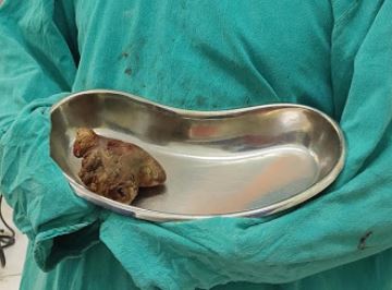
Renal calculi are a common cause of blood in the urine (hematuria) and pain in the abdomen, flank, or groin. They occur in one in 11 people at some time in their lifetimes, with men affected 2 to 1 over women. Development of the stones is related to decreased urine volume or increased excretion of stone-forming components such as calcium, oxalate, uric acid, cystine, xanthine, and phosphate. Calculi may also be caused by low urinary citrate levels or excessive urinary acidity. This activity reviews the cause, pathophysiology, and presentation of renal calculi and highlights the role of the interprofessional team in its management.
Objectives:
- Describe the types of renal calculi.
- Review the presentation of renal calculi.
- Summarize the treatment options for renal calculi.
- Outline the importance of improving care coordination among interprofessional team members to improve outcomes for patients affected by renal calculi.
Introduction
Renal calculi are a common cause of blood in the urine (hematuria) and pain in the abdomen, flank, or groin. They occur in one in 11 people at some time in their lifetimes with men affected 2 to 1 over women. Development of the stones is related to decreased urine volume or increased excretion of stone-forming components such as calcium, oxalate, uric acid, cystine, xanthine, and phosphate. Calculi may also be caused by low urinary citrate levels or excessive urinary acidity.[1][2][3]
Renal calculi present with excruciating pain and most patients present to the emergency department in agony. A single event does not cause kidney failure but recurrent renal calculi can damage the tubular epithelial cells, which can lead to functional loss of the renal parenchyma.
Etiology
Urolithiasis occurs when solutes crystallize out of urine to form stones. Urolithiasis may occur due to anatomic features leading to urinary stasis, low urine volume, dietary factors (e.g., high oxalate or high sodium), urinary tract infections, systemic acidosis, medications, or uncommonly genetic factors such as cystinuria. The most common cause of stone disease is inadequate hydration and subsequent low urine volume. The other four most common factors contributing to urinary stone formation are hypercalciuria, hyperoxaluria, hyperuricosuria, and hypocitraturia.[4][5]
The four major types of renal calculi include
- Calcium stones (due to hyperparathyroidism, renal calcium leak, hyperoxaluria, hypomagnesemia, and hypocitraturia)
- Uric acid stones are associated with a pH of less than 5, a high intake of purine foods (fish, legumes, meat), or cancer. These stones may also be associated with gout.
- Struvite stones (caused by gram negative-urease positive organisms that breakdown urea into ammonia. Common organisms include pseudomonas, proteus, and klebsiella. E coli is not associated with struvite stones)
- Cystine stones are due to an intrinsic metabolic defect causing the failure of the renal tubules to reabsorb cystine, lysine, ornithine, and arginine.
Many drugs are known to cause stones and include the following:
- Atazanavir
- Indinavir
- Triamterene
- Guaifenesin
- Overuse of silicate
- Sulfonamide
There also appears to be a genetic association to renal calculi. In some families, there may be mutations that cause a defect in the renal tubular handling of calcium and other substrates.
Epidemiology
Overall urinary stone prevalence in the United States has increased from 3.8% in 1970 to 8.8% in 2010. For patients with a history of a previous urinary stone, recurrence rates approach 50% at ten years. There is traditionally a high incidence of urinary stones in the Southeastern and South Central United States, termed the “Stone Belt,” which probably reflects the hot weather climate and relative dehydration that occurs in these areas. Before the development of modern urologic techniques for treatment, mortality from untreated staghorn (infection) calculi was 27%. Currently, mortality from stone disease is rare, although there is still a significant rate (28%) of renal deterioration with certain stone types, particularly staghorn (struvite or infection) stones.[6][7]
Pathophysiology
Most urinary stones start as Randall's plaque at the junction of the nephron's collecting tubule and the renal pelvis in the papilla. These plaques start suburothelial and then gradually grow until they break through the urothelium into the renal pelvis. They form an anchored lithogenic nidus for stone formation. Once in continuous contact with urine, layers of calcium oxalate typically start to form on the calcium phosphate nidus (all Randall's plaques are composed of calcium phosphate). Calcium oxalate stones tend to form when the urinary pH is under 7.2, while calcium phosphate will form in more alkaline urine.
Hyperparathyroidism and similar metabolic disturbances like renal tubular acidosis typically form stones primarily or significantly composed of calcium phosphate. Overly acidic urine is the primary cause of uric acid stones (not hyperuricosuria).[8][9]
Most renal calculi are made of calcium, followed by urate crystals. Supersaturation of the urine is the common denominator in all cases of renal calculi. In some cases, calcium oxalate stones may deposit in the renal papilla. Calcium phosphate stones usually precipitate in the basement membrane of the thin loop of Henle and may erode into the interstitium. The colicky pain s usually due to the dilatation and spasm of the ureter.
Natural urinary stone inhibitors include:
- Water (urinary volume)
- Citrate
- Tamm-Horsfall protein
- Nephrocalcin
- Uropontin
- Glycosaminoglycans
History and Physical
Patients with a stone disease will most commonly present with acute, severe flank pain that will often radiate to the abdomen and the groin, testicle, or labia. It is often sharp and severe in nature. It may also be colicky. The pain is often associated with nausea and vomiting due to the embryological origins of the urogenital tract.
Renal colic usually peaks within 90 to 120 minutes, and the pain radiation follows dermatomes T10 to S4. The first phase may wake the patient up from sleep, and the pain is steady, followed by waves of excruciating pain. The second phase is characterized by constant pain and may last 3 to 4 hours. The third phase is associated with mild pain relief, but waves of pain may persist. This phase may last 4 to 16 hours.
Patients may also present with fever, chills, or other systemic signs of sepsis if infected. This condition, called pyonephrosis or obstructive pyelonephritis, is potentially severe and life-threatening, requiring emergency decompression surgery.[10]
Patients often present with hematuria, as 85% of patients demonstrate at least microscopic hematuria on urinalysis.
The physical exam may reveal costovertebral tenderness and hypoactive bowel sounds. The testis and pubic area may also be tender to touch. Fever is rarely seen in renal colic, but the presence of fever, pyuria, and leucocytosis may be indicative of pyelonephritis.
High-risk factors include:
- Bone disorders
- Chronic diarrhea, malabsorption
- Diabetes, obesity (especially in women)
- Family history of kidney stones
- Gastrointestinal disease
- GI bypass surgery (especially Rous-en-Y)
- Gout
- Hyperparathyroidism
- Prior stones
- Renal tubular acidosis
- Sarcoidosis
Evaluation
A urinalysis should be obtained on every patient with a suspected kidney stone. Hematuria is usually present, but up to 15% of kidney stone patients will not demonstrate even microscopic hematuria. The presence of urinary crystals may suggest urolithiasis. Positive nitrites, leukocytes, and bacteria suggest infection, which should be cultured and treated aggressively.[11]
A KUB can be obtained to screen for the presence of significant nephrolithiasis but may often miss small stones, calculi hidden by overlying bowel, or uncalcified. Ultrasound may be very useful for assessing obstruction and resultant hydronephrosis, especially in pregnancy, where x-ray studies are discouraged. It can also be used to measure the resistive index, which can suggest ureteral obstruction.
Resistive Index = (peak systolic velocity - end-diastolic velocity)/peak systolic velocity
Values of 0.70 or less are considered normal, while higher values suggest obstructive uropathy. Bilateral high resistive indices suggest medical renal disease, while a unilateral high resistive index (0.75 or higher) suggests an obstruction, such as from a stone. Once a ureteral stone has been identified, the lower the resistive index, the more likely the stone will pass spontaneously. [12]
Ultrasound can also identify uric acid and other non-calcific stones if they are large enough (usually greater than 4 mm), but it can also miss the presence of stones less than 5 mm.
The most sensitive and reliable test to diagnose urolithiasis is a non-contrast abdominal and pelvic CT scan, which will also provide information regarding obstruction with resultant hydronephrosis or concerns for infection.[13][14][15][16][17] Other labs to obtain would include a WBC with differential and a urine culture if the patient is febrile or has a urinalysis suggestive of a possible infection. The initial use of IV contrast for CT scans in patients with abdominal pain is not recommended. In many cases, an atypical abdominal pain will ultimately turn out to be a kidney stone that has moved or the presence of a urological anatomical variant such as a horseshoe kidney.
Even without IV contrast, in most cases, the correct diagnosis can be easily made. If contrast is absolutely necessary, doing the non-contrast study first eliminates urinary stones from consideration. Certainly, if the urinalysis is abnormal for blood or possible infection, a non-contrast abdominal and pelvic CT should be performed before using contrast, which will make identifying any urinary stones far more difficult. If this recommendation is not followed, sooner or later, contrast will be given to a patient who will ultimately be diagnosed with urinary stones. Obscuring urinary stones with IV contrast can make it much more difficult to determine their size, number, or shape, complicating decisions on optimum treatment and possible surgery.[13][14][15][16][17]
A simultaneous KUB should be done if the CT is positive for stones. This will provide information useful in tracking or following the progress of the stone, its degree of calcification, and its shape, which cannot always be identified from the CT scan alone.[18]
Treatment / Management
Many stones may be watched conservatively as an outpatient, with intervention planned as an outpatient. Smaller stones (less than 5 mm) have a greater chance (90%) of passing on their own with medical expulsion therapy (usually tamsulosin, nifedipine, or alfuzosin). Any hint of a urinary tract infection should be treated aggressively with antibiotics.[19][20][21]
Acute management requires IV hydration, analgesia, and antiemetic medications. Studies show that desmopressin can lower the pain of renal calculi. Anecdotal reports indicate that using calcium channel blockers or alpha-blockers can provide pain relief due to the relaxation of the ureter and helps the passage of the stone distally. The urine should be strained for stones.
There are several cases where urgent intervention is required.
- An obstructing stone in a patient with a urinary tract infection, fever, or sepsis. (This is called pyonephrosis or obstructive pyelonephritis and requires urgent surgical decompression by urology or interventional radiology)
- Nausea or pain uncontrolled with outpatient management
- An obstructing stone in a solitary kidney
- Any degree of simultaneous bilateral obstruction can easily lead to renal failure.
- Any degree of obstruction with a rising creatinine
In the case of urinary tract infection or urosepsis with an obstructing stone, the obstruction should first be relieved with either a ureteral double J stent or nephrostomy tube placement. The decision of which treatment modality is most appropriate should be made by urology. In general, the more severely ill the patient, the greater the benefit from a nephrostomy tube. Definitive stone management can then occur once the infection is no longer active. Morbidly obese patients and those who cannot be safely taken off of their blood thinners may require a double J stent, regardless.
Electively, stones can be surgically managed in several ways.
- Extracorporeal shockwave lithotripsy (ESWL) can be used to break up stones anywhere in the urinary tract but is primarily used in the kidney and upper ureter.[22]
- Ureteroscopy with laser lithotripsy can be used to manage stones endoscopically and is preferred for ureteral stones, especially in the lower ureter.[23]
- For large (greater than 2 cm) stones in the renal pelvis, percutaneous nephrolithotomy can be performed.
- Robotic-assisted laparoscopic pyelolithotomy with intracorporeal pyeloscopy can be used in complicated stone cases in horseshoe kidneys.[24]
Once the patient has had their acute stone episode treated, it is recommended to evaluate the patient for the underlying cause for their stone episode, particularly if he or she has had stones in the past. This would involve obtaining a basic metabolic panel and a 24-hour urine collection for stone prevention analysis. Patients must understand that this represents their commitment to follow a long-term course of therapy for stone prevention and that no treatment plan is foolproof. An occasional stone may still be produced but is much less likely on prophylaxis.
Physicians evaluating 24-hour kidney stone results should not only look at the normal ranges but also at what may be optimal. For example, optimal 24-hour urinary calcium should be no more than 250 mg, oxalate less than 25 mg, citrate more than 600 mg, urinary volume more than 2,000 ccs, and urinary uric acid at 600 mg or less. While these levels may not be realistically obtainable in every patient, they are used as goals for treatment where the intention is to get as many chemistry levels optimal as possible, even if they are all technically normal.[25][26][27]
Analysis of 24-hour urine tests is often considered complicated but is actually quite simple for the vast majority of patients.
- Hypercalcemic patients should have a parathyroid hormone level check for possible hyperparathyroidism.
- Hydration should be optimized with a goal of at least 2 liters of urine production a day.
- Thiazides are used for hypercalciuria.
- Potassium citrate is used for hypocitraturia and aciduria.
- Allopurinol will help lower high urinary uric acid levels.
Appropriate thiazide use has been shown to lower hypercalciuric calcium stone disease from 2.94 to 0.05 stones per year (P<0.001) while long-term use of potassium citrate can diminish calcium stone production by 80%.[28][29] Limiting sodium intake is important to allow thiazide to perform its hypocalciuric action. A daily increase of 100 mEq of sodium will raise urinary calcium excretion by 50 mg.
Essentially, just three drugs. A companion StatPearls reference article, "24 Hour Urine Testing for Nephrolithiasis: Guide to Interpretation," by Leslie S and Bashir K, is recommended for more details on 24-hour urine interpretation and preventive therapy.[30]
Admission is recommended in the following situations:
- Inadequate pain relief with oral analgesics
- Patient with a transplanted kidney and renal calculi
- Presence of renal calculi and pyelonephritis
- Obstructed pyelonephritis (pyonephrosis)
Dissolution therapy does not work for calcium stones but may be used to manage uric acid and cystine stones. Uric acid calculi can be dissolved by making the urine consistently alkaline with potassium citrate and/or sodium bicarbonate.[31] (Potassium citrate is usually preferred due to its lower sodium load.) In addition, allopurinol can be used to reduce renal uric acid excretion.[32] Thiazide diuretics are recommended for hypercalciuric patients with recurrent calcium stones but cannot dissolve existing calculi.[30] Cystine stones can be managed with D-penicillamine, aggressive fluid intake, and optimal alkalinization therapy.[33]
Differential Diagnosis
- Appendicitis
- Cholecystitis
- Acute epididymitis
- Diverticulitis
- Hernia
- PID
Staging
European Guidelines for Patients with Renal Calculi
- Check urine for hematuria, pH, and bacteria.
- Obtain a urine culture.
- Order BUN and serum creatinine.
- Order serum calcium, uric acid, sodium, and potassium levels.
- Order a complete CBC and CRP.
- Obtain a coagulation profile in case surgical intervention is necessary.
- Obtain a non-contrast CT scan.
Prognosis
Close to 80% to 90% of renal calculi pass spontaneously. About 3% of patients need admission because of pain, inability to pass the stone, or dehydration. A few patients may develop urinary tract obstruction and an upper urinary tract infection. This can result in urosepsis or pyelonephritis. Most of these patients require an urgent procedure to bypass the stone until the infection is resolved, at which time an elective procedure can be performed to remove the stone.
The recurrence rate of renal calculi has been reported to be about 50% within 5 years. Individuals with ongoing malignancy or metabolic disorders are at a higher risk for recurrence. The key for all patients with renal calculi is to stay hydrated; no medical therapy is successful without adequate hydration and sufficient urinary fluid output.
Complications
- Abscess
- Urosepsis
- Ureteral scarring or perforation
- Urine extravasation
- Kidney atrophy in chronic cases\
- Renal failure
Deterrence and Patient Education
Patients should generally avoid diets high in calcium while limiting excessive salt and meat animal protein intake. A low oxalate diet is also recommended. Overly restrictive dietary calcium is not recommended as this may lead to a lack of intestinal oxalate binding and hyperoxaluria.[34]
Pearls and Other Issues
- Ultimately, the success of any kidney stone preventive treatment program will depend on the patient's willingness to follow a long-term course of treatment that will involve some level of dietary modifications, medications, personal sacrifice, and lifestyle change with no obvious immediate or noticeable benefits.
- Patients on treatment may still make stones, albeit fewer than otherwise.
- Patients may cheat on their therapies from time to time, and since they do not seem to pay any immediate price or penalty, many will revert to their previous diets and behaviors.
- Patients may also develop an over-reliance on drug therapy so they can minimize the dietary changes requested.
Enhancing Healthcare Team Outcomes
Most patients with kidney stones, even those with multiple recurrences, are unaware of the availability of 24-hour urine testing and the potential benefits of preventive measures based on this testing. Successful kidney stone preventive programs require high levels of patient compliance, motivation, and discipline for their efficacy. An interprofessional team of nurses, nurse practitioners, physician assistants, and physicians should educate patients about preventive therapy. Still, only those individuals who are strongly motivated are likely to have long-term success.[27]
The health care team involved in the care of nephrolithiasis patients has an absolute obligation to inform patients of the existence of such programs, particularly in cases of multiple stone recurrences, solitary kidneys, high surgical risk factors, or those in the pediatric age group. [35][36]
Clinicians looking after patients with renal calculi should educate them about the importance of hydration; failing to do so will mean low effectiveness of medical therapy. Patients with recurrent renal calculi should be referred to a specialist for workup to rule out an anatomical or metabolic problem. Only through open communication between the team members can the morbidity of renal calculi be lowered.