Continuing Education Activity
Incision and drainage (I and D) is a widely used procedure in various care settings including emergency departments and outpatient clinics. It is the primary treatment for skin and soft tissue abscesses, with or without adjunctive antibiotic therapy. This activity reviews the incision and drainage procedure, risks and benefits. The role of an interprofessional team will be discussed.
Objectives:
- Identify the anatomical considerations of incision and drainage.
- Describe the technique of incision and drainage.
- Outline the appropriate evaluation of the potential complications of incision and drainage.
- Describe interprofessional team contributions to the comprehensive management of patients with skin and soft tissue abscesses.
Introduction
Incision and drainage (I&D) is a widely used procedure in various care settings, including emergency departments and outpatient clinics. It is the primary treatment for skin and soft tissue abscesses, with or without adjunctive antibiotic therapy. This activity will focus specifically on its use in the management of cutaneous abscesses. Based on 2013 data from the CDC, cutaneous abscesses accounted for about 2% of all presentations to the emergency department. The same data reports that 0.9% of all patients who presented to the emergency department underwent incision and drainage. In the pediatric population, the incidence of skin and soft tissue infection (SSTI) has increased, and hospitalization due to SSTI doubled in the last 20 years.[1] As such, being well versed in I&D is essential for clinicians in both the adult and pediatric care settings.
Anatomy and Physiology
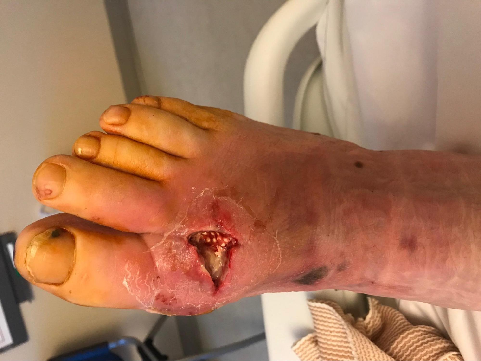
Cutaneous abscesses are localized collections of pus that occur within the dermis and subcutaneous space. They occur virtually anywhere on the body; however, common locations for an abscess to develop are the groin, buttocks, axillae, and extremities.
In most cases, a cutaneous abscess can be diagnosed clinically on the basis of physical examination alone. The classic characteristics of an abscess are erythema, induration, tenderness to palpation, and fluctuance. Care must be taken to differentiate between cellulitis and abscess, as the treatment for cellulitis is antibiotic therapy without drainage. Typically an abscess will be fluctuant on exam, whereas this is not a feature of cellulitis. In cases of equivocal clinical findings, ultrasonography can be used to assess for the presence of an abscess, in addition to providing information on size and location.
Studies have been done to compare the use of physical exam and ultrasonography in the detection of an abscess. A prospective study in the pediatric population revealed that in cases when abscesses could not be diagnosed clinically, bedside ultrasound had higher sensitivity and specificity than the exam alone.[2] In the same study, there was no difference in sensitivity and specificity between exam and ultrasound when the abscess was diagnosed clinically. In terms of differences in outcomes (i.e., treatment failure), there is a study that assessed treatment failure rates ten days after I&D.[3] Patients who were diagnosed by ultrasound in addition to physical exam had lower treatment failure rates ten days after I&D in comparison to patients who were evaluated by exam alone. This suggests that performing an ultrasound to evaluate for abscess improves treatment outcomes; this is, of course, reliant on the availability of experienced technicians and radiologists.
Indications
Most patients with an abscess should have incision and drainage performed, as antibiotic therapy alone is not sufficient for treatment. In cases of small fluid collections, conservative management with antibiotics, in addition to the manual expression of pus can be considered.
Contraindications
Possible contraindications to bedside incision and drainage include large and deep abscesses, the presence of a pulsatile mass at the site of infection, proximity to the vasculature and nervous structures, the presence of a foreign body, and particular locations of an abscess.
Certain locations on the body make bedside incision and drainage more technically difficult to perform, whether due to the inherent sensitivity of the area (e.g., the palms, soles, and face) or associated complications. Examples of locations that warrant evaluation by surgery due to a high potential for complications include perirectal and periareolar abscesses, which could be complicated by fistula formation. An otolaryngologist should evaluate neck abscesses that could potentially have developed from preexisting cystic lesions.
Consider also the need for prophylactic antibiotics in patients with abnormal or artificial heart valves as this may delay the procedure. Additionally, it is prudent to inquire about underlying bleeding disorders and to determine a history of allergy to lidocaine, epinephrine, or latex if using latex gloves.
Equipment
Generally, the following sterile equipment is required for a safe and successful procedure: cleansing agent (typically povidone-iodine or chlorhexidine), a local injectable anesthetic agent (1% or 2% lidocaine, lidocaine with epinephrine, or bupivacaine), 5 to 10 mL syringe with 25 to 40 gauge needle, 4x4 gauze, scalpel blade with a handle, curved hemostat, normal saline solution with large syringe/splash guard/bowl for irrigation, packing material (iodoform or plain gauze), swabs for wound culture (if desired), scissors and tape for dressing the wound.[4]
Incision and drainage is a painful procedure that, in addition to local anesthetic, may also require oral or even parenteral analgesia.
Personnel
The procedure is relatively simple and is often performed by a single clinician.
In certain situations, the presence of support staff may be warranted. In the pediatric population, child life services can play a critical role in facilitating a successful procedure. Child life specialists are professionals whose role is to provide emotional support to children and families in a health care setting. A recent study published in pediatric emergency medicine evaluated the impact of child life on the emotional response of pediatric patients who underwent laceration repair in the emergency department.[5] Comparing children who underwent the procedure without the presence of these specialists with children assisted by child life, those with support were found to have less emotional distress during the procedure.
Preparation
Informed consent from the patient or the patient’s legal guardian must be obtained before the procedure. Risks including bleeding, pain and possible scar formation should be relayed. The verification of tetanus immunization status is also an important step in preparation.
The clinician performing the procedure should follow universal precautions that include wearing a gown, gloves, and a facemask or goggles for protection. Although I&D is not considered a sterile procedure (given that the area of interest is already infected), it is prudent to practice sterile precautions.
Technique or Treatment
Holding the scalpel with a steady grip, an incision is made directly over the center of the abscess until pus is expressed. The incision should be made parallel to skin tension lines in order to prevent scar tissue formation. A curved hemostat can then be used for blunt dissection to further disrupt loculations within the infected cavity. Manual expression can be used to facilitate drainage as well. After the abscess is drained, the wound should be copiously irrigated with sterile normal saline solution. Wound packing is not recommended for abscesses that are 5 cm or less in diameter, as it has not been shown to affect outcomes and may contribute to increased pain.[6][7] Furthermore, packing has not been shown to reduce the risk of abscess recurrence.[8]
The next step is to cover the site with sterile dressing and tape. A follow-up visit is advised 2 to 3 days after the procedure for removal of the packing. Wounds are then left to close by secondary intention.
An alternative to I&D is needle aspiration, though this is much less commonly used, given that it is both more invasive and less effective than I&D. In a randomized clinical control trial comparing outcomes with I&D to ultrasound-guided needle aspiration, the overall success of producing purulent drainage with needle aspiration was 26% compared to an 80% success rate in patients who underwent I&D.[9]
Another alternative to conventional incision and drainage is the loop drainage technique, which may reduce pain and scarring at the site of infection. Studies suggest that loop drainage is associated with a lower failure rate than conventional therapy, although it is not yet a widely used procedure.[10]
Complications
Typically I&D is well tolerated with pain being the most common complication. Inadequately drained abscesses can lead to the extension of the infection into adjacent tissues and worsening of clinical status.
Clinical Significance
As stated, incision and drainage is a common procedure in a variety of care settings. It is the standard of treatment for subcutaneous abscesses, with or without adjunctive antibiotic therapy.
Enhancing Healthcare Team Outcomes
The successful treatment of skin abscesses is not limited to clinician proficiency in performing incision and drainage. Often patients will understandably have questions regarding disease recurrence and prevention, and there is a significant amount of research to address these topics.
Although bacterial etiology was not discussed in this article, skin and soft tissue infections are many times caused by Staphylococcus aureus, with the prevalence of MRSA increasing. IDSA (Infectious Diseases Society of America) guidelines on the management of skin and soft tissue infections due to MRSA discuss measures to prevent recurrence. These guidelines are derived from important research performed by not only physicians but other integral members of the healthcare team as well. There have been multiple publications by nursing on the subject of recurrence prevention.[11][12] The results of these studies support IDSA guidelines of proper hand hygiene and post-drainage MRSA decolonization with mupirocin. It is important to recognize that standards of care are the result of interprofessional efforts.