Raccoon Sign


Definition/Introduction

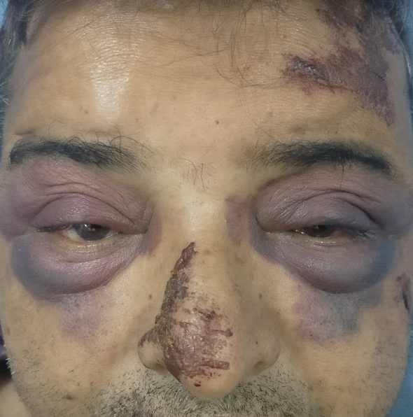
The 'Raccoon sign' comprises unilateral or bilateral progressive periorbital ecchymosis associated with edema.[1] It is also referred to as the 'raccoon eyes' and the 'panda sign.' The pooling of blood around the eyes is most commonly associated with fractures of the base of the anterior cranial fossa. This finding is not observed immediately following the injury and is typically delayed by 1 to 3 days.[2] If it presents bilaterally, this finding is highly predictive of an anterior skull base fracture. Classically, the tarsal plate will be spared in this condition. Further extravasation of the blood beyond the periorbital region is limited owing to the orbital septum inserted into the tarsal plate.[3]

When a skull base fracture is present, the raccoon sign is present in 50 to 60% of such cases.[4] This sign is a reliable bedside clinical marker and is usually associated with anterior skull base fractures alongside the involvement of the frontal and the orbital bones. Thin-cuts computed tomogram skull (less than 5 mm) bone window imaging is needed to identify such fractures in some cases of these cohorts.

Issues of Concern

If the raccoon sign is present in a trauma victim, one should be aware of the following associated injuries and complications it harbingers such as:

  1. Cerebrospinal fluid (CSF) rhinorrhea
  2. Injury to eyeballs
  3. Injury to cranial nerves I, II, III, IV, and VI
  4. Intracranial misplacement of the nasogastric or nasotracheal tube
  5. Diffuse axonal injury
  6. Insult to the hypothalamic-pituitary axis (HPA) causing endocrinopathies
  7. Maxillofacial injuries
  8. Cervical spine injury
  9. Pneumocephalus
  10. Later stage meningitis

Clinical Significance

The various etiologies behind the causation of the 'Raccoon sign' can be summarized as follows:

  1. Traumatic
    1. Minor events like the plucking of the eyebrows
    2. Non-accidental injuries
    3. Thoracic trauma and crush injuries
    4. Following vigorous sneezing, coughing, or vomiting (probably due to periorbital venous hemorrhage)
    5. Posttraumatic orbital emphysema (due to fracture of orbital wall and entrapment of air from paranasal sinuses)[4][5]
  2. Vascular
    1. Subarachnoid hemorrhage (SAH) following the rupture of an ophthalmic artery aneurysm
    2. Venous sinus thrombosis (commonly secondary to cavernous sinus thrombosis)
    3. Giant cell arteritis
    4. Trigeminal autonomic cephalalgia
    5. Benign intracranial hypertension
    6. Acute migraine (due to vasodilation and release of vasoactive substances, including heparin)[6][7]
  3. Infectious
    1. Severe pediatric adenovirus infection
    2. Periorbital leukoderma
    3. Frontal sinus mucocele[8][9]
  4. Immune-mediated
    1. The neonatal lupus erythematosus (vasculitis may lead to facial and periorbital purplish-red plaques)
    2. Chronic atypical neutrophilic dermatosis with lipodystrophy and elevated temperature (CANDLE) syndrome (due to a predilection to involve periorbital skin)
    3. Lichen planus pigmentosus (hyperpigmentation due to pigment incontinence)
    4. Sweet syndrome or acute febrile neutrophilic dermatosis (skin disease characterized by the sudden onset of fever, an elevated white blood cell count, and tender, red, well-demarcated papules, and plaques that show dense infiltrates by neutrophil granulocytes on histologic examination)
      • Raccoon sign in this condition results from secondary vascular damage caused by prolonged exposure to matrix metalloproteinases.
      • Toxic products of neutrophils in the infiltrate may be responsible for vascular damage leading to erythrocyte extravasation.[10]
  5. Metabolic
    1. Amyloidosis (most commonly light chain (AL) type) is due to increased vascular fragility resulting from amyloid deposition; this is one of the most common causes of raccoon eyes other than trauma; the finding presents in about one-fifth of patients with AL amyloidosis.
    2. Myxedema[11][12]
  6. Malignancies
    1. Hematological malignancies (blastic plasmacytoid dendritic cell neoplasm, lymphoblastic lymphoma, and acute myeloid leukemia)
    2. Metastatic neuroblastoma (secondary to tumoral obstruction of the palpebral vessels)and neuroblastoma (observed in 5.4% of cases)
    3. Orbital metastasis of solid malignancies
    4. Kaposi sarcoma
    5. Multiple myeloma (infiltration of amyloid proteins in the capillaries leads to increased fragility, which can burst under minor stress and is usually painful)[13][14]
  7. Genetic
    1. Hemophilia
  8. Iatrogenic (perioperative raccoon eyes)
    1. Usually observed among patients with systemic amyloidosis of AL type
    2. Post-rhinoplasty (due to a bleeding disorder)
    3. Post-endoscopic retrograde cholangiopancreatography (ERCP), prone positioning, consumption of steroids, and Valsalva maneuver might cause increased venous pressure, which in turn lead to the rupture of fragile orbital vessels in an elderly patient)[15][16]

Nursing, Allied Health, and Interprofessional Team Interventions

If a nurse encounters a patient with a raccoon sign, they should immediately ask the patient about a history of trauma, CSF rhinorrhea, and visual difficulties. One should avoid inserting a nasogastric tube in such patients as inadvertent intracranial misplacement may occur. It is better to place the orogastric tube instead if intubation is necessary.

The periorbital ecchymosis perse resolves within 2 to 3 weeks following spontaneous gradual degradation of the blood products.


(Click Image to Enlarge)
Raccoon sign
Raccoon sign
Contributed by Sunil Munakomi, MD

(Click Image to Enlarge)
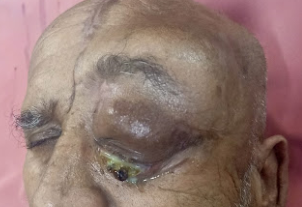
Left eye proptosis, periorbital edema and late echymosis due to orbital involvement of lymphoma
Contributed by Sunil Munakomi, MD

(Click Image to Enlarge)
Raccoon sign
Raccoon sign
Contributed by Sunil Munakomi, MD
Details

Author

Joe M. Das

Editor:

Sunil Munakomi

Updated:

4/10/2023 3:18:35 PM

References


[1]

Timmerman R. Images in clinical medicine. Raccoon eyes and neuroblastoma. The New England journal of medicine. 2003 Jul 24:349(4):e4     [PubMed PMID: 12878754]


[2]

Solai CA, Domingues CA, Nogueira LS, de Sousa RMC. Clinical Signs of Basilar Skull Fracture and Their Predictive Value in Diagnosis of This Injury. Journal of trauma nursing : the official journal of the Society of Trauma Nurses. 2018 Sep/Oct:25(5):301-306. doi: 10.1097/JTN.0000000000000392. Epub     [PubMed PMID: 30216260]


[3]

McPheeters RA, White S, Winter A. Raccoon eyes. The western journal of emergency medicine. 2010 Feb:11(1):97     [PubMed PMID: 20411091]


[4]

Herbella FA, Mudo M, Delmonti C, Braga FM, Del Grande JC. 'Raccoon eyes' (periorbital haematoma) as a sign of skull base fracture. Injury. 2001 Dec:32(10):745-7     [PubMed PMID: 11754879]


[5]

Satyarthee GD, Sharma BS. Posttraumatic orbital emphysema in a 7-year-old girl associated with bilateral raccoon eyes: Revisit of rare clinical emergency, with potential for rapid visual deterioration. Journal of pediatric neurosciences. 2015 Apr-Jun:10(2):166-8. doi: 10.4103/1817-1745.159199. Epub     [PubMed PMID: 26167226]


[6]

Aalbers M, van Dijk JMC. Teaching NeuroImages: Raccoon eye in subarachnoid hemorrhage. Neurology. 2019 Mar 26:92(13):e1534-e1535. doi: 10.1212/WNL.0000000000007180. Epub     [PubMed PMID: 30910950]


[7]

DeBroff BM, Spierings EL. Migraine associated with periorbital ecchymosis. Headache. 1990 Apr:30(5):260-3     [PubMed PMID: 2354948]


[8]

Chuang Yu, Chiu CH, Wong KS, Huang JG, Huang YC, Chang LY, Lin TY. Severe adenovirus infection in children. Journal of microbiology, immunology, and infection = Wei mian yu gan ran za zhi. 2003 Mar:36(1):37-40     [PubMed PMID: 12741731]


[9]

Mehta AA, Wagner LH, Blace N. Spontaneous upper eyelid ecchymosis: A rare presenting sign for frontal sinus mucocele. Orbit (Amsterdam, Netherlands). 2017 Jun:36(3):183-187. doi: 10.1080/01676830.2017.1280058. Epub 2017 Mar 10     [PubMed PMID: 28282265]


[10]

Wisuthsarewong W, Soongswang J, Chantorn R. Neonatal lupus erythematosus: clinical character, investigation, and outcome. Pediatric dermatology. 2011 Mar-Apr:28(2):115-21. doi: 10.1111/j.1525-1470.2011.01300.x. Epub 2011 Mar 1     [PubMed PMID: 21362029]


[11]

Matsuura H, Anzai Y, Kuninaga N, Maeda T. Raccoon Eye Appearance: Amyloidosis. The American journal of medicine. 2018 Jul:131(7):e305. doi: 10.1016/j.amjmed.2018.02.009. Epub 2018 Mar 14     [PubMed PMID: 29550357]


[12]

de Moura CG, Cruz CM, de Souza SP. Raccoon sign. Arthritis and rheumatism. 2013 Mar:65(3):692. doi: 10.1002/art.37793. Epub     [PubMed PMID: 23203502]


[13]

Liu YT, Yang MH, Cao LZ, Huang YH, Xie M, Yang LC, Yang H, Tang X. [Painless skin nodules and ecchymosis in a school-aged girl]. Zhongguo dang dai er ke za zhi = Chinese journal of contemporary pediatrics. 2015 Oct:17(10):1131-6     [PubMed PMID: 26483238]


[14]

Inokuchi R, Tagami S, Maehara H. An elderly woman with bilateral raccoon eyes. Emergency medicine journal : EMJ. 2016 Nov:33(11):781. doi: 10.1136/emermed-2015-205071. Epub     [PubMed PMID: 28319930]


[15]

Weingarten TN, Hall BA, Richardson BF, Hofer RE, Sprung J. Periorbital ecchymoses during general anesthesia in a patient with primary amyloidosis: a harbinger for bleeding? Anesthesia and analgesia. 2007 Dec:105(6):1561-3, table of contents     [PubMed PMID: 18042847]


[16]

Nasiri J, Zamani F. Periorbital Ecchymosis (Raccoon Eye) and Orbital Hematoma following Endoscopic Retrograde Cholangiopancreatography. Case reports in gastroenterology. 2017 Jan-Apr:11(1):134-141. doi: 10.1159/000456657. Epub 2017 Mar 17     [PubMed PMID: 28611566]

Level 3 (low-level) evidence