Continuing Education Activity
Lymphedema is a chronic disease marked by the increased collection of lymphatic fluid in the body, causing swelling, which can lead to skin and tissue changes. The chronic, progressive accumulation of protein-rich fluid within the interstitium and the fibro-adipose tissue exceeds the capacity of the lymphatic system to transport the fluid. Swelling associated with lymphedema can occur anywhere in the body, including the arms, legs, genitals, face, neck, chest wall, and oral cavity. There are many psychological, physical, and social sequelae related to a diagnosis of lymphedema. This activity reviews the presentation of lymphedema and highlights the role of the interprofessional team in its management.
Objectives:
- Describe the pathophysiology of lymphedema.
- Review the presentation of lymphedema.
- Summarize the treatment options for lymphedema.
- Review the importance of improving care coordination among interprofessional team members to improve outcomes for patients affected by lymphedema.
Introduction
Lymphedema is a chronic disease marked by the increased collection of lymphatic fluid in the body, causing swelling, which can lead to skin and tissue changes. The chronic, progressive accumulation of protein-rich fluid within the interstitium and the fibro-adipose tissue exceeds the capacity of the lymphatic system to transport the fluid. Swelling associated with lymphedema can occur anywhere in the body, including the arms, legs, genitals, face, neck, chest wall, and oral cavity. There are many psychological, physical, and social sequelae related to a diagnosis of lymphedema. Lymphedema is classified as either (genetic) primary lymphedema or (acquired) secondary lymphedema.
The lymphatic vessels transport lymph. Lymph is composed of white blood cells, triglycerides, bacteria, cell debris, water, and protein. It has a composition comparable to blood plasma. The lymph drainage system is complex and comprises initial lymphatics (lymph capillaries), pre-collectors, collectors, lymphatic trunks, and lymph nodes. Topographically, the lymph system is distinguished as superficial (subcutaneous) and deep (subfascial). The superficial system drains the skin and subcutis areas. The deep system drains muscles, joints, tendon sheaths, and nerves. Both systems are connected via the perforating vessels, which conduct lymph fluid from the subfascial areas to the surface.
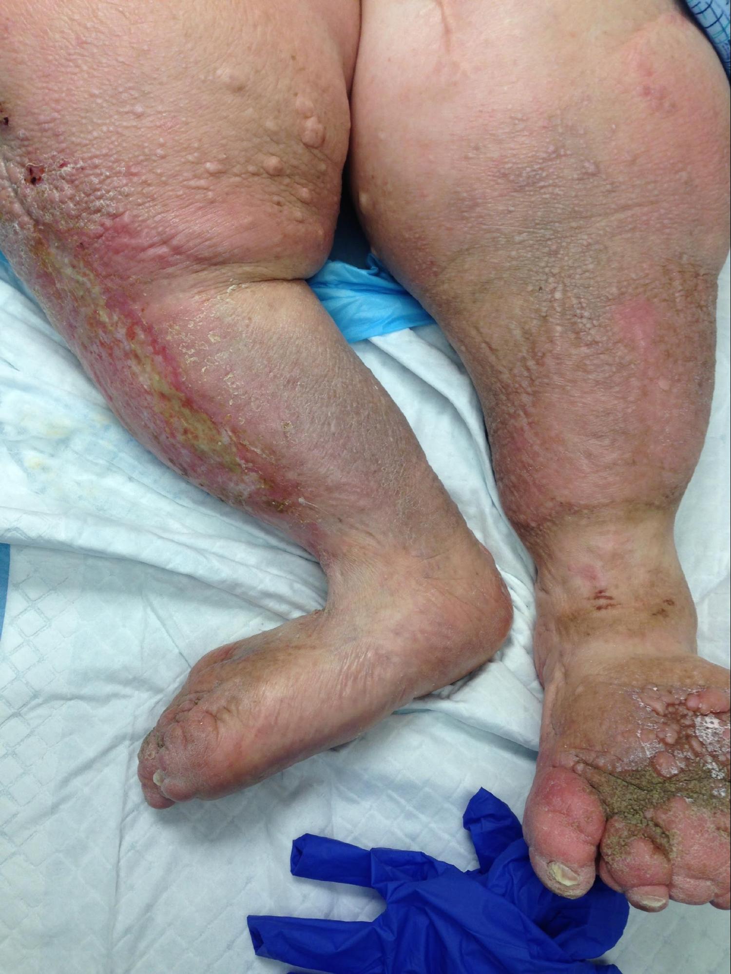
Signs and symptoms of lymphedema include distal swelling in the extremities, including the arms, hands, legs, feet; swelling proximally in the breast, chest, shoulder, pelvis, groin, genitals, face/intraoral tissues; restricted range of motion in the joints because of swelling and tissue changes; skin discoloration; pain and altered sensation; limb heaviness; and difficulty fitting into clothing.
Etiology
Primary lymphedema is an inherited or congenital condition that causes a malformation of the lymphatics system, most often because of genetic mutation. Primary lymphedema can be subdivided into 3 categories: 1) congenital lymphedema, present at birth or recognized within two years of birth; 2) lymphedema praecox, occurring at puberty or the beginning of the third decade; or 3) lymphedema tarda, which begins after 35 years of age.
Secondary lymphedema results from insult, injury, or obstruction to the lymphatic system. While the most common cause of lymphedema worldwide is filariasis caused by infection by Wuchereria bancrofti, in developed countries, most secondary lymphedema cases are due to malignancy or related to the treatment of malignancy. This includes surgical excision of lymph nodes, local radiation treatment, or medical therapy. Breast cancer is the most common cancer associated with secondary lymphedema in developed countries.
Epidemiology
Primary lymphedema is rare, affecting 1 in 100,000 individuals. Secondary lymphedema is the most common cause of the disease and affects approximately 1 in 1000 Americans.
The identification of the incidence and prevalence of lymphedema is complex. Lymphedema is remarkably prevalent, but the population implications of lymphatic dysfunction are not well studied. Prevalence estimates for lymphedema are relatively high, yet its prevalence is likely underestimated. There is an absence of prospectively designed and rigorously performed relevant epidemiologic studies that limit the true prevalence of the disease.[1]
The incidence of lymphedema is most widely studied in the oncologic population. One in 5 women who survive breast cancer will develop lymphedema.[2] In head and neck cancer, lymphatic and soft tissue complications can develop throughout the first 18 months post-treatment, with greater than 90% of patients experiencing some form of internal, external, or combined lymphedema. Over half of those patients developing fibrosis.[3] In one recent study, 37% of women treated for gynecological cancer had measurable evidence of lymphedema within 12 months post-treatment.[4] In the gynecologic oncologic population, more extensive lymph node dissection, receipt of chemotherapy and radiation therapy, increasing body mass index, insufficient levels of physical activity, a diagnosis of vulvar/vaginal cancer, and presence of pre-treatment lymphedema were identified as potential risk factors to lymphedema development.[4]
Pathophysiology
Primary lymphedema is associated with dysplasia of the lymphatic system and can also develop with conditions of other vascular abnormalities, including Klippel-Trenaunay-Weber syndrome,[5], and Turner syndrome.[6] Primary lymphedema is marked by hyperplasia, hypoplasia, or aplasia of the lymphatic vessels.
Secondary lymphedema develops due to damage or dysfunction of the normally functioning lymphatic system. Although cancer treatments, including oncologic surgical procedures such as axillary lymph node dissection and excision in breast cancer and radiation treatment, are the most common cause of lymphedema in the United States, filariasis is the most common cause of secondary lymphedema globally.[7] Filariasis is the direct infestation of lymph nodes by the parasite, Wuchereria bancrofti. The spread of the parasite by mosquitos affects millions of people in the tropic and subtropic regions of Asia, Africa, the Western Pacific, and Central and South America.
Oncologic surgical procedures such as sentinel lymph node biopsy and radical dissection that require excision of regional lymph nodes or vessels can lead to the development of secondary lymphedema. Other surgical procedures linked to secondary lymphedema development include peripheral vascular surgery, burn scar excision, vein stripping, and lipectomy.
Nonsurgical causes of lymphedema include recurrent tumors or malignancy that have metastasized to the lymph nodes; obstructive lesions within the lymphatic system; infected and/or traumatized lymphatic vessels; scar tissue obliterating the lumen of the lymphatic vessels. Edema from deep venous thrombosis (DVT) or nonobstructive causes of chronic venous insufficiency at the extremities may lead to secondary lymphedema.
Although there is no definitive cure for lymphedema, with proper diagnosis and management, its progression and potential complications can successfully be managed.[8]
Histopathology
There is no pathognomonic histologic finding for lymphedema but may include:
- Dermal edema (earlier stages)
- Hyperkeratosis
- Epidermal papillomatosis and hyperplasia
- Telangiectatic: dermal vessels with a thickened wall
- Thickened upper dermis with fibrillar collagen and an increased number of fibroblasts
History and Physical
A thorough history and physical is paramount in differentiating primary from secondary lymphedema as the 2 are very similar. Primary lymphedema is congenital, and secondary lymphedema results from insult, injury, or obstruction. A detailed history of duration, distribution, infections, foreign travel, cancer, liver dysfunction, cardiac dysfunction, and prior surgery, especially with lymph node dissection, is critical.
In its early stages, lymphedema often resembles general edema and is frequently dismissed as a simple swelling or edema. In lymphedema, the elevation of the extremity or diuretic therapy is inadequate and does not resolve the swelling. During the beginning stages, pitting is clear, the skin is soft, and limb elevation assists in resolving the edema. As the disease progresses, pitting ceases to be visible, the skin hardens, and elevation does not relieve the swelling.[9] Lymphedema can be stigmatizing and cause the patient, significant emotional distress. The risk of developing secondary lymphedema is ongoing, and the lymphedema symptoms may not develop until many years later.
History
- Thorough family history is essential if primary lymphedema is suspected. It is important to evaluate if other family members, usually from an earlier generation, suffer or have suffered from swollen feet, ankles, and legs due to an “unknown cause.”
- Cancer
- Injuries
- Severe burns
Signs and Symptoms
- Edema, especially of an extremity
- Hyperkeratosis: skin becomes scaly and thickens
- Lymphangioma: small blisters and bumps develop on the skin
- Lymphorrhea: lymph fluid leaks from the skin
Physical Exam
Skin
- Dryness, increased thickness, hyperkeratosis, lymphangiomas (blisters containing lymph fluid), abnormally warm or hot, unusually dark skin, and any nodules are all lymphedema indicators.
- Papillomatosis, a cobblestone appearance of the skin, may also be present; it results from dilated and distended lymph vessels enveloped in fibrotic tissue.[10]
- Hyperkeratosis
- Lymphangioma
- Lymphorrhea
- Positive Stemmer's sign: inability to pinch a fold of skin at the root of the second toe suggests lymphedema[10]
Extremity
- Size: Less than a 20% difference in the affected extremity is considered mild or moderate, and greater than 20% is considered severe.[11]
Evaluation
Lymphedema is often confused with other causes of extremity edema and enlargement. Understanding the risk factors and physical examination signs of lymphedema can accurately diagnose patients about 90% of the time. Correct diagnosis is imperative so patients can be managed appropriately.[12] Diagnosis is suspected by evaluating the history and physical examination. Lymphoscintigraphy confirms the diagnosis.[13]
Blood, urine, or tissue studies are not needed to make the diagnosis. These tests might help to define the underlying causes of lower extremity edema when the etiology is unclear. If a renal or hepatic cause is suspected, liver function, blood urea nitrogen (BUN)/creatinine levels, and urinalysis results should be checked. Neoplastic markers may be checked if suspected. Complete blood count (CBC) with differential should be checked if an infection is considered.
Imaging is unnecessary to make the diagnosis but can be used as confirmation, assessment of the extent of involvement, and help determine therapeutic intervention.
Lymphoscintigraphy is a procedure that uses a small amount of radioactive protein-dye is injected into the web space between the first and second digits of the affected limb. The limb is imaged with a gamma camera to observe the dye as it moves through the lymphatic system. Images showing dye outside the lymphatic structures suggest edema of lymphatic origin.[10]
Newer technologies include 3-dimensional magnetic resonance imaging (MRI), computerized tomography (CT), ultrasound, and bioelectrical impedance analysis. Ultrasound is useful to exclude other etiologies like DVT, venous insufficiency and can also help in identifying tissue changes and masses that might be the cause of lymphatic compression. CT and MRI can investigate soft tissue edema with good sensitivity and specificity, but they are relatively expensive.[14]
Treatment / Management
Lymphedema is a progressive disease, and early diagnosis and treatment are paramount. Therefore, it is critical to diagnose and treat both mild and early onset cases to halt the progression of this lifelong and often debilitating condition. For patients to improve their knowledge base and learn helpful evidence-based management and coping strategies, patients must be referred to a specialist holding certification in lymphedema treatment and management. This specialist may be a physician, an occupational therapist, or a physical therapist.
Therapy
- Decongestive lymphedema therapy (DLT): Is the primary treatment for moderate-to-severe lymphedema and mobilizes lymph and dissipates fibrosclerotic tissue. [15]
- Manual lymph drainage (MLD): Light lymph massage designed to increase lymph flow
- Compression: Helps with drainage, but the improper application can result in skin irritation and increase the risk of infection
- Skincare: Fastidious skin care is essential to prevent secondary skin infections
- Exercise: Light exercise promotes lymph drainage and protein absorption via muscle contraction.
- Drug therapy: Adjunctive only for pain control or secondary infection
- Surgery
- Debulking is often ineffective[16]
- Microsurgical techniques
- Vascularized Lymph Node Transfer (VLNT)
- Lymphaticovenous Anastomoses (LVA): VLNT and LVA are microsurgical procedures that can improve the patient's physiologic drainage of the lymphatic fluid and eliminate the need for compression garments in some patients. These procedures have better results when performed when a patient's lymphatic system has less damage.
- Suction-Assisted Protein Lipectomy (SAPL): Is more effective in later stages of lymphedema and allow removal of lymphatic solids and fatty deposits that are poor candidates for conservative lymphedema therapy or VLNT or LVA surgeries[17][18]
Differential Diagnosis
- Congestive heart failure
- Glomerulonephritis
- Nephrotic syndrome
- Hypoproteinemia
- Drug reactions
- Cirrhosis of the liver
- Pretibial myxedema
- Constrictive pericarditis
- Lower limb dependency syndrome
- Lipedema
- Bilateral chronic venous insufficiency
- Malignancy
- DVT
- Malignant lymphedema
- Postoperative complications following surgery
- Cellulitis
- Baker cyst
- Cyclical and idiopathic edema[19]
- Arthritis in children is associated with lower limb swelling, but the underlying mechanism is unknown.[20]
Staging
Lymphedema Stages
Stage 0 (Latency stage)
- The patient is considered “at-risk” for lymphedema development due to injury to the lymphatic vessels but does not present with outward signs of edema.
- Includes patients with breast cancer who have undergone sentinel lymph node biopsy and or radiation but have not yet developed swelling.
- Lymphatic transport capacity has been reduced, which predisposes the patient to lymphatic overload and resultant edema.
Stage 1 (Spontaneous)
- Reversible
- Has pitting edema
- Swelling at this stage is soft and may respond to elevation
Stage 2 (Spontaneously irreversible)
- Has tissue fibrosis/induration
- Swelling does not respond to elevation
- Skin and tissue thickening occurs as the limb volume increases
- Pitting may be present but may be difficult to assess due to tissue and or skin fibrosis
Stage 3 (Lymphostatic elephantiasis)
- Show the following:
- Pitting edema
- Fibrosis
- Skin changes
- During this stage, papillomas may form, infections/cellulitis may occur, and the skin becomes dry
The Stemmer sign may not be present in Stages 1 or 2.
Prognosis
A cure is rarely achieved once lymphedema occurs. Meticulous treatment and preventive measures can help lessen symptoms, slow or stop disease progression, and prevent complications. Patients with chronic lymphedema for ten years have a 10% risk of developing lymphangiosarcoma. This tumor is highly aggressive, requires radical amputation of the involved extremity, and has a very poor prognosis. Five-year survival is less than 10%.[21]
Complications
Complications of lymphedema also include:
- Cellulitis: often recurrent
- Lymphangitis
- Superficial bacterial and fungal infections
- Lymphangio-adenitis
- Deep vein thrombosis (DVT)
- Severe functional impairment
- Psychosocial dysfunction
- Cosmetic embarrassment
- Amputation
- Complications following surgery are common and include:
- Partial wound separation
- Seroma
- Hematoma
- Skin necrosis
Consultations
- Oncology for neoplasm
- Infectious disease for recurrent cellulitis or complex infections
- Referral for rehabilitation services with a certified lymphedema therapist (CLT)
Deterrence and Patient Education
Education
- Self MLD
- Infection prevention
- Exercise
- Weight control
- Use of compression garments
- Avoid venipuncture in the affected extremity
- Avoidance of other constricting items; do not take BP measurements on the affected extremity
Support
There are numerous support groups and resources that can be found on the National Lymphedema Network website.
Enhancing Healthcare Team Outcomes
Lymphedema is quite common, but the diagnosis and treatment are complex. The majority of patients with lymphedema first present to the primary care provider. These professionals should be aware of the diagnosis of lymphedema and know the causes. While most cases are benign, the condition may be caused by an underlying malignancy.
An interprofessional team is recommended to achieve the best lymphedema management and care. The first step is recognition and early diagnosis, often by a primary care physician or nurse practitioner. Since most cases are related to neoplasm and post-surgical issues, surgeons and oncologists with a solid foundation in lymphedema recognition, prevention and, treatment are essential. The addition of a certified lymphedema therapist rounds out the team.
The pharmacist should educate the patient on compression stockings or garments. Irrespective of treatment, compression stockings make a major difference in the swelling. Many patients do become anxious or depressed because of poor aesthetics, and hence, a mental health nurse should counsel them.
There is no cure for lymphedema, and treatment is lifelong. Depending on which extremity is involved, the condition can affect function and quality of life. Rigid adherence to compression stockings is mandatory to obtain relief from the pain and swelling. In addition, skin dryness and pruritus also need to be addressed. All patients should be seen by a wound care nurse if there is tissue breakdown. At this point, the chances of healing are small, and daily wound dressings are necessary.