Congenital Diaphragmatic Hernia

Earn CME/CE in your profession:


Continuing Education Activity

Congenital diaphragmatic hernia is a condition resulting from the herniation of abdominal contents into the thoracic cavity resulting in lung hypoplasia and altered pulmonary vascular development. This activity reviews the pathogenesis, evaluation, and management of congenital diaphragmatic hernia. It highlights the role of an interprofessional team in the short term and long term management of survivors with this condition.

Objectives:

  • Describe the pathophysiology of congenital diaphragmatic hernia.
  • Review the various methods to assess the fetal lung volume and severity of diaphragmatic hernia in the antenatal period.
  • Outline the management of infants with congenital diaphragmatic hernia.
  • Describe the importance of having an interprofessional approach to improve the short term and long term outcomes of survivors with congenital diaphragmatic hernia.

Introduction

Congenital diaphragmatic hernia (CDH) is a condition resulting from a developmental defect in the diaphragm leading to protrusion of abdominal contents into the thoracic cavity. Depending on the location of the defect in the diaphragm, the hernias can be classified into different types. Bochdalek hernias result from a defect in the postero-lateral part of the diaphragm and are the most common type (70% to 75%), with the majority occurring on the left side and less frequently on the right side. Morgagni hernias result from a defect in the anteromedial part of the diaphragm (20% to 25%), and central hernias account for 2% to 5%. Bilateral defects are very rare and associated with a poor prognosis. Acquired diaphragmatic hernia, which is most often a result of trauma to the diaphragm, and is not discussed here.

Etiology

The etiology of congenital diaphragmatic hernia remains unclear. It is thought to be multifactorial with genetic, environmental, and/or nutritional factors playing a role. CDH can be an isolated anomaly or associated with anomalies in other organ systems. Multiple chromosomal aberrations (deletions, aneuploidies, etc.) and single-gene mutations (GATA4, LRP2, etc.) are associated with CDH. The most frequent aneuploidies associated with CDH include trisomy 18, trisomy 13, trisomy 21, and less frequently Turner syndrome (45, X). Some of the syndromes associated with CDH include Pallister-Killian syndrome, 8p23.1 deletion syndrome, Fryns syndrome, Cornelia de Lange syndrome. Exposure to teratogenic agents like mycophenolate mofetil, allopurinol, and lithium during pregnancy is reported to be associated with CDH.[1] Recently, studies indicate disturbances in the retinoid-signaling pathway as a potential cause of CDH.[2][3][4]

Epidemiology

The incidence of CDH is approximately 1 to 4/10,000 live births and varies across the population.[5][6][7][8][9] There is a slightly higher male prevalence reported in one study, although multiple other studies did not observe this association.[5]

Pathophysiology

The septum transversum and pleuroperitoneal membranes are the main components in the development of the diaphragm, which is completed by the 12th week of gestation. Any disturbance in the formation of the pleuroperitoneal membranes can result in diaphragmatic discontinuity and congenital diaphragmatic hernia. The associated herniation of the abdominal viscera into the thoracic cavity can interfere with normal lung development and forms the basis for the two main pathological findings noted in CDH- lung hypoplasia and abnormal pulmonary vascular development. Lung hypoplasia is reflected by a marked decrease in the airway generations, terminal bronchioles, and alveoli. The hypoplasia is noted bilaterally with the lung on the same side of the defect affected more severely than the other lung. There is also an abnormal vascular remodeling of the pulmonary vasculature resulting in the thickening of the arterial medial walls with the potential to develop persistent pulmonary hypertension (PH). Left ventricular hypoplasia and dysfunction, which can worsen the PH, are also noted in CDH. Another hypothesis for the occurrence of CDH is that the pulmonary hypoplasia is the primary causal factor resulting in herniation of abdominal viscera into the thoracic cavity. Irrespective of the cause, pulmonary hypoplasia, abnormal pulmonary vasoreactivity, and PH are the major contributors to the morbidity and mortality associated with CDH.

History and Physical

With the advances in prenatal imaging, about two-thirds of the cases of congenital diaphragmatic hernia are detected in the antenatal period. The presentation in the postnatal period depends on the size of the defect. Large diaphragmatic hernias usually present at birth with respiratory distress, cyanosis, decreased breath sounds on the affected side, displaced heart sounds, and scaphoid abdomen. Small hernias may have a delayed presentation with mild respiratory distress and/or feeding problems. As discussed earlier, CDH can be part of a genetic syndrome or associated with involvement of other organ systems with resulting congenital heart defects, neural tube defects, intestinal anomalies, renal anomalies, etc.

Evaluation

More than half of the cases of congenital diaphragmatic hernia are suspected prenatally during the routine anomaly scan done between 18 to 24 weeks of gestation. Visualization of abdominal contents in the thorax and mediastinal shift to the contralateral side of the diaphragmatic defect on ultrasound examination are characteristic features of CDH. However, mild cases may not be identified until later if the defect is small, and there is an absence of herniation of abdominal contents into the thoracic cavity. A right-sided CDH with the herniated liver can be difficult to diagnose as the echogenicity of the liver is similar to that of the lung tissue on ultrasound. In such instances, a color Doppler ultrasound examination helps identify the presence of ductus venosus and intrahepatic vessels in the thoracic cavity. Fetal magnetic resonance imaging (MRI) is increasingly being used in the assessment of the severity of CDH and other associated anomalies. The degree of lung hypoplasia, the position of the liver, and co-existing anomalies determine the survival chances and prognosis in CDH. The lung area to head ratio is a marker of the degree of lung volume. It is calculated by measuring the lung area of the contralateral lung divided by the fetal head circumference. Metkus et al. reported from a cohort of fetuses with CDH diagnosed before 25 weeks gestation that a lung-to-head circumference ratio (LHR)>1.35 was associated with 100% survival and an LHR <0.6 with no survival.[10] However, with the increasing survival of infants with CDH, this ratio is less predictive of mortality and more predictive of morbidity. Observed to the expected lung-to-head ratio (O/E LHR) for the gestational age might be a better predictor of survival as the measurements used to calculate LHR vary across the gestational age with the lung growth being four times higher than head growth during pregnancy.[11] Reports from the antenatal CDH registry show that an O/E LHR less than 15 is associated with 100% mortality. In contrast, the predicted survival is more than 75% in cases where the O/E LHR is more than 45 in isolated left-sided CDH.[12][13] Assessment of total lung volume and observed to expected total fetal lung volume (O/E TFLV) on MRI imaging are also noted to be of prognostic value.[14] In a recent meta-analysis by Oluyomi-Obi et al., it was shown that O/E LHR, O/E TFLV, and liver herniation are good predictors of mortality in CDH.[15] The outcome of polyhydramnios may complicate the pregnancy due to compression of the esophagus and impairment of fetal swallowing. This can result in the premature delivery of the fetus, adding to the morbidity and mortality associated with CDH. Antenatal corticosteroid administration may be considered due to this risk of preterm delivery. Genetic evaluation is recommended in all cases of CDH, given the increased risk of chromosomal abnormalities and associated genetic syndromes.

Treatment / Management

Antenatal Management

Once the diagnosis of congenital diaphragmatic hernia is confirmed, it is recommended to have close monitoring to check fetal well-being. In cases of moderate to severe CDH, selective centers in the U.S. are offering fetal therapy for eligible patients. In this therapy, the fetal trachea is occluded by an inflated balloon under endoscopic visualization around 26 to 30 weeks gestation. This results in the accumulation of the lung fluid and subsequent stretch and growth of the lung. Tracheal occlusion has been shown to affect surfactant production by decreasing the number of type 2 pneumocytes. Hence, the balloon is retrieved around 33 to 34 weeks gestation to allow for some surfactant production. There is an increased risk of preterm delivery associated with this therapy, but studies have shown a better survival rate in the fetal surgery group compared to the control group.[16][17][18] All the infants undergoing fetal surgery will still require a postnatal repair of the CDH. Two multinational, randomized controlled trials (Tracheal Occlusion To Accelerate Lung Growth Trial -TOTAL and Fetoscopic Endoluminal Tracheal Occlusion-FETO) are ongoing to evaluate the benefit of tracheal occlusion in fetuses with moderate and severe CDH.

Postnatal Management

The delivery of infants with CDH is not recommended before the completion of 37 weeks gestation. In a study by Hutcheon et al., it was noted that the mortality of infants with CDH delivered at 40 weeks was significantly lesser than those delivered at 37 weeks.[19] Since the overall outcomes of infants vary within the ‘term (37 to 40 weeks)’ gestation with better prognosis seen with advancing gestation, it is preferable to wait until 39 weeks of gestation as long as no other complications are necessitating an early delivery.[20][21] Delivery at a tertiary center with expertise in the management of CDH and easy access to extracorporeal membrane oxygenation (ECMO) therapy is strongly recommended. 

Delivery Room Management

In cases of prenatally diagnosed CDH, it is recommended to place a nasogastric tube soon after delivery for decompression of the stomach and intestines. If the infant has respiratory distress, avoidance of bag-mask ventilation, and prompt intubation should be performed. The rest of the delivery room management should be in accordance with the current neonatal resuscitation program (NRP) guidelines. An emerging area of research is the delivery room management of these infants with an intact umbilical cord, assuming that this will allow the better cardiopulmonary transition. Small pilot studies have shown that resuscitation in CDH infants with an intact cord is safe and feasible.[22][23]. Trials evaluating the benefit of delayed cord clamping in such infants are also ongoing. Further randomized trials are needed before such approaches are recommended.

Ventilator Management

Avoidance of ventilator-induced lung injury with gentle ventilation strategies is the cornerstone of respiratory management in infants with CDH. Maintaining the preductal saturations in the 85%-95% range with a peak inspiratory pressure < 25 cm H2O, positive end-expiratory pressure of 3-5 cm H2O, and targeting PCO2 in the 45-60 mmHg range is a reasonable strategy to mitigate lung injury. High-frequency ventilation should be considered if adequate ventilation/oxygenation cannot be achieved using a conventional ventilator. In a multicenter randomized trial (VICI trial) comparing conventional mechanical ventilation to high-frequency oscillatory ventilation as the initial mode in infants with CDH, it was shown that the infants in the conventional mechanical ventilation group had shorter ventilation time and lesser need for ECMO. Still, there was no significant difference in the primary outcome of BPD/death between the groups.[24] Surfactant administration was not shown to be beneficial in term and preterm infants with CDH.[25][26] Thus, its routine use is not recommended in term infants and has to be considered on a case by case basis in preterm infants.

Management of PH

The abnormal remodeled pulmonary vasculature in infants with CDH contributes to the development of PH. PH is suspected based on clinical findings of hypoxemia, cyanosis, differential cyanosis with preductal oxygen saturations higher than postductal saturations if there is right to left shunting across the PDA. It is recommended to obtain an echocardiogram on suspicion of PH or within 48 hours to assess the pulmonary vascular pressures. Typical findings on echocardiogram in PH include right ventricular dysfunction, pulmonic and tricuspid valve regurgitation, and bidirectional or right-to-left ductal shunting. Management of PH includes optimization of ventilatory settings to target adequate ventilation and oxygenation, maintaining systemic arterial blood pressures in the normal range with the use of vasopressors if needed, and the use of pulmonary vasodilators to increase pulmonary blood flow as needed.

Inhaled nitric oxide (iNO), which is a selective pulmonary vasodilator, is the first line of choice in the medical management of PH in infants > 34 weeks gestation. Despite the NINOS trial not demonstrating a reduced need for ECMO/Death with the use of iNO in infants with CDH, iNO continues to be used in the management of PH in CDH. [27] Milrinone, a phosphodiesterase-3 inhibitor, is helpful in infants with co-existing left ventricular dysfunction. Other drugs such as sildenafil, bosentan, PGE1, PGI2 are also used to treat PH with variable success. ECMO is considered to be the last resort option for eligible infants who are unresponsive to conventional medical therapy. Eligible criteria for initiation of ECMO are available in the consensus guidelines published by the CDH Euro consortium.[28]

Surgical Treatment

CDH was treated for many years as an emergent surgical diagnosis, but mortality rates were high secondary to PH.[29] The practice of rushing infants directly from the delivery room to the operating room was brought into question, and research demonstrated no clinical benefit.[30] Today, surgical repair is usually delayed at least 48 to 72 hours after birth to allow time for the adaptation of the pulmonary vasculature and improvement of PH. For children in whom PH is severe, the surgery is delayed further, until the PH is adequately controlled. The survival rate of infants with CDH decreases to around 50% if ECMO is required. The timing of surgical repair once ECMO is initiated is highly variable. Bleeding complications are higher following CDH on ECMO compared to repair after decannulation. However, some centers infuse aminocaproic acid and modify anticoagulation protocols at the time of operation to combat the bleeding risks, and report fewer circuit complications and earlier decannulation when surgical repair is performed on ECMO.[31] 

The surgical approach to CDH has historically been through a subcostal abdominal incision. Minimally invasive surgery has become increasingly popular over time, and many CDH repairs in stable children are now done thoracoscopically.[32] Minimally invasive techniques may lessen postoperative pain and avoid complications associated with thoracotomy and laparotomy. Carbon dioxide insufflation during these procedures may cause intraoperative hypercapnia and acidosis, but the clinical ramifications of these changes are unclear.[33] When the diaphragmatic defect is large, a primary repair may be inadvisable or impossible. The gap may be bridged with a prosthetic patch, biologic patch, or abdominal or thoracic muscle flap. All of these techniques are associated with a risk of future CDH recurrence.

Readers are also encouraged to refer to standard practice guidelines and other comprehensive review articles on the management of infants with CDH.[28][34][35][36]

Differential Diagnosis

The differential diagnosis of congenital diaphragmatic hernia includes other thoracic lesions such as congenital cystic adenomatoid malformation, bronchopulmonary sequestration, bronchogenic cysts, bronchial atresia, teratomas. The appearance of intraabdominal contents within the thorax on x-ray differentiates CDH from other lesions. Antenatally, the presence of a stomach bubble and intestinal peristalsis noted within the thorax on ultrasonogram points towards a diagnosis of CDH. Diaphragmatic eventration in which the diaphragm is intact but thin and elevated can be difficult to differentiate from CDH prenatally.

Prognosis

Prematurity, the size of the defect, degree of lung hypoplasia and pulmonary hypertension, and associated anomalies are some of the main factors that determine the morbidity and mortality associated with Congenital diaphragmatic hernia. With advances in ventilation strategies, management of pulmonary hypertension, and refinement of surgical techniques, the outcomes of infants with CDH have improved over the past few years. The reported overall survival rates in CDH range between 60% to 70%.[37][38][39]

Complications

Infants with congenital diaphragmatic hernia have a multitude of long term complications that persist beyond the period of infancy. Respiratory complications include chronic lung disease, home oxygen requirement, aspiration pneumonia, pulmonary hypertension, and obstructive airway disease. Gastrointestinal complications such as oral feeding aversion, gastroesophageal reflux, and growth failure are common. Neurocognitive delays, behavioral disorders, sensorineural hearing loss are also noted. Hernia recurrence is another risk that can present months to years later, especially in infants with large defects. Orthopedic deformities such as pectus, asymmetry of the chest wall, scoliosis have also been described in a significant number of infants post CDH repair.[40] All these clinical issues have shown to impact the quality of life of the affected children.[41] However, in a recent study from Sweden, it was noted that the health-related quality of life in children born with CDH is good overall.

Deterrence and Patient Education

Patients with congenital diaphragmatic hernia require long term periodic follow up due to the associated complications, as mentioned above. Patients and their parents should be educated about the need for a regular follow up not only with the pediatrician for the preventive health care visits but also with subspecialists to limit the disability resulting from the complications. A detailed list of recommendations for follow-up needed for these infants is summarized in a guideline from the American Academy of Pediatrics.[40]

Enhancing Healthcare Team Outcomes

The management of infants with congenital diaphragmatic hernia requires the services of an interprofessional team. After the diagnosis in the antenatal period, parents should be allowed to discuss the care with a group, including maternal-fetal medicine, pediatric surgery, neonatology, and social work as appropriate. Genetic evaluation and counseling are recommended for the identification of risk in future pregnancies. Following the repair in the postnatal period, a standardized and interprofessional follow-up to provide surveillance, screening, and clinical care is recommended to improve outcomes.


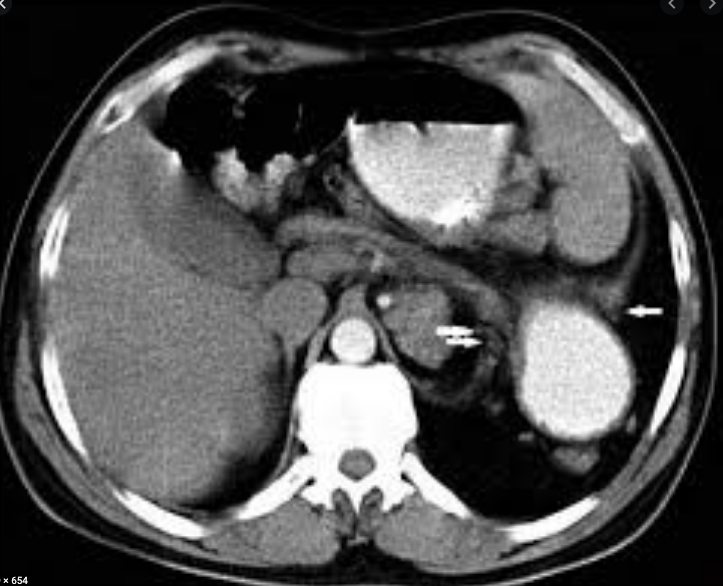
(Click Image to Enlarge)
delayed diaphragmatic hernia on CT imaging
Contributed by Mark Pellegrini (Public Domain)
Details

Updated:

8/8/2023 12:13:52 AM

References


[1]

Kosiński P, Wielgoś M. Congenital diaphragmatic hernia: pathogenesis, prenatal diagnosis and management - literature review. Ginekologia polska. 2017:88(1):24-30. doi: 10.5603/GP.a2017.0005. Epub     [PubMed PMID: 28157247]


[2]

Montedonico S, Nakazawa N, Puri P. Congenital diaphragmatic hernia and retinoids: searching for an etiology. Pediatric surgery international. 2008 Jul:24(7):755-61. doi: 10.1007/s00383-008-2140-x. Epub 2008 Apr 10     [PubMed PMID: 18401587]


[3]

Coste K, Beurskens LW, Blanc P, Gallot D, Delabaere A, Blanchon L, Tibboel D, Labbé A, Rottier RJ, Sapin V. Metabolic disturbances of the vitamin A pathway in human diaphragmatic hernia. American journal of physiology. Lung cellular and molecular physiology. 2015 Jan 15:308(2):L147-57     [PubMed PMID: 25416379]


[4]

Clugston RD, Zhang W, Alvarez S, de Lera AR, Greer JJ. Understanding abnormal retinoid signaling as a causative mechanism in congenital diaphragmatic hernia. American journal of respiratory cell and molecular biology. 2010 Mar:42(3):276-85. doi: 10.1165/rcmb.2009-0076OC. Epub 2009 May 15     [PubMed PMID: 19448158]

Level 3 (low-level) evidence

[5]

Dott MM, Wong LY, Rasmussen SA. Population-based study of congenital diaphragmatic hernia: risk factors and survival in Metropolitan Atlanta, 1968-1999. Birth defects research. Part A, Clinical and molecular teratology. 2003 Apr:67(4):261-7     [PubMed PMID: 12854661]


[6]

McGivern MR, Best KE, Rankin J, Wellesley D, Greenlees R, Addor MC, Arriola L, de Walle H, Barisic I, Beres J, Bianchi F, Calzolari E, Doray B, Draper ES, Garne E, Gatt M, Haeusler M, Khoshnood B, Klungsoyr K, Latos-Bielenska A, O'Mahony M, Braz P, McDonnell B, Mullaney C, Nelen V, Queisser-Luft A, Randrianaivo H, Rissmann A, Rounding C, Sipek A, Thompson R, Tucker D, Wertelecki W, Martos C. Epidemiology of congenital diaphragmatic hernia in Europe: a register-based study. Archives of disease in childhood. Fetal and neonatal edition. 2015 Mar:100(2):F137-44. doi: 10.1136/archdischild-2014-306174. Epub 2014 Nov 19     [PubMed PMID: 25411443]


[7]

Gallot D, Boda C, Ughetto S, Perthus I, Robert-Gnansia E, Francannet C, Laurichesse-Delmas H, Jani J, Coste K, Deprest J, Labbe A, Sapin V, Lemery D. Prenatal detection and outcome of congenital diaphragmatic hernia: a French registry-based study. Ultrasound in obstetrics & gynecology : the official journal of the International Society of Ultrasound in Obstetrics and Gynecology. 2007 Mar:29(3):276-83     [PubMed PMID: 17177265]


[8]

Burgos CM, Frenckner B. Addressing the hidden mortality in CDH: A population-based study. Journal of pediatric surgery. 2017 Apr:52(4):522-525. doi: 10.1016/j.jpedsurg.2016.09.061. Epub 2016 Sep 23     [PubMed PMID: 27745705]


[9]

Balayla J, Abenhaim HA. Incidence, predictors and outcomes of congenital diaphragmatic hernia: a population-based study of 32 million births in the United States. The journal of maternal-fetal & neonatal medicine : the official journal of the European Association of Perinatal Medicine, the Federation of Asia and Oceania Perinatal Societies, the International Society of Perinatal Obstetricians. 2014 Sep:27(14):1438-44. doi: 10.3109/14767058.2013.858691. Epub 2013 Nov 29     [PubMed PMID: 24156638]


[10]

Metkus AP, Filly RA, Stringer MD, Harrison MR, Adzick NS. Sonographic predictors of survival in fetal diaphragmatic hernia. Journal of pediatric surgery. 1996 Jan:31(1):148-51; discussion 151-2     [PubMed PMID: 8632269]


[11]

Jani J, Nicolaides KH, Keller RL, Benachi A, Peralta CF, Favre R, Moreno O, Tibboel D, Lipitz S, Eggink A, Vaast P, Allegaert K, Harrison M, Deprest J, Antenatal-CDH-Registry Group. Observed to expected lung area to head circumference ratio in the prediction of survival in fetuses with isolated diaphragmatic hernia. Ultrasound in obstetrics & gynecology : the official journal of the International Society of Ultrasound in Obstetrics and Gynecology. 2007 Jul:30(1):67-71     [PubMed PMID: 17587219]


[12]

Jani J, Nicolaides KH, Benachi A, Moreno O, Favre R, Gratacos E, Deprest J. Timing of lung size assessment in the prediction of survival in fetuses with diaphragmatic hernia. Ultrasound in obstetrics & gynecology : the official journal of the International Society of Ultrasound in Obstetrics and Gynecology. 2008 Jan:31(1):37-40     [PubMed PMID: 18069722]


[13]

Shue EH, Miniati D, Lee H. Advances in prenatal diagnosis and treatment of congenital diaphragmatic hernia. Clinics in perinatology. 2012 Jun:39(2):289-300. doi: 10.1016/j.clp.2012.04.005. Epub     [PubMed PMID: 22682380]

Level 3 (low-level) evidence

[14]

Büsing KA, Kilian AK, Schaible T, Endler C, Schaffelder R, Neff KW. MR relative fetal lung volume in congenital diaphragmatic hernia: survival and need for extracorporeal membrane oxygenation. Radiology. 2008 Jul:248(1):240-6. doi: 10.1148/radiol.2481070952. Epub     [PubMed PMID: 18566176]


[15]

Oluyomi-Obi T, Kuret V, Puligandla P, Lodha A, Lee-Robertson H, Lee K, Somerset D, Johnson J, Ryan G. Antenatal predictors of outcome in prenatally diagnosed congenital diaphragmatic hernia (CDH). Journal of pediatric surgery. 2017 May:52(5):881-888. doi: 10.1016/j.jpedsurg.2016.12.008. Epub 2016 Dec 21     [PubMed PMID: 28095996]


[16]

Jani JC, Nicolaides KH, Gratacós E, Valencia CM, Doné E, Martinez JM, Gucciardo L, Cruz R, Deprest JA. Severe diaphragmatic hernia treated by fetal endoscopic tracheal occlusion. Ultrasound in obstetrics & gynecology : the official journal of the International Society of Ultrasound in Obstetrics and Gynecology. 2009 Sep:34(3):304-10. doi: 10.1002/uog.6450. Epub     [PubMed PMID: 19658113]


[17]

Belfort MA, Olutoye OO, Cass DL, Olutoye OA, Cassady CI, Mehollin-Ray AR, Shamshirsaz AA, Cruz SM, Lee TC, Mann DG, Espinoza J, Welty SE, Fernandes CJ, Ruano R. Feasibility and Outcomes of Fetoscopic Tracheal Occlusion for Severe Left Diaphragmatic Hernia. Obstetrics and gynecology. 2017 Jan:129(1):20-29. doi: 10.1097/AOG.0000000000001749. Epub     [PubMed PMID: 27926636]

Level 2 (mid-level) evidence

[18]

Al-Maary J, Eastwood MP, Russo FM, Deprest JA, Keijzer R. Fetal Tracheal Occlusion for Severe Pulmonary Hypoplasia in Isolated Congenital Diaphragmatic Hernia: A Systematic Review and Meta-analysis of Survival. Annals of surgery. 2016 Dec:264(6):929-933     [PubMed PMID: 26910202]

Level 1 (high-level) evidence

[19]

Hutcheon JA, Butler B, Lisonkova S, Marquette GP, Mayer C, Skoll A, Joseph KS. Timing of delivery for pregnancies with congenital diaphragmatic hernia. BJOG : an international journal of obstetrics and gynaecology. 2010 Dec:117(13):1658-62     [PubMed PMID: 21125710]


[20]

Zhang X, Kramer MS. Variations in mortality and morbidity by gestational age among infants born at term. The Journal of pediatrics. 2009 Mar:154(3):358-62, 362.e1. doi: 10.1016/j.jpeds.2008.09.013. Epub 2008 Oct 31     [PubMed PMID: 18950794]


[21]

Sengupta S, Carrion V, Shelton J, Wynn RJ, Ryan RM, Singhal K, Lakshminrusimha S. Adverse neonatal outcomes associated with early-term birth. JAMA pediatrics. 2013 Nov:167(11):1053-9. doi: 10.1001/jamapediatrics.2013.2581. Epub     [PubMed PMID: 24080985]


[22]

Lefebvre C, Rakza T, Weslinck N, Vaast P, Houfflin-Debarge V, Mur S, Storme L, French CDH Study Group. Feasibility and safety of intact cord resuscitation in newborn infants with congenital diaphragmatic hernia (CDH). Resuscitation. 2017 Nov:120():20-25. doi: 10.1016/j.resuscitation.2017.08.233. Epub 2017 Aug 30     [PubMed PMID: 28860014]

Level 2 (mid-level) evidence

[23]

Foglia EE, Ades A, Hedrick HL, Rintoul N, Munson DA, Moldenhauer J, Gebb J, Serletti B, Chaudhary A, Weinberg DD, Napolitano N, Fraga MV, Ratcliffe SJ. Initiating resuscitation before umbilical cord clamping in infants with congenital diaphragmatic hernia: a pilot feasibility trial. Archives of disease in childhood. Fetal and neonatal edition. 2020 May:105(3):322-326. doi: 10.1136/archdischild-2019-317477. Epub 2019 Aug 28     [PubMed PMID: 31462406]

Level 2 (mid-level) evidence

[24]

Snoek KG, Capolupo I, van Rosmalen J, Hout Lde J, Vijfhuize S, Greenough A, Wijnen RM, Tibboel D, Reiss IK, CDH EURO Consortium. Conventional Mechanical Ventilation Versus High-frequency Oscillatory Ventilation for Congenital Diaphragmatic Hernia: A Randomized Clinical Trial (The VICI-trial). Annals of surgery. 2016 May:263(5):867-74. doi: 10.1097/SLA.0000000000001533. Epub     [PubMed PMID: 26692079]

Level 1 (high-level) evidence

[25]

Van Meurs K, Congenital Diaphragmatic Hernia Study Group. Is surfactant therapy beneficial in the treatment of the term newborn infant with congenital diaphragmatic hernia? The Journal of pediatrics. 2004 Sep:145(3):312-6     [PubMed PMID: 15343181]


[26]

Lally KP, Lally PA, Langham MR, Hirschl R, Moya FR, Tibboel D, Van Meurs K, Congenital Diaphragmatic Hernia Study Group. Surfactant does not improve survival rate in preterm infants with congenital diaphragmatic hernia. Journal of pediatric surgery. 2004 Jun:39(6):829-33     [PubMed PMID: 15185206]


[27]

. Inhaled nitric oxide and hypoxic respiratory failure in infants with congenital diaphragmatic hernia. The Neonatal Inhaled Nitric Oxide Study Group (NINOS). Pediatrics. 1997 Jun:99(6):838-45     [PubMed PMID: 9190553]


[28]

Snoek KG, Reiss IK, Greenough A, Capolupo I, Urlesberger B, Wessel L, Storme L, Deprest J, Schaible T, van Heijst A, Tibboel D, CDH EURO Consortium. Standardized Postnatal Management of Infants with Congenital Diaphragmatic Hernia in Europe: The CDH EURO Consortium Consensus - 2015 Update. Neonatology. 2016:110(1):66-74. doi: 10.1159/000444210. Epub 2016 Apr 15     [PubMed PMID: 27077664]

Level 3 (low-level) evidence

[29]

Puligandla PS, Grabowski J, Austin M, Hedrick H, Renaud E, Arnold M, Williams RF, Graziano K, Dasgupta R, McKee M, Lopez ME, Jancelewicz T, Goldin A, Downard CD, Islam S. Management of congenital diaphragmatic hernia: A systematic review from the APSA outcomes and evidence based practice committee. Journal of pediatric surgery. 2015 Nov:50(11):1958-70. doi: 10.1016/j.jpedsurg.2015.09.010. Epub 2015 Sep 21     [PubMed PMID: 26463502]

Level 1 (high-level) evidence

[30]

Nio M, Haase G, Kennaugh J, Bui K, Atkinson JB. A prospective randomized trial of delayed versus immediate repair of congenital diaphragmatic hernia. Journal of pediatric surgery. 1994 May:29(5):618-21     [PubMed PMID: 8035269]

Level 1 (high-level) evidence

[31]

Kays DW. ECMO in CDH: Is there a role? Seminars in pediatric surgery. 2017 Jun:26(3):166-170. doi: 10.1053/j.sempedsurg.2017.04.006. Epub 2017 Apr 25     [PubMed PMID: 28641755]


[32]

Tsao K, Lally PA, Lally KP, Congenital Diaphragmatic Hernia Study Group. Minimally invasive repair of congenital diaphragmatic hernia. Journal of pediatric surgery. 2011 Jun:46(6):1158-64. doi: 10.1016/j.jpedsurg.2011.03.050. Epub     [PubMed PMID: 21683215]


[33]

Pierro A. Hypercapnia and acidosis during the thoracoscopic repair of oesophageal atresia and congenital diaphragmatic hernia. Journal of pediatric surgery. 2015 Feb:50(2):247-9. doi: 10.1016/j.jpedsurg.2014.11.006. Epub 2014 Nov 7     [PubMed PMID: 25638611]


[34]

Canadian Congenital Diaphragmatic Hernia Collaborative, Puligandla PS, Skarsgard ED, Offringa M, Adatia I, Baird R, Bailey M, Brindle M, Chiu P, Cogswell A, Dakshinamurti S, Flageole H, Keijzer R, McMillan D, Oluyomi-Obi T, Pennaforte T, Perreault T, Piedboeuf B, Riley SP, Ryan G, Synnes A, Traynor M. Diagnosis and management of congenital diaphragmatic hernia: a clinical practice guideline. CMAJ : Canadian Medical Association journal = journal de l'Association medicale canadienne. 2018 Jan 29:190(4):E103-E112. doi: 10.1503/cmaj.170206. Epub     [PubMed PMID: 29378870]

Level 1 (high-level) evidence

[35]

Chandrasekharan PK, Rawat M, Madappa R, Rothstein DH, Lakshminrusimha S. Congenital Diaphragmatic hernia - a review. Maternal health, neonatology and perinatology. 2017:3():6. doi: 10.1186/s40748-017-0045-1. Epub 2017 Mar 11     [PubMed PMID: 28331629]


[36]

Kumar VH. Current Concepts in the Management of Congenital Diaphragmatic Hernia in Infants. The Indian journal of surgery. 2015 Aug:77(4):313-21. doi: 10.1007/s12262-015-1286-8. Epub 2015 May 30     [PubMed PMID: 26702239]


[37]

Harting MT, Lally KP. The Congenital Diaphragmatic Hernia Study Group registry update. Seminars in fetal & neonatal medicine. 2014 Dec:19(6):370-5. doi: 10.1016/j.siny.2014.09.004. Epub 2014 Oct 11     [PubMed PMID: 25306471]


[38]

Hagadorn JI, Brownell EA, Herbst KW, Trzaski JM, Neff S, Campbell BT. Trends in treatment and in-hospital mortality for neonates with congenital diaphragmatic hernia. Journal of perinatology : official journal of the California Perinatal Association. 2015 Sep:35(9):748-54. doi: 10.1038/jp.2015.46. Epub 2015 May 7     [PubMed PMID: 25950919]


[39]

Barrière F,Michel F,Loundou AD,Fouquet V,Kermorvant E,Blanc S,Carricaburu E,Desrumaux A,Pidoux O,Arnaud A,Berte N,Blanc T,Lavrand F,Levard G,Rayet I,Samperiz S,Schneider A,Marcoux MO,Winer N,Chaussy Y,Datin-Dorriere V,Ballouhey Q,Binet A,Muszynski C,Breaud J,Garenne A,Storme L,Boubnova J, One-Year Outcome for Congenital Diaphragmatic Hernia: Results From the French National Register. The Journal of pediatrics. 2018 Feb;     [PubMed PMID: 29212620]


[40]

American Academy of Pediatrics Section on Surgery, American Academy of Pediatrics Committee on Fetus and Newborn, Lally KP, Engle W. Postdischarge follow-up of infants with congenital diaphragmatic hernia. Pediatrics. 2008 Mar:121(3):627-32. doi: 10.1542/peds.2007-3282. Epub     [PubMed PMID: 18310215]


[41]

Michel F, Baumstarck K, Gosselin A, Le Coz P, Merrot T, Hassid S, Chaumoître K, Berbis J, Martin C, Auquier P, PACA Group Research for Quality of Life of children with a congenital diaphragmatic hernia. Health-related quality of life and its determinants in children with a congenital diaphragmatic hernia. Orphanet journal of rare diseases. 2013 Jun 20:8():89. doi: 10.1186/1750-1172-8-89. Epub 2013 Jun 20     [PubMed PMID: 23786966]

Level 2 (mid-level) evidence