Continuing Education Activity
Tinea cruris, also known as jock itch, is an infection involving the genital, pubic, perineal, and perianal skin caused by pathogenic fungi known as dermatophytes. The evaluation and treatment of tinea cruris are discussed in the activity. This activity reviews the role of the healthcare team in improving care for patients with this condition. The activity will also highlight evidence-based therapeutics and lifestyle modifications to avoid common mistakes and mistreatments associated with the management of dermatophyte infections.
Objectives:
- Identify the etiology of tinea cruris.
- Outline the evaluation of tinea cruris.
- Review the treatment options available for tinea cruris.
- Describe interprofessional team strategies to improve the management of patients affected by tinea cruris.
Introduction
Tinea cruris, also known as jock itch, is an infection involving the genital, pubic, perineal, and perianal skin caused by pathogenic fungi known as dermatophytes.[1][2][3][4][5] These dermatophytes affect keratinized structures such as hair and the epidermis' stratum corneum resulting in a characteristic rash. Intertriginous areas are hospitable environments for fungus, with sweating, maceration, and alkaline pH being responsible for the groin's predilection for infection.[1][3][4][5]
While tinea infections are often classified by the location of the body affected, they are also organized according to the responsible organism's primary source and mode of transmission.[3] Geophilic, zoophilic, and anthropophilic fungi are found in and transmitted by soil, animals, and humans, respectively.[1][3][6][7] Autoinfection of dermatophytes is also possible and especially crucial in tinea cruris as foot-to-groin spread can occur.[1]
Etiology
Tinea cruris is caused by dermatophytes belonging to three genera, Trichophyton, Epidermophyton, and Microsporum.[5] Trichophyton rubrum has been isolated most commonly and remains the most frequent cause of tinea cruris worldwide; however, most studies do recognize the increasing prevalence of Trichophyton mentagrophytes and other organisms in certain regions.[1][2][3][4][7] Several risk factors have been identified that predispose an individual to tinea cruris, including excessive perspiration, occlusive clothing, improper hygiene, diabetes mellitus, immunocompromise, and lower socioeconomic status.[1][2][3][6]
Athletes, especially those involved in contact sports, may be more likely to contract tinea infections.[1] Genetics can also make a patient more susceptible to dermatophytes.[3][6] Of all these factors, perspiration appears to be the most influential variable in the development of infection.[1][8] In India, an area affected disproportionately often with dermatophytes, a study was conducted in response to the increasing frequency of and decreased treatment efficacy for local tinea infections.[9] Diabetes mellitus, family members with tinea, and personal history of cooking food were found to be positively associated with chronic and relapsing disease.[9]
Epidemiology
Cutaneous mycoses, including tinea cruris, affect 20 to 25 percent of the world's population.[3][10] Developing and tropical countries have an increased prevalence of dermatophyte infections secondary to high temperatures and increased humidity.[1][11] In the United States, there have been an estimated 29.4 million cases of superficial fungal infections and over 51 million reported physician visits.[6] Adolescent and adult males comprise the majority of patients seen for tinea cruris and are affected by the disorder with increased frequency.[1][2][11] Worldwide increases in the occurrence of dermatophytoses and the discovery of recalcitrant infections have caused global concern.[3][4][5][9]
Pathophysiology
A simplified explanation of the complex and not well-understood pathophysiology of dermatophytes includes the organism's use of proteinases to digest keratin found in the skin's stratum corneum.[6][12][7]
Histopathology
While not always necessary for diagnosis, a potassium hydroxide (KOH) preparation may reveal histology consistent with dermatophyte infections.[1][2][3] Characteristic findings include branching or non-branching septate hyphae and possible arthroconidiospores.[2][3]
History and Physical
Patients with tinea cruris present complaining of a pruritic rash involving the groin.[1][2] The area may be irritated and painful if maceration is present, and secondary infections may result in inflammation and discomfort.[2] Duration of symptoms, previous occurrences, similar rashes in other locations, and past treatments should be elucidated. Individuals should be questioned about any history of diabetes, immunocompromise, renal disease, or hepatic dysfunction.[13] Clinicians should inquire about excessive sweating, wardrobe changes, and personal hygiene habits. A review of the patient's environmental and occupation exposures, including people, pets, animals, and contaminated soil, may be contributory.[13]
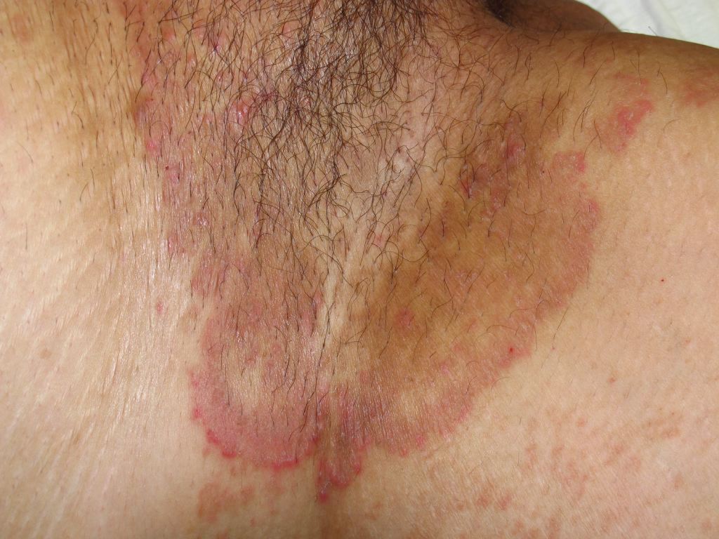
On physical examination, an erythematous, scaly, annular plaque with a raised leading edge and central clearing can be visualized, extending anywhere from the groin, upper thigh, and perineum to the perianal region.[1][2]
Evaluation
In most cases, tinea cruris can be diagnosed clinically; however, several tests exist to investigate a rash of the groin with unknown etiology.[1][2][3] Potassium hydroxide (KOH) preparations, skin biopsy with periodic acid-Schiff (PAS) stain, and fungal cultures on Sabouraud’s agar media can be utilized when the diagnosis is in question or the case of recurrent or recalcitrant episodes.[1][2][3]
In general, the sample should be obtained from the leading edge of the lesion to ensure an adequate collection of infected scales.[1] Potassium hydroxide (KOH) mounts are generally procured with a scalpel scraping technique; however, new studies have indicated that utilizing a cellophane adhesive tape method may simplify the collection process, facilitate transportation, provide a higher quality sample, and improve slide preservation time.[14]
Treatment / Management
Antifungals utilized in treating dermatophytoses, including tinea cruris, target the synthesis of ergosterol, a vital component of fungal plasma membranes.[4] Management strategies are similar worldwide; however, some countries have specific guidelines based on their region's fungal profile.[6] Topical therapies are effective and usually preferred.[2][4][6] Allylamines (terbinafine, butenafine, naftifine) and azoles (clotrimazole, miconazole, sulconazole, oxiconazole, econazole, ketoconazole) are the mainstays of topical treatment regimens. They are generally prescribed once or twice daily for two to four weeks.[1][3][6]
Deciding which agent to use should be based on patient compliance, cost, and medication accessibility, as there is insufficient data to directly compare the effectiveness of individual drugs and classes.[8][15][16][13] Allylamines have a potentially shorter treatment timeline, have demonstrated lower relapse rates, and their metabolism is independent of the cytochrome p450 system.[8][13] Azoles are not as costly as allylamines but often require a longer treatment duration.[8] One newer topical azole, luliconazole, requires only once daily application for one week and may improve patient compliance through a more convenient dosing schedule.[6] Ciclopirox olamine is an older topical preparation with a unique mechanism of action compared to the commonly used allylamines and azoles.[17] Recent studies have demonstrated a number of benefits to ciclopirox therapy; however, it remains an underutilized antifungal medication.[17]
Oral preparations exist to manage tinea cruris and are indicated for chronic, recurrent, and recalcitrant disease.[1][2][4][6] Extensive or diffuse rashes and patients with immunocompromise may also require systemic treatments.[4] Stratum corneum penetration and concentration maintenance, keratin adherence, patient tolerance, and a minimal drug interaction profile are hallmarks of the ideal systemic medication for dermatophytoses.[4] Oral terbinafine and itraconazole have favorable characteristics for dermatophyte management and are the most often prescribed.[4][15][16][13] Fluconazole has demonstrated efficacy in treating tinea cruris; however, it is not preferred due to its poor keratin adherence and prolonged treatment duration.[4] Griseofulvin has similar pharmacokinetic limitations as fluconazole, and it is more appropriately utilized in the management of tinea capitis than tinea cruris.[1][4][6] Due to its potential for hepatotoxicity, oral ketoconazole is no longer recommended for dermatophytoses.[1][2][4][15][16] Topical antifungal therapies may be used as an adjunct in patients requiring systemic treatment. Topical and oral antibiotics can be administered when a secondary bacterial infection is present.[3][13]
A commonly used alternative treatment known as Whitfield's ointment has insufficient evidence of benefit.[18] Nystatin, a frequently utilized treatment for cutaneous candida infections, is ineffective for managing dermatophytoses such as tinea cruris.[2] Combined topical corticosteroid and antifungal therapy remain controversial. Some studies have demonstrated improved cure rates with concomitant steroid and antifungal topical applications; however, these results were based on low-quality evidence.[18] While steroids may improve acute inflammation and itching, they may also fortify dermatophyte's plasma membranes rendering antifungal medications less effective.[4] Steroids also activate fungal metabolism and can potentially facilitate the worsening of the primary infection.[4] Tinea incognito is another possible complication of steroid administration where the typical presentation of tinea is masked, and diagnosis is delayed.[1] Currently, topical steroids are not recommended as part of an evidence-based tinea cruris treatment regimen.[2][3][2][18][13]
Differential Diagnosis
The differential diagnosis for tinea cruris includes several other dermatologic conditions affecting the groin with similar presentations. Candidiasis, erythrasma, psoriasis, and seborrheic dermatitis exhibit comparable signs and symptoms and are most commonly confused with the fungal groin infection. Unlike tinea cruris, candidal intertrigo frequently affects women, and the rash may involve the scrotum and penis in males.[1][2] Satellite lesions and erythema without central clearing are indicative of candida as opposed to tinea. The rash of erythrasma lacks an active border and demonstrates coral-red fluorescence on Wood lamp examination. Psoriasis will likely manifest in other areas in addition to the crural region. Seborrheic dermatitis presents with greasy scales on an erythematous base.[2]
Prognosis
Patients with tinea cruris who undergo an appropriate treatment course experience cure rates ranging from 80 to 90 percent.[19]
Complications
Failure of therapy and recurrence are the most likely complications of tinea cruris. They have been attributed to reinfection from close contacts, autoinfection from separate body locations, infection by uncommon species such as zoonoses, misdiagnosis, drug resistance, and non-adherence to the management plan.[3] Steroid use may suppress the physical signs of tinea cruris, making the diagnosis more difficult. Also, chronic application can result in skin atrophy and telangiectasias.[4] Secondary bacterial infection is another potential complication of tinea cruris.[3] Majocchi’s granuloma is an uncommon complication of cutaneous fungal infections in which dermatophytes disseminate into the subcutaneous tissue secondary to skin breakdown, immunosuppression, or topical steroid use resulting in a deep, inflammatory disease.[3][20] A dermatophytid reaction may result in an allergic response at a separate location from the original tinea site.[21]
Consultations
Consultation with dermatology or infectious disease may be warranted in recurrent or recalcitrant cases.[3]
Deterrence and Patient Education
Patient education should focus on non-pharmacologic measures to treat and prevent recurrences of tinea cruris. Loose-fitting, non-restrictive garments should be encouraged, and clothing should not be donned until the underlying skin is completely dried.[3] Because autoinfection originating from tinea pedis may occur, patients should avoid walking barefoot, and protective footwear should be used in public facilities.[3][13] Identification and treatment of potentially infected contacts, whether human or animal, should be undertaken.[13] Self-treatment with over-the-counter antifungals and steroids should be discouraged as this may result in resistant or chronic infections and can hinder a clinician's ability to make an accurate and timely diagnosis.[4]
Enhancing Healthcare Team Outcomes
Tinea cruris is a prevalent pathology with a worldwide distribution and an extensive history of affecting human populations. Despite our familiarity with the condition, there has been limited research specific to this subset of dermatophyte infections. Renewed interest has emerged with the development of recalcitrant infections and concern over fungal resistance. Self-treatment with easy-to-access over-the-counter topical antifungal and steroid preparations have been implicated as a probable cause of the observed decrease in treatment efficacy. In the context of fungal resistance, newly developed formulations such as luliconazole and underutilized, older agents like ciclopirox may be beneficial.
With regard to corticosteroids, there is conflicting data on the appropriateness of their use for tinea cruris. Current management principles and guidelines label them as mistreatment; however, continued investigations of their utility are underway. If a practitioner believes topical steroids may benefit a patient, close supervision should be maintained throughout the treatment course with continued consideration of known adverse event potential for the individual and public health outcomes.
With an understanding of the changing landscape of this common condition, and by implementing an interprofessional healthcare team approach including primary care clinicians (including PAs and NPs), dermatologists, infectious disease specialists, pharmacists, and nursing, patient care and public health may be improved through targeted, conscientious mycological treatment, patient education, and antifungal stewardship. [Level 2] This interprofessional paradigm should lead to the best outcomes with minimal to no adverse events. [Level 5]