Mallory Bodies


Introduction

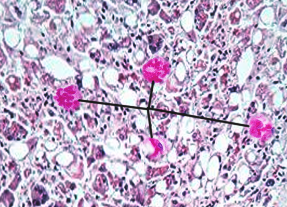
Mallory bodies (MBs), also known as Mallory-Denk bodies (MDBs), are cytoplasmic hyaline inclusions of hepatocytes, once thought to be specific for alcoholic hepatitis. MBs are not seen in other liver diseases, which include nonalcoholic steatohepatitis (NASH), cholestatic liver diseases, primary biliary cirrhosis (PBC), and hepatocellular carcinoma (HCC).[1][2] In 1911, Frank Burr Mallory discovered these bodies while examining the hepatocytes of patients with alcoholic hepatitis. In 1975, Helmut Denk found the first animal model of MBs by feeding mice griseofulvin, which allowed further research on MBs, resulting in the renaming of MDBs.[1] See Image. Mallory Bodies.

Issues of Concern

The development of MDBs indicates that intracellular protein quality has failed.[3]

Causes

In general, MDB formation presents in liver diseases. Those include hepatitis B and C, alcoholic liver disease, NAFLD, HCC, PBC, chronic cholestasis, focal nodular hyperplasia, Wilson disease, and copper toxicosis.[4]

However, it also presents in glucocorticoid therapy, intestinal bypass surgery for obesity, Weber-Christian disease, von Gierke disease, radiation pneumonitis, asbestosis, amiodarone, a beta lipoproteinemia, porphyria cutanea tarda, antitrypsin deficiency, Indian childhood cirrhosis, perhexiline maleate hepatitis, cirrhosis, 2′3′-dideoxyinosine diethylaminoetheoxyhex-estriol-induced hepatitis, hepatic adenoma, sclerosing hyaline necrosis in Bloom syndrome, congenital fibrosis.[5]

Rarely do MDBs present in nonhepatic cells, but examples do exist, which present in asbestosis, renal cell carcinoma, and type 2 pneumocytes.[1]

Anatomical Pathology

P62 (a sequestosome), ubiquitin, and intermediate filament proteins keratins 8 and 18 (K8/18) are the significant elements that make up a Mallory body.[1] In normal conditions, K8 and 18 are present in a 1 to 1 ratio.[6] Protein misfolding, proteasome overload, a ratio of K8 greater than K18, and including transamidation of K8 contribute to MDB formation [7]. K8 is insoluble and not easily amenable to degradation, thus resulting in Mallory-Denk bodies. While overexpression of K18 inhibits MDB formation.[1] K8 is more likely to change its helical shape to cross-beta sheets resulting in Mallory-Denk body formation. Those that do not have K8 do not make MDB thus linking cross-beta sheets as a necessity for MDB development.[8]

Clinical Pathology

Cytoplasmic inclusion bodies, intracytoplasmic hyaline bodies (IHB), and MDBs can be present in HCC. MDB was linked to the steatohepatitis variant of HCC, whereas intracytoplasmic hyaline bodies (IHBs) were not. However, the presence of IHB correlates with shorter survival than MDBs.[9] MDBs are distinguishable from other cytoplasmic inclusions such as IHBs. For example, MDBs consists of ubiquitin, p62, and keratin, whereas IHBs only consist of ubiquitin and p62. IHBs have intracytoplasmic hyaline bodies been found to be a morphological precursor to MDBs.[1]

Morphology

MDBs are cytoplasmic inclusions that are predominantly filamentous, ranging from a diameter of 3 to 24 nm versus 10 nm of classical IF.[1] Mallory-Denk bodies can be classified as type I (parallels filaments), II (randomly orient filaments), or III (granular and amorphous). Type II occurs in the periphery while type III occurs around the center. MDB occur in ballooned hepatocytes. They are visible via hematoxylin-eosin stain, however immunohistochemical staining to CK or ubiquitin is more sensitive. Pericellular fibrosis and neutrophil tend to surround the hepatocytes with MDB are called satellitosis.[1][10] Distribution of MDB in the cells suggests different stages of their formation. For example, small cytoplasmic globular structures are early, large para-nuclear inclusions are mature, and the ones located in the periphery are considered old.[1] MDB can be found in different zones of the liver and varies depending on the disease process. In primary biliary cholangitis and Wilson disease, it is seen in zone 1, whereas in ASH and NAFLD it is present in zone 3.[4]

Mechanisms

As Denk had used griseofulvin in his study to create MDB, recent studies have used diethyl-1, 4-dihydro-2, 4, 6-trimethyl-3, 5-pyridine dicarboxylate (DDC).[11] DDC induced MDB faster than griseofulvin. They have proposed that there are three mechanisms in the formation of MDB.

Before MDB occur, ballooning of hepatocytes occurs first, which are the response of oxidative stress, such as abnormal protein (heat shock proteins) or fat result in water accumulation into the hepatic cytoplasm.[4]  HSP formation indicated cellular dysfunction

The first mechanism is that epigenetics (acetylation, methylation, and ubiquitination of histones) contribute to hepatocyte memory during MDB formation.[12]

The second pathway is the shift from the 26s proteasome to the immunoproteasome. The 26S proteasome plays a part in degradation intracellular (cytosolic, nuclear and membrane) proteins.[13]  The way DDC induces MDB is that it changes 26s proteasome to immunoproteasome, thus resulting in protein accumulation.[7] When chaperones (i.e., heat shock protein 70), proteins that refold misfolded proteins, become flawed, inclusion bodies such as MDB form. Alcohol and DDC cause chaperons to become defected. Misfolded proteins are targeted by ubiquitination, which signals the molecule via p62 for proteasomal and autophagic degradation.[1]

The third is the chronic activation of the Toll-like signaling pathways which stimulate proinflammatory and cell growth pathways. IFN-gamma and TNF-alpha stimulate TLR receptors, which cause up-regulation of growth factor resulting in the proliferation of MDB forming cells. Drugs as well can create a shift to the formation of the immunoproteasome rather than the 26s proteasome.[7]

DDC stimulates tumor necrosis factor-alpha (TNF-alpha) and interferon-gamma (IFN-gamma) expression, which in turn activates the toll-like receptor (TLR). Proinflammatory cytokines TNF-alpha and IFN-gamma via TLR signaling cause up-regulation of the immunoproteasome and down-regulation of the 26s proteasome which results in undigested proteins and MDB formation.[7]

Clinicopathologic Correlations

Clinical findings of ASH (jaundice, leukocytosis, fever) may demonstrate classical histological finding of steatosis; however few or no MDB.  The opposite may be true, as well.[1] MDB is noted to be fewer and less developed in NASH, which tends to be less severe of the two. NASH in children often do not have MDB, which suggests aging may play a role as well.[1] MDB as a NAFLD marker depends on the histological scoring system. Matteoni et al. emphasized MDB while Kleiner et al., which is a more accepted scoring system, did not.[1] Some recommend for KRT8/K18 ratio to be a biomarker for HCC.[6] In alcohol using patient, a sensitive and specific test to detect MDB is Ub/immunostaining.[4]

Inclusions do occur in the liver, muscle, and neural tissues which suggest a common intracellular network of protein synthesis and degradation along with responsiveness to stressors. MBD can be considered a protective mechanism of cell injury or rather a step in the pathogenesis of liver damage.[4]

Clinical Significance

Abnormal protein folding is known to cause other diseases, including Alzheimer disease.  In a recent literature review,[5] there is common ground in the mechanism involved Alzheimer disease and MDB formation. Due to its similarities, the proposal was that future studies should test betaine or SAMe (methyl donors) effect on Alzheimer disease, as they have shown to prevent MDB formation. Beta sheets which are present in amyloid deposits are also present in MDB.[1]

Denk, in his publication, notes that MDB formation occurs secondary to abnormal hyperkeratosis, which has links to Vitamin A deficiency.  In his animal study, Vitamin A level was decreased from 30% at day 12 of treatment with Griseofulvin down to13% at 60 days.[14]

In patients with alcoholic liver disease, about 70 to 75% have MDB.  However, in patients with NAFLD, MDB ranges from 7% to 90%.[4] The wide range of MDB in NAFLD is likely the result of not having the specific amount which qualifies as excessive alcohol use, which would categorize the patient as alcoholic rather than non-alcoholic.

In severe alcoholic hepatitis, non-responders to corticosteroids had high histopathological findings of ballooning degeneration and MDB suggesting that histopathology findings can identify those who may respond to corticosteroids.[15] Studies have shown that autophagy contributes to MDB degradation along with other intracellular compartments.[16][17][18] Fenofibrate, a fibric acid derivative that lowers cholesterol, prevented MDB formation by preventing disruption of intermediate filament.[19]

Methyl donors like Betaine and SAMe prevent MDB formation. SAMe prevents demethylation of histones, which occurs with DDC while Betaine prevents MDB formation by preventing the changes in methionine metabolism and by betaine-homocysteine methyltransferase (BHMT) methionine increases from homocysteine.[12][7][20] TLR and p62 pathways are prevented by betaine and SAMe as well.


(Click Image to Enlarge)
<p>Mallory Bodies

Mallory Bodies. Mallory bodies, also known as Mallory-Denk bodies, are cytoplasmic hyaline inclusions of hepatocytes, once thought to be specific for alcoholic hepatitis.


Contributed by O Chaigasame, MD

Details

Author

Emir Kurtovic

Editor:

Faten Limaiem

Updated:

9/12/2022 9:18:33 PM

References


[1]

Zatloukal K, French SW, Stumptner C, Strnad P, Harada M, Toivola DM, Cadrin M, Omary MB. From Mallory to Mallory-Denk bodies: what, how and why? Experimental cell research. 2007 Jun 10:313(10):2033-49     [PubMed PMID: 17531973]


[2]

Aishima S, Fujita N, Mano Y, Iguchi T, Taketomi A, Maehara Y, Oda Y, Tsuneyoshi M. p62+ Hyaline inclusions in intrahepatic cholangiocarcinoma associated with viral hepatitis or alcoholic liver disease. American journal of clinical pathology. 2010 Sep:134(3):457-65. doi: 10.1309/AJCP53YVVJCNDZIR. Epub     [PubMed PMID: 20716803]


[3]

Afifiyan N, Tillman B, French BA, Sweeny O, Masouminia M, Samadzadeh S, French SW. The role of Tec kinase signaling pathways in the development of Mallory Denk Bodies in balloon cells in alcoholic hepatitis. Experimental and molecular pathology. 2017 Oct:103(2):191-199. doi: 10.1016/j.yexmp.2017.09.001. Epub 2017 Sep 19     [PubMed PMID: 28935395]


[4]

Basaranoglu M, Turhan N, Sonsuz A, Basaranoglu G. Mallory-Denk Bodies in chronic hepatitis. World journal of gastroenterology. 2011 May 7:17(17):2172-7. doi: 10.3748/wjg.v17.i17.2172. Epub     [PubMed PMID: 21633525]


[5]

French SW, Mendoza AS, Peng Y. The mechanisms of Mallory-Denk body formation are similar to the formation of aggresomes in Alzheimer's disease and other neurodegenerative disorders. Experimental and molecular pathology. 2016 Jun:100(3):426-33. doi: 10.1016/j.yexmp.2016.03.010. Epub 2016 Apr 9     [PubMed PMID: 27068270]


[6]

Golob-Schwarzl N, Bettermann K, Mehta AK, Kessler SM, Unterluggauer J, Krassnig S, Kojima K, Chen X, Hoshida Y, Bardeesy NM, Müller H, Svendova V, Schimek MG, Diwoky C, Lipfert A, Mahajan V, Stumptner C, Thüringer A, Fröhlich LF, Stojakovic T, Nilsson KPR, Kolbe T, Rülicke T, Magin TM, Strnad P, Kiemer AK, Moriggl R, Haybaeck J. High Keratin 8/18 Ratio Predicts Aggressive Hepatocellular Cancer Phenotype. Translational oncology. 2019 Feb:12(2):256-268. doi: 10.1016/j.tranon.2018.10.010. Epub 2018 Nov 12     [PubMed PMID: 30439626]


[7]

French SW, Bardag-Gorce F, Li J, French BA, Oliva J. Mallory-Denk body pathogenesis revisited. World journal of hepatology. 2010 Aug 27:2(8):295-301. doi: 10.4254/wjh.v2.i8.295. Epub     [PubMed PMID: 21161012]


[8]

Mahajan V, Klingstedt T, Simon R, Nilsson KP, Thueringer A, Kashofer K, Haybaeck J, Denk H, Abuja PM, Zatloukal K. Cross β-sheet conformation of keratin 8 is a specific feature of Mallory-Denk bodies compared with other hepatocyte inclusions. Gastroenterology. 2011 Sep:141(3):1080-1090.e1-7. doi: 10.1053/j.gastro.2011.05.039. Epub 2011 May 27     [PubMed PMID: 21699779]


[9]

Aigelsreiter A, Neumann J, Pichler M, Halasz J, Zatloukal K, Berghold A, Douschan P, Rainer F, Stauber R, Haybaeck J, Denk H, Lackner C. Hepatocellular carcinomas with intracellular hyaline bodies have a poor prognosis. Liver international : official journal of the International Association for the Study of the Liver. 2017 Apr:37(4):600-610. doi: 10.1111/liv.13325. Epub 2017 Jan 12     [PubMed PMID: 27885796]


[10]

Denk H, Stumptner C, Zatloukal K. Mallory bodies revisited. Journal of hepatology. 2000 Apr:32(4):689-702     [PubMed PMID: 10782920]


[11]

Yuan QX, Marceau N, French BA, Fu P, French SW. Mallory body induction in drug-primed mouse liver. Hepatology (Baltimore, Md.). 1996 Sep:24(3):603-12     [PubMed PMID: 8781332]


[12]

Bardag-Gorce F, Oliva J, Villegas J, Fraley S, Amidi F, Li J, Dedes J, French B, French SW. Epigenetic mechanisms regulate Mallory Denk body formation in the livers of drug-primed mice. Experimental and molecular pathology. 2008 Apr:84(2):113-21. doi: 10.1016/j.yexmp.2007.12.004. Epub 2008 Jan 11     [PubMed PMID: 18281034]


[13]

Livneh I, Cohen-Kaplan V, Cohen-Rosenzweig C, Avni N, Ciechanover A. The life cycle of the 26S proteasome: from birth, through regulation and function, and onto its death. Cell research. 2016 Aug:26(8):869-85. doi: 10.1038/cr.2016.86. Epub 2016 Jul 22     [PubMed PMID: 27444871]


[14]

Denk H, Franke WW, Eckerstorfer R, Schmid E, Kerjaschki D. Formation and involution of Mallory bodies ("alcoholic hyalin") in murine and human liver revealed by immunofluorescence microscopy with antibodies to prekeratin. Proceedings of the National Academy of Sciences of the United States of America. 1979 Aug:76(8):4112-6     [PubMed PMID: 386356]


[15]

Shasthry SM, Rastogi A, Bihari C, Vijayaraghavan R, Arora V, Sharma MK, Sarin SK. Histological activity score on baseline liver biopsy can predict non-response to steroids in patients with severe alcoholic hepatitis. Virchows Archiv : an international journal of pathology. 2018 Apr:472(4):667-675. doi: 10.1007/s00428-018-2330-4. Epub 2018 Mar 7     [PubMed PMID: 29516163]


[16]

Harada M. Autophagy is involved in the elimination of intracellular inclusions, Mallory-Denk bodies, in hepatocytes. Medical molecular morphology. 2010 Mar:43(1):13-8. doi: 10.1007/s00795-009-0476-5. Epub 2010 Mar 26     [PubMed PMID: 20340001]


[17]

Harada M, Hanada S, Toivola DM, Ghori N, Omary MB. Autophagy activation by rapamycin eliminates mouse Mallory-Denk bodies and blocks their proteasome inhibitor-mediated formation. Hepatology (Baltimore, Md.). 2008 Jun:47(6):2026-35. doi: 10.1002/hep.22294. Epub     [PubMed PMID: 18454506]


[18]

Ke PY. Diverse Functions of Autophagy in Liver Physiology and Liver Diseases. International journal of molecular sciences. 2019 Jan 13:20(2):. doi: 10.3390/ijms20020300. Epub 2019 Jan 13     [PubMed PMID: 30642133]


[19]

Nikam A, Patankar JV, Somlapura M, Lahiri P, Sachdev V, Kratky D, Denk H, Zatloukal K, Abuja PM. The PPARα Agonist Fenofibrate Prevents Formation of Protein Aggregates (Mallory-Denk bodies) in a Murine Model of Steatohepatitis-like Hepatotoxicity. Scientific reports. 2018 Aug 28:8(1):12964. doi: 10.1038/s41598-018-31389-3. Epub 2018 Aug 28     [PubMed PMID: 30154499]


[20]

Oliva J, Bardag-Gorce F, Li J, French BA, Nguyen SK, Lu SC, French SW. Betaine prevents Mallory-Denk body formation in drug-primed mice by epigenetic mechanisms. Experimental and molecular pathology. 2009 Apr:86(2):77-86. doi: 10.1016/j.yexmp.2008.11.002. Epub 2008 Nov 24     [PubMed PMID: 19073172]