Definition/Introduction
Trendelenburg sign is a physical examination finding seen when assessing for any dysfunction of the hip. A positive Trendelenburg sign usually indicates weakness in the hip abductor muscles consisting of the gluteus medius and gluteus minimus. A positive sign is defined by a contralateral pelvic drop during a single leg stance. Named after a German surgeon, Friedrich Trendelenburg, the Trendelenburg sign was first described in 1897 to identify hip abductor weakness in individuals with congenital dislocation of the hip and progressive muscular atrophy.[1]
Anatomy/Physiology
Gluteus medius and minimus muscles are the primary abductors of the hip. These muscles are essential in providing stability of the hip and pelvis to maintain a mid-line center of gravity. Both muscles receive their innervation from the superior gluteal nerve arising from contributions from the L4-S1 nerve roots.[2] The muscle function can become compromised through several etiologies, some of which are listed below. A few etiologies include damage to the nerve resulting in hip dislocation, diseases such as poliomyelitis, avulsion of muscle ligaments from their distal attachment to the femur, fractures, etc.[3]
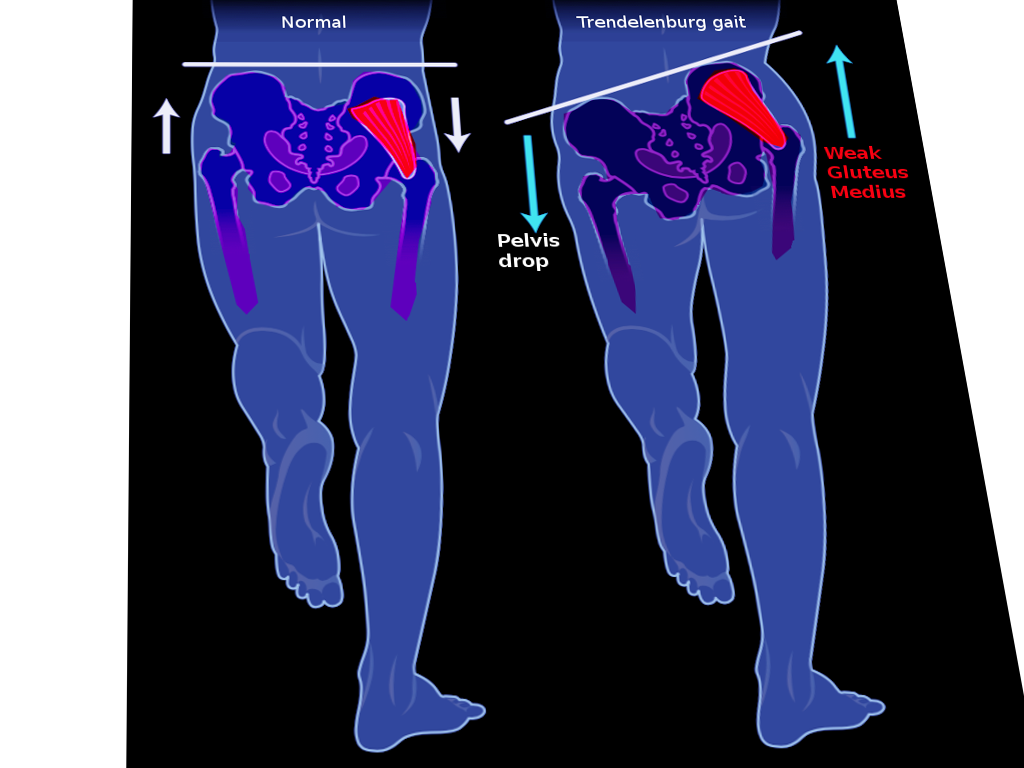
The Trendelenburg sign is most apparent during the gait cycle (see Illustration. Trendelenburg Gait). When the leg supports the weight of the body on the lesioned side, the pelvis rises ipsilaterally. This presentation is more accurately a dipping of the pelvis towards the contralateral side. Because the pelvis cannot be maintained in a level plane by the lesioned abductors, the patient falls towards the good side and simultaneously leans the torso towards the lesioned side in an attempt to maintain balance. This type of gait is known as the Trendelenburg gait.
Methods of Evaluation
Prerequisites of Testing
- The patient can stand on the affected side for more than 30 seconds
- The patient can understand commands
- The patient has intact coordination
- The patient has free abduction movement; no fixed adduction deformity
Standing Test
The patient stands on the affected leg in a single leg stance for up to 30 seconds. The provider stands behind the patient at the hip level and places their hands on the iliac crests on either side of the pelvis observing to see if it stays level during the single-leg stance. Repeat the test on the opposite side. A positive Trendelenburg sign is when the pelvis drops on the unaffected side.[4]
Gait Test
The examiner asks the patient to walk a short distance. In a normal gait, the body shifts the weight to the stance leg, allowing the center of gravity to shift as well, which stabilizes the body. In a patient with positive Trendelenburg sign constituting of abductor weakness, when they lift the unaffected leg, the shift does not occur; therefore, the patient is unable to maintain balance, leading to instability.
Bottom Line
The Trendelenburg sign alone cannot be used to diagnose hip conditions but is essentially a physical exam assessment used to aid in the diagnosis of various hip pathologies. A health care provider should still complete a full history and physical and order appropriate lab and imaging tests to assist in the final diagnosis contributing to a positive Trendelenburg sign. Other diagnostic tests can include X-rays, ultrasonography, computed tomography scans, and magnetic resonance imaging to diagnose the primary condition.
Issues of Concern
The Trendelenburg sign is useful in assessing different hip dysfunctions, but the interpretation of the results remains controversial as there is no clearly established standard for a positive test. According to Bailey et al., only two authors have objectively defined the point at which pelvic drop can be considered a positive Trendelenburg test result: Asayama et al. established a positive Trendelenburg test result as a pelvic tilt angle of greater than 2 degrees, while Westhoff et al. (2005) considered “positive” to be a pelvic drop in the non-stance limb during the single-stance phase of over 4 degrees and/or a maximum pelvic drop in the stance phase exceeding 8 degrees (Bailey et al., 2009).[1][5] In recent years, the Hardcastle modification of the Trendelengberg test (described above) can help to eliminate false positives.
False positives can occur with:
- Obesity
- Fixed adduction deformity
- Medialization of lower limb axis, such as coxa vara, genu varum, and malunited supracondylar femur with varus deformity
- Non-intact quadratus lumborum (pulls the pelvis on the unaffected side up if it is not intact)
- Pain
- Poor balance
- Lack of cooperation or understanding
- Costo-pelvic impingement
- Scoliosis
- Pelvic drop can occur even in healthy individuals with normal abductor mechanism when the abductor muscles are not working adequately.
False negatives can occur with[6]:
- Use of supra-pelvic muscles
- Use of psoas and rectus femoris
- Wide lateral translocation of the trunk to allow balance over the hip as a fulcrum
- In patients with early stages of osteonecrosis, despite having an abductor mechanism defect, Trendelenburg sign and, therefore, gait remain masked.
Clinical Significance
The Trendelenburg sign is useful in identifying weak hip abductors as well as assessing other mechanical, neurological, and spinal disorders. Especially in the post-operative setting, a positive Trendelenburg sign may identify the source of patient complications. Particularly in the posterior approach to the hip, the gluteus medius is in danger of being detached from its attachment at the greater trochanter. Therefore orthopaedic surgeons must be careful in their surgical approach to avoid this complication.
A positive Trendelenburg test is a finding in the following [7]:
- Any condition that brings the origin and insertion of gluteus medius together:
- Subluxation or dislocation of the hip
- Coxa vara
- Greater trochanter fractures
- Slipped femoral capital epiphysis
- Legg-Calve-Perthes disease
- Abductor paralysis or weakness:
- Polio
- Root-lesion
- Post-operative nerve damage
- Muscle-wasting disease
- Hemiplegic cerebral palsy
- Muscular dystrophy
- Any painful hip disorder which results in gluteal inhibition
- Others
- Lower back pain
- Osteoarthritis of the hip
- Greater trochanter fractures
- Femoral neck fractures
- Short leg syndrome
- The initial time following total hip replacement
- Cleidocranial dysostosis
Nursing, Allied Health, and Interprofessional Team Interventions
A patient-centered approach to care is necessary to manage and treat a patient with a positive Trendelenburg sign. A team of health care workers, including the patient’s primary care physician, specialists like orthopaedics and neurologists, nursing, physical therapists, and/or chiropractors, all working in an interdisciplinary manner with the patient can help find the root cause and treat the patient. Often patients will need to follow up with their physical therapists and other doctors managing their condition on a long term basis as it takes time to make a full recovery depending on the diagnosis.
Management
Medical management of a positive Trendelenburg sign depends on the underlying cause. If the positive Trendelenburg sign is the result of pain, providers might suggest pain control through the use of over the counter nonsteroidal anti-inflammatory drugs (NSAIDs). In severe cases, doctors might prescribe steroids such as cortisone injections to help reduce pain. If the patient has short leg syndrome, patients might benefit from a shoe lift to minimize the waddling gait. Other times the clinician might suggest a harness for developmental dysplasia of the hip or cast for Legg Calve Perthes disease. Physical therapists will often recommend various exercises to strengthen the hip abductors so that the patient can maintain a midline center of gravity preventing the side-to-side motion during their gait cycle. Osteopathic manipulative treatment could result in improved gait and range of motion for individuals with somatic dysfunctions, short-leg syndrome, and lower back pain contributing to positive Trendelenburg sign.[8] Biofeedback through the use of electromyography can help patients understand how their muscles respond to specific movements and adjust their gait accordingly. Essentially this will allow patients to control their muscle movements and increase their range of motion.[9]