Mitral Valve Repair

Earn CME/CE in your profession:


Continuing Education Activity

Mitral valve repair is commonly used to treat mitral valve pathology due to mitral regurgitation. Mitral regurgitation (MR) is classified as primary or secondary, depending on whether the lesion is located at the mitral valve apparatus or due to left ventricular changes. This activity reviews and highlights the role of an interprofessional care team in managing patients who undergo mitral valve repair.

Objectives:

  • Identify the relevant anatomy, indications, and contraindications for mitral valve repair.
  • Describe the equipment, personnel, preparation, and technique in regards to mitral valve repair.
  • Review appropriate evaluation of the potential complications and clinical significance of mitral valve repair.
  • Summarize interprofessional team strategies for improving care coordination and communication to advance mitral valve repair and improve outcomes.

Introduction

Although the standard of care for mitral valve (MV) pathology due to degenerative changes is surgical repair, patient outcomes depend on multiple factors including pre-operative status, the severity of mitral regurgitation (MR), the technique of repair and surgeon and center experience. If MV repair is carried out in a timely fashion, the operative risk is low, and life expectancy is close to that of similar sex-aged matched controls. In high-risk patients, the choice amongst surgical, percutaneous, and conservative approaches can be challenging but should have as its basis patient comorbidities and surgical expertise. Mitral valve repair (MVr) surgery has some advantages over mitral valve replacement (MVR), although patient-specific factors must be taken into consideration. Of note, close to 50% of patients with severe mitral valve pathology are not candidates for surgical intervention due to age or other comorbidities.[1]

Up to 2 to 3% of adults in the United States are afflicted with degenerative mitral valve disease.[2] Patients with degenerative MV pathology who develop symptoms of MR have a poor prognosis, with annual mortality rates of up to 34%.[3] Mitral stenosis (MS), primarily caused by rheumatic heart disease (RHD), is commonly treated by percutaneous balloon mitral valvuloplasty or MVR. Repair is usually not feasible in these patients with rheumatic mitral disease. MR is classified as primary or secondary, depending on whether the lesion is located at the mitral valve apparatus or due to left ventricular changes, respectively. While severe primary MR still receives treatment with surgical intervention, percutaneous techniques for repair and replacement are also gaining traction. MVR may be considered in patients with MR caused by papillary muscle rupture, degenerative and ischemic MR, or in patients with a failed repair undergoing reoperation.[4]

Anatomy and Physiology

The mitral valve apparatus has a complex anatomy, including an annulus, two leaflets, three types of chordae tendinae, and two papillary muscles.  The MV annulus is a saddle-shaped structure in continuity with the aortic valve. The annulus is most vulnerable to dilatation at its insertion on the posterior leaflet because it is the thinnest at this junction. There are two MV leaflets; the anterior is tall and narrow, and the posterior is shorter and broader. These two leaflets meet at their respective commissures, known as the anterolateral and posteromedial commissures. Each leaflet divides into three scallops, 1 to 3, from lateral to medial, and are designated A1, A2, A3 (anterior) and P1, P2, P3 (posterior).[5] The anterior leaflet occupies two-thirds of the valvular area and one-third of the annular area, whereas the posterior leaflet comprises two-thirds of the annular circumference. The leaflets have a normal line of coaptation from 7 to 9 mm, allowing for a range of normal physiologic pressures and volumes. The posterior, or mural leaflet, is more vulnerable to prolapse because it is attached to the free wall of the ventricle, leaving it exposed to the recurrent stress of ventricular contraction.[6]

Fibrous structures, known as the chordae tendinae, attach the MV leaflets to the papillary muscles. There are three types of chordae based on their level of insertion: primary, secondary, and tertiary. Primary chordae insert on the leaflet’s free margin and aid in the prevention of leaflet prolapse.  Secondary chordae insert on the leaflet’s rugged surface, and tertiary on the basal portion of the leaflet. There are two papillary muscles known as anterolateral and posteromedial. The posteromedial muscle receives vascular supply solely by the posterior descending artery, while the supply to anterolateral is by both the left anterior descending artery and the left circumflex artery.[5] The aforementioned elements of the mitral valve apparatus work in concert to maintain valvular competence and enable proper blood flow from the left atrium (LA) to the left ventricle (LV). A normal MV area is 4 to 6 cm. In MR patients, the LV is commonly thin, dilated, dysfunctional, and arrhythmogenic.

Indications

Rheumatic heart disease commonly causes MS, thought to be related to an exaggerated immune response secondary to cross-reactivity between streptococcal antigen and MV tissue.[7] Changes associated with rheumatic mitral disease include a “fish-mouth” appearance of the MV orifice, commissural fusion, leaflet thickening, and shortening and fusion of chordae tendinae. These changes lead to the subsequent hockey-stick appearance of the anterior leaflet on echocardiography. Other etiologies of MS include mitral annular calcification (MAC) common in elderly patients or those with advanced renal disease, radiation valvulitis, systemic inflammatory disorders, obstructing lesions, infectious vegetations, and congenital valvular abnormalities.[8] Progressive MS, especially a valve area less than 2 cm, leads to a diastolic pressure gradient between the LA and LV, increasing LA pressures and potentially reducing forward flow. Tachycardia should be avoided in patients with MS, as it reduces diastolic filling time, leading to an increased transmitral gradient. Increased LA pressures can lead to atrial enlargement with increased risk of thromboembolism and arrhythmias, most commonly atrial fibrillation (AF). Elevated pulmonary back pressure can subsequently cause pulmonary edema and hypertension, precipitating right ventricular (RV) failure and possible tricuspid regurgitation. Decreased forward flow can produce poor LV filling and reduced cardiac output (CO).

Clinicians can best accomplish quantification of MS with echocardiography, based on mean diastolic transmitral pressure gradient, MV area, and LA and right-sided chamber sizes and pressures.[9] The mean diastolic pressure gradient gets calculated with continuous-wave Doppler from an apical four-chamber view. The Doppler reveals diastolic transmitral flow via a velocity measurement, which then converts into a pressure with Bernoulli’s simplified equation to discern the mean MV gradient. The definition of severe MS is a mean gradient over 10 mmHg, moderate MS is 5 to 10 mmHg, and mild MS is below 5 mmHg. The transmitral gradient is under strong influence from heart rate and forward blood flow. According to the 2014 valvular heart disease guidelines and the 2017 guideline update, they consider MS to be very severe if MVA less than or equal to 1 cm squared, and severe if it is less than or equal to 1.5 cm squared.

MR can be defined as primary or secondary depending on whether the abnormality is located at the level of the mitral valve apparatus or the left ventricle, respectively. In developing countries, mitral valve prolapse is the most common cause of MR requiring surgical repair, while degenerative disease is most common in the United States.[10] Mitral valve prolapse is a systolic leaflet displacement of greater than or equal to 2 mm above the mitral annulus plane in a long-axis view. Barlow's disease results in abnormal accumulation of mucopolysaccharides and fibroelastic deficiency, leading to loss of mechanical integrity of the MV. Primary MR can also result from infective endocarditis, MAC, RHD, connective tissue disorders, congenital malformations, and drug use. Secondary MR, also known as functional MR, is due to LV remodeling or dyssynchrony, mitral annular dilatation, and impaired LV contractility. Secondary causes of MR can subdivide into ischemic and non-ischemic. Ischemic MR is caused by LV dysfunction secondary to coronary artery disease (CAD) and foreshadows a poor prognosis in terms of survival and development of heart failure. Non-ischemic MR results from the various cardiomyopathies (dilated, restrictive, and hypertrophic) as well as secondary to AF with subsequent annular dilatation.

MR leads to LA and LV volume overload, depending on the time course of the MR (acute vs. chronic) and the magnitude of the regurgitant volume. Acute MR leads to a sudden increase in preload and LV filling pressures that can cause pulmonary edema. Cardiac output also becomes reduced since blood flow now gets directed to the LA, which can precipitate cardiogenic shock; this is a common presentation in patients with infective endocarditis, chordae tendinae rupture, or papillary muscle rupture following myocardial infarction (MI). Chronic MR divides into three stages: a compensated stage where most patients are asymptomatic, a transitional stage with LV remodeling, and a decompensated stage marked by insidious symptom development. Chronic MR also leads to LA enlargement, which increases the risk for atrial arrhythmias and thromboemboli. Carpentier developed a functional classification of MR based on leaflet motion.[11]

Echocardiography is the modality of choice for the categorization of MR. Color Doppler is useful in assessing the jet area, as well as its ratio to the LA area. A jet over 40% of the LA area suggests severe MR. In severe MR, measured peak mitral inflow velocity is usually more than 120 cm/sec, and diastolic reversal of pulmonary venous flow is present.[11] Worsening MR leads to systolic flow reversal along with a blunting of the systolic component. In patients with severe MR and preserved EF, prompt surgical intervention is paramount to prevent adverse LV remodeling. Beta natriuretic peptide (BNP), global longitudinal strain, exercise capacity, and right ventricular systolic pressure are important prognostic indicators in such patients.[12] According to the American College of Cardiology/American Heart Association (ACC/AHA) guidelines, the following are indications for surgery in severe primary MR: chronic severe primary MR, with repair preferable over replacement if durable repair can be achieved (class I).[13]  

Contraindications

Patients with severe MR must be evaluated by a cardiac surgeon with experience in mitral valve surgery. The standard pre-operative evaluation must be undertaken to determine surgical candidacy, including assessment of coronary arteries, medical co-morbidities, and prior surgical history. Patients with aortic calcification, RV dysfunction, or severe MAC are considered relative contraindications. Society of Thoracic Surgeons (STS) risk calculation can be performed to aid with determination of surgical risk, including that of mortality and major morbidity. Severe LV dysfunction is also a relatively contraindication as repair of the mitral valve leads to a competent valve, thereby increasing afterload on the LV. Thus, the pre-operative LV function and ejection fraction usually overestimates the true left ventricular function in cases of severe MR since the regurgitant valve acts as a "pop-off" valve. Severe emphysema, restrictive lung disease, and pulmonary hypertension (PHT) are also commonly seen comorbidities with severe longstanding MR and pose relative contraindication to surgery.[14]

Equipment

MVr requires cardiopulmonary bypass and, in the vast majority of cases, requires full cardiac arrest. Standard equipment will include instrumentations necessary for open cardiac operations. Transesophageal echocardiography is mandatory for intraoperative evaluation of repair and ventricular function.

Personnel

MVr requires standard open cardiac surgical staff, which includes at a minimum: cardiac surgeon experienced in mitral valve surgery, cardiac anethesiologist, perfusionist, surgical assistant, surgical scrub nurse or technician, and circulating nurse. Postoperatively, care of these patients requires a critical care intensivist, cardiologist, intensive care and ward nursing, physical therapy, and social workers.

Preparation

Preoperative assessment of patients before cardiac surgery requires an appraisal of the patient and procedural risks, including a thorough history, focused physical examination, and review of diagnostic studies and pertinent consultant preoperative evaluations. Significant cardiovascular risk factors include myocardial ischemia, ventricular dysfunction with heart failure, and atherosclerotic disease of the carotid arteries or proximal aorta. The etiology of ventricular dysfunction is essential to establish optimal perioperative hemodynamic parameters. Patients can be stratified into low, intermediate, or high risk depending on their presentation. Low-risk patients are those with angina without evidence of preoperative MI scheduled for elective surgery. Intermediate risk patients are those with an acute MI who are hemodynamically stable but are usually hospitalized and might be receiving heparin and antiplatelet medications. High-risk patients are those with the highest risk of morbidity and mortality, presenting as hemodynamically unstable following an acute MI.[15] Preoperative risk evaluation of patients with CHF includes assessment of LV and RV dysfunction. An anesthetic plan should consist of appropriate intraoperative monitoring modalities, optimal anesthetic induction agents, vasoactive infusions, and the need for mechanical support. Pulmonary hypertension, defined as mean pulmonary artery pressure (PAP) greater than 25 mmHg at rest, significantly increases morbidity and mortality risk.[15]

Perioperative stroke is common in patients with severe atherosclerosis of the proximal aorta or the carotid arteries. Other comorbidities that require investigation include diabetes, hypertension, previous cerebrovascular event, peripheral vascular disease, and history of smoking-related pulmonary pathology. Perioperative stroke risk is reducible by ensuring that the patient is appropriately taking aspirin, beta-blockers, and statins. Patients with a very high stroke risk should have an evaluation by a peripheral vascular surgeon before surgery. During cardiac surgery, thrombi or atheromatous debris can dislodge from aortic plaques during clamping and unclamping of the aorta, or by turbulent high-velocity blood flow via cannulae.[16]

Noncardiac risk factors that cannot be modified include female gender, advanced age, NYHA IV functional status, preexisting renal insufficiency, prior transient ischemic attack (TIA), anemia, and tobacco use. Preexisting renal insufficiency can lead to acute kidney injury (AKI) in about 1 to 2% of patients, which significantly increases mortality.[17] These patients should avoid exposure to nephrotoxic agents, and have their volume status optimized. Reduced cardiac output or hypotension needs to promptly treated. Anemia is a common perioperative finding in cardiac surgical patients and is independently associated with increased transfusion risk.[17] In diabetic patients, hypoglycemia and marked hyperglycemia should be strictly avoided to prevent perioperative complications. Hypertensive patients should have chronically administered oral antihypertensive agents continued up to the time of surgery, excluding angiotensin-converting enzyme (ACE) inhibitors and angiotensin II receptor blockers (ARBs).

Patients with chronic obstructive pulmonary disease (COPD) should be at their optimal baseline level of pulmonary function before the intervention. Smokers should receive counsel about preoperative cessation.[18] Ensuring optical preoperative nutrition and postoperative rehabilitation programs has improved functional capacity in elderly patients. For elective surgical procedures, prolonged aortic cross-clamp time and total CPB time confer a higher risk. The mortality risk also directly correlates with the procedural volume or experience of both the hospital and the cardiac surgeon.[19] Medical management of MV pathology includes diuretics and rate-controlling agents, such as beta-blockers, for symptomatic relief. Patients with AF should receive anticoagulation with warfarin. Vasodilators are frequently used to reduce arterial resistance and improve CO, which increases the MV closing force and reduces backflow. Although acute MR reduction is effective with vasodilators, sustained improvement can be challenging.

Technique or Treatment

Standard MVr is performed utilizing full cardiopulmonary bypass (CPB) and ischemic arrest. There are numerous possible surgical approaches, including median sternotomy, right thoracotomy, and robot-assisted. Regardless of incisional approach taken, the core principles of mitral valve repair remain the same: the goal is to create a competent mitral valve with good coaptation depth, ring annuloplasty, and avoidance of systolic anterior motion. We describe here the surgical techniques for MVr via median sternotomy. This approach is necessary for patients requiring additional concomitant procedures including coronary artery bypass grafting (CABG), ascending aortic intervention, or additional/multiple valve intervention.

Cannulation for CPB is achieved via the ascending aorta and bicaval venous cannulation. Cardioplegic arrest is achieved via the antegrade and retrograde routes. After aortic crossclamping and adequate diastolic cardioplegic arrest, the interatrial groove (Sondergaard's or Waterston's) is exposed. Left atriotomy is created away from the pulmonary veins. Alternatively, a transseptal approach can be performed by right atriotomy and incising the septum by the fossa ovalis. Appropriate retractors are placed for exposure. The valve is then inspected systematically using saline injection as well as visual inspection of each segment. Repair technique will depend on findings of valve pathology. Isolated P2 prolapse, for instance, can be repaired either by triangular vs. quadrangular resection and ring annuloplasty, or by creating neo-chordae to the appropriate papillary muscle. We recommend ring annuloplasty for all repairs. Annuloplasty sutures are placed around the annulus, and the anterior leaflet height is sized. Depending on MR pathology (primary vs. secondary, ischemic vs. non-ischemic), the ring can be true-sized or undersized. It is important to test the valve for coaptation depth with the ideal depth being about 1 cm. It is also important to avoid excessive undersizing of the ring as this may cause systolic anterior motion (SAM).

Alain Carpentier’s technique for correction of MR includes leaflet repair with quadrangular resection and rigid annuloplasty to correct annular dilatation, developed through autopsy and pathology studies of the MV.[20] Lawrie et al. described a functional correction of MR, which spares valve leaflets and chordae during the repair.[26] A flexible annuloplasty ring corrects annular dilatation, and proprietary fabric artificial chordae are used to repair prolapse and realign leaflets.[21] In patients with degenerative MR, the MV annulus can double in size leading to flattening of the valvular orifice and reduced leaflet edge apposition. With inadequate leaflet coaptation, ventricular systole adds additional stress to the leaflet bodies and marginal chordae, leading to further dysfunction and failure.

With Lawrie’s technique, avoidance of reoperation and significant recurrent MR as assessed by echocardiography at ten years is 90.1% and 93.9%, respectively, according to reports. Also, if the ventricular filling gets optimized through adequate preload and afterload, atrioventricular dyssynchrony is avoided, and hypercontractility is limited, there is almost no postoperative systolic anterior motion (SAM).[22] Braunberger et al.reported long-term results of valve repair in nonrheumatic MR.[29] In patients with isolated posterior leaflet prolapse, 10- and 20-year freedom from reoperation was 98.5% and 96.9%, respectively. In patients with isolated anterior prolapse, it was 86.2% and 82.2%, respectively. In patients with bileaflet prolapse, freedom from reoperation was 88.1% and 82.6%, respectively. These data confirm the results of Carpentier’s repair and its stability over an extended period.

Minimally invasive mitral valve surgery (MIMVS) can be divided into two groups: partial sternotomy and right thoracotomy, including the open and video-assisted methods, with or without robotic assistance.[27][28] MIMVS has been shown to reduce surgical trauma by avoidance of a full sternotomy incision. MIMVS approaches will require different cannulation techniques, generally involving the femoral vessels. Sündermann et al. showed equivalent short-term and mid-term outcomes with MIMVS, as compared to conventional surgery, regarding the incidence of stroke, mortality, and durability of repair.[30] Patients who undergo MIMVS have reduced blood loss, need for blood transfusion, mechanical ventilation time, and intensive care unit stay, as well as a quicker resumption of regular activity.[23] Iribarne et al. reported that MIMVS is associated with lower hospital costs.[32] Due to these highly optimistic results, there has been an increase in the proportion of MIMVS from 10% in 2004 to 20% in 2008. The least invasive approach for MIMVS is robotically assisted, without thoracotomy or significant rib spreading, but it also has higher operative costs. This method allows excellent three-dimensional visualization of the valvular and subvalvular apparatus via EndoWrist technology, which permits complex surgical maneuvers.

An innovative approach for the treatment of degenerative MV disease due to posterior leaflet prolapse is trans-apical beating heart MV repair with polytetrafluoroethylene (PTFE) chordae implantation. CPB is avoided as a small left anterolateral thoracotomy incision is made at the fourth or fifth intercostal space to access the cardiac apex. The Neochordae device used for this procedure allows the physician to grasp and pierce the leaflet while pulling the PTFE cord through the prolapsing segment. The neochordae get externalized at the level of the cardiac apex and titrated to maximal coaptation on echocardiography with the resolution of MR. Most patients were found to be stable at early follow-up. This technique is utilized to treat MR due to prolapsing lesions early in the history of the disease, with little or no annular dilatation and limited LV remodeling.[24] Trans-catheter techniques are utilized in the presence of prolapse to target both the leaflets and the annulus to achieve favorable long-term results. Excluding ring annuloplasty correlates with poor long term outcomes. Transcatheter methods should be utilized only in patients that are appropriate for such interventions.

Complications

Recurrent MR is the most common complication following primary MVr. Intraoperative assessment via TEE of the repaired valve is crucial, as it can aid in immediate assessment of the valve. If there is persistent mild or greater MR, the valve must be re-explored and either re-repaired or replaced. This decision is critical in cases of impaired LV function, as these patients may not tolerated a long repeat ischemic/arrest period. MVr long term durability is more difficult to predict. Flameng et al. [25] reported that only 50% of patients remain free from MV incompetence seven years following repair. A study by Chikwe et al. [26] demonstrated that mitral valve repair did not confer a long term survival benefit in patients over the age of 60 who required concomitant CABG surgery. Fifty-nine octogenarians undergoing MV surgery for nonrheumatic disease demonstrated similar outcomes between repair and replacement.[27] A Society for Thoracic Surgeons (STS) national database analysis included 8523 MVrs and 3520 MVRs and concluded that there was operative mortality reduction in the MVr cohort as compared to MVR with and without chordal preservation.[28]

Following successful repair, patients with PHT/AF have worse long-term survival and event-free survival, as well as increased compromise of MV repair.[29] Based on findings by the Mayo Clinic group, postoperative AF occurs in up to 24% of patients previously in sinus rhythm, particularly those with LA enlargement, and is associated with increased mortality.[30] To aid with this complication, there has been a recent trend to perform surgical ablation of AF during mitral valve repair. Up to 32.2% of patients presenting for repair have AF, and concomitant AF ablation took place in 61.5% of patients according to the STS database.[31] Gillinov et al. reported that the addition of AF ablation to MV surgery increased the rate of freedom from AF at one year (63.2% vs. 39.4%) with similar mortality in both groups. Still, pacemaker implantation increased in the ablation group.[32]

SAM of the mitral valve can result if there is a discrepancy between the annular area and leaflet tissue following repair. SAM is the result of anterior MVP into the left ventricular outflow tract (LVOT) during systole. It most commonly occurs with an undersized annuloplasty ring or excess leaflet tissue.[33] SAM can lead to residual MR and LVOT obstruction, both of which are observable on intraoperative transesophageal echocardiography (TEE). If SAM is seen intraoperatively following the repair, the ventricular filling should immediately be optimized, atrioventricular pacing should be implemented to improve atrioventricular synchrony, and ventricular hypercontractility should lessen. Postoperative beta-blockers are useful in this regard. David and colleagues report freedom from MR ranging from 65 to 80% at 12 years postoperatively, depending on which MV leaflet suffered prolapse.[34] In patients with Barlow disease, Jouan et al. describe a 9.8% recurrence rate of moderate or severe MR.[35]

Clinical Significance

With advanced age, degenerative MV disease is the most common cause of MR. MVr is the gold standard for degenerative MR and is the recommendation of the current guidelines for the management of valvular heart disease. The ACC/AHA guidelines recommend that surgery take place before LV dysfunction (class IIa) in experienced centers. Mitral regurgitation imposes significant volume overload on the LV, leading to permanent structural and functional deterioration of the myocardium and subsequent heart failure. Timely correction of MR is paramount to the preservation of cardiac function. MIMVS and transcatheter MVr technologies have been instrumental in allowing repair of MV pathology in high-risk patients who are not appropriate for open-heart surgery. 

Enhancing Healthcare Team Outcomes

MR is the second most common valvular heart disease after aortic stenosis in the general population and requires an interprofessional healthcare team for management. In industrialized countries, the most common etiology is degenerative MV disease leading to prolapse caused by either myxomatous degeneration or fibroelastic deficiency. MVr is preferred over MVR if complete and durable repair can be achieved. While the natural history of this valvular pathology is poor, leading to eventual LV decompensation, an appropriate and timely repair can be lifesaving and prolongs life expectancy to that of the healthy age-matched population. Mortality is increased in patients with symptoms of CHF and reduced EF. Transcatheter techniques may be challenging in patients with certain anatomical limitations, including calcified leaflets and advanced disease. Repair is not always effective, and patient selection is imperative to prevent the recurrence of MR.

The success of repair depends partially on the center and the surgeon’s level of experience. Intraoperative collaboration with cardiac anesthesia and TEE is critical. Current guidelines propose that these procedures should take place at “Heart Valve Centers of Excellence” (HVCE) that offer comprehensive options for diagnosis and management of the valvular disease.[36] An HVCE should deliver superior quality of care due to a larger volume of repair procedures, advanced imaging techniques, and greater transparency regarding patient outcomes.[37] An annual surgical volume of fewer than 25 cases per year correlates with lower repair rate, increased one-year mortality, and a higher incidence of subsequent reoperation.[38]

An interprofessional team dedicated to these patients should include cardiologists, anesthesiologists, nurse anesthetists, and intensivists. Cardiology specialty nursing should be available to assist at every step, both perioperatively as well as during the procedures, working in collaboration with the clinicians and specialists to provide monitoring and patient and family education. Perioperative evaluation should consist of high-quality TEE with 3-dimensional technology. Goals for repair should include below 1% mortality for isolated repair, a near 100% repair rate, and less than 5% repair failure at a five-year follow-up. Centers should be involved in research and innovation of techniques for procedural improvement. A successful HVCE should adhere to international guidelines, engage in the appropriate and timely referral of patients, partake in evaluation and enhancement of patient outcomes, and participate in regional or national outcome registries. With an interprofessional team approach, including specialists, clinicians, nurses, and pharmacists, patient results can improve, and adverse events kept to a minimum, resulting in a better quality of life and better outcomes. [Level 5] 

Nursing, Allied Health, and Interprofessional Team Interventions

The role of the nurse is limited to the care of the postoperative patient. This includes:

  • Educating the patient on the course of the illness
  • Encourage incentive spirometry
  • Ambulate
  • Eat a healthy diet
  • Remain compliant to medications-esp if an oral anticoagulant has been prescribed
  • Maintain a healthy body weight
  • Be aware of antibiotic precautions when undergoing invasive tests or procedures

Nursing, Allied Health, and Interprofessional Team Monitoring

After surgery, the nursing responsibilities include:

  • Monitor vital signs
  • Monitor Ins and Outs
  • Check daily labs and x-ray
  • Monitor output from the mediastinal tubes
  • Assess neurovitals
  • Check and clean the wounds
  • Dispense medications as ordered


(Click Image to Enlarge)
Insufficient mitral valve (the valve no longer closes).
Contributed by Bruno Bordoni, PhD.

(Click Image to Enlarge)
Mitral Valve, Repair Table 01
Contributed by Irim Salik, MD

(Click Image to Enlarge)
Mitral Valve, Repair Table 02
Contributed by Irim Salik, MD

(Click Image to Enlarge)
<p>Mitral Valve, Transverse View. The image shows the anatomy of the mitral valve.</p>

Mitral Valve, Transverse View. The image shows the anatomy of the mitral valve.


Contributed by S Bhimji, MD


(Click Image to Enlarge)
Mitral valve anatomy
Image courtesy S Bhimji MD
Details

Author

Irim Salik

Editor:

Jason Widrich

Updated:

7/24/2023 11:37:14 PM

References


[1]

Saccocci M, Taramasso M, Maisano F. Mitral Valve Interventions in Structural Heart Disease. Current cardiology reports. 2018 May 17:20(6):49. doi: 10.1007/s11886-018-0982-y. Epub 2018 May 17     [PubMed PMID: 29770888]


[2]

Chiu P, Goldstone AB, Woo YJ. Would evolving recommendations for mechanical mitral valve replacement further raise the bar for successful mitral valve repair? European journal of cardio-thoracic surgery : official journal of the European Association for Cardio-thoracic Surgery. 2018 Oct 1:54(4):622-626. doi: 10.1093/ejcts/ezy284. Epub     [PubMed PMID: 30165483]


[3]

Enriquez-Sarano M, Avierinos JF, Messika-Zeitoun D, Detaint D, Capps M, Nkomo V, Scott C, Schaff HV, Tajik AJ. Quantitative determinants of the outcome of asymptomatic mitral regurgitation. The New England journal of medicine. 2005 Mar 3:352(9):875-83     [PubMed PMID: 15745978]


[4]

LaPar DJ, Kron IL. Should all ischemic mitral regurgitation be repaired? When should we replace? Current opinion in cardiology. 2011 Mar:26(2):113-7. doi: 10.1097/HCO.0b013e3283439888. Epub     [PubMed PMID: 21245751]

Level 3 (low-level) evidence

[5]

Harb SC, Griffin BP. Mitral Valve Disease: a Comprehensive Review. Current cardiology reports. 2017 Aug:19(8):73. doi: 10.1007/s11886-017-0883-5. Epub     [PubMed PMID: 28688022]


[6]

Salem A, Abdelgawad AME, Elshemy A. Early and Midterm Outcomes of Rheumatic Mitral Valve Repair. The heart surgery forum. 2018 Aug 14:21(5):E352-E358. doi: 10.1532/hsf.1978. Epub 2018 Aug 14     [PubMed PMID: 30311884]


[7]

Shuhaiber J, Anderson RJ. Meta-analysis of clinical outcomes following surgical mitral valve repair or replacement. European journal of cardio-thoracic surgery : official journal of the European Association for Cardio-thoracic Surgery. 2007 Feb:31(2):267-75     [PubMed PMID: 17175161]

Level 2 (mid-level) evidence

[8]

Zhou YX, Leobon B, Berthoumieu P, Roux D, Glock Y, Mei YQ, Wang YW, Fournial G. Long-term outcomes following repair or replacement in degenerative mitral valve disease. The Thoracic and cardiovascular surgeon. 2010 Oct:58(7):415-21. doi: 10.1055/s-0029-1240925. Epub 2010 Oct 4     [PubMed PMID: 20922625]


[9]

Castleberry AW, Williams JB, Daneshmand MA, Honeycutt E, Shaw LK, Samad Z, Lopes RD, Alexander JH, Mathew JP, Velazquez EJ, Milano CA, Smith PK. Surgical revascularization is associated with maximal survival in patients with ischemic mitral regurgitation: a 20-year experience. Circulation. 2014 Jun 17:129(24):2547-56. doi: 10.1161/CIRCULATIONAHA.113.005223. Epub 2014 Apr 17     [PubMed PMID: 24744275]


[10]

Baumgartner H, Hung J, Bermejo J, Chambers JB, Evangelista A, Griffin BP, Iung B, Otto CM, Pellikka PA, Quiñones M, American Society of Echocardiography, European Association of Echocardiography. Echocardiographic assessment of valve stenosis: EAE/ASE recommendations for clinical practice. Journal of the American Society of Echocardiography : official publication of the American Society of Echocardiography. 2009 Jan:22(1):1-23; quiz 101-2. doi: 10.1016/j.echo.2008.11.029. Epub     [PubMed PMID: 19130998]


[11]

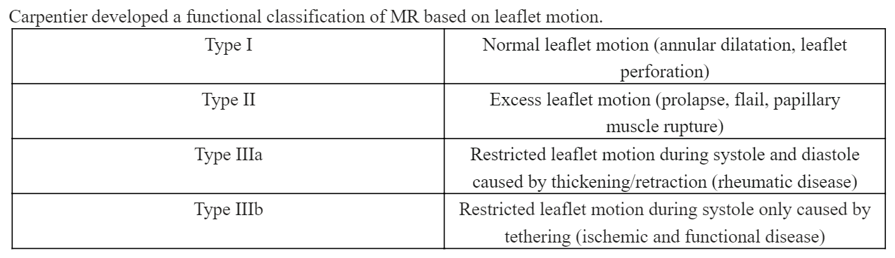
Carpentier A, Relland J, Deloche A, Fabiani JN, D'Allaines C, Blondeau P, Piwnica A, Chauvaud S, Dubost C. Conservative management of the prolapsed mitral valve. The Annals of thoracic surgery. 1978 Oct:26(4):294-302     [PubMed PMID: 380485]


[12]

Mentias A, Naji P, Gillinov AM, Rodriguez LL, Reed G, Mihaljevic T, Suri RM, Sabik JF, Svensson LG, Grimm RA, Griffin BP, Desai MY. Strain Echocardiography and Functional Capacity in Asymptomatic Primary Mitral Regurgitation With Preserved Ejection Fraction. Journal of the American College of Cardiology. 2016 Nov 1:68(18):1974-1986. doi: 10.1016/j.jacc.2016.08.030. Epub 2016 Aug 31     [PubMed PMID: 27591831]


[13]

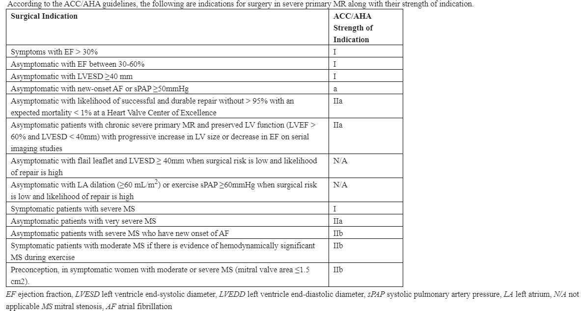
Nishimura RA, Otto CM, Bonow RO, Carabello BA, Erwin JP 3rd, Fleisher LA, Jneid H, Mack MJ, McLeod CJ, O'Gara PT, Rigolin VH, Sundt TM 3rd, Thompson A. 2017 AHA/ACC Focused Update of the 2014 AHA/ACC Guideline for the Management of Patients With Valvular Heart Disease: A Report of the American College of Cardiology/American Heart Association Task Force on Clinical Practice Guidelines. Journal of the American College of Cardiology. 2017 Jul 11:70(2):252-289. doi: 10.1016/j.jacc.2017.03.011. Epub 2017 Mar 15     [PubMed PMID: 28315732]

Level 1 (high-level) evidence

[14]

Wu AH, Aaronson KD, Bolling SF, Pagani FD, Welch K, Koelling TM. Impact of mitral valve annuloplasty on mortality risk in patients with mitral regurgitation and left ventricular systolic dysfunction. Journal of the American College of Cardiology. 2005 Feb 1:45(3):381-7     [PubMed PMID: 15680716]


[15]

Chevalier P, Burri H, Fahrat F, Cucherat M, Jegaden O, Obadia JF, Kirkorian G, Touboul P. Perioperative outcome and long-term survival of surgery for acute post-infarction mitral regurgitation. European journal of cardio-thoracic surgery : official journal of the European Association for Cardio-thoracic Surgery. 2004 Aug:26(2):330-5     [PubMed PMID: 15296892]


[16]

Gatti G, Morra L, Castaldi G, Maschietto L, Gripshi F, Fabris E, Perkan A, Benussi B, Sinagra G, Pappalardo A. Preoperative Intra-Aortic Counterpulsation in Cardiac Surgery: Insights From a Retrospective Series of 588 Consecutive High-Risk Patients. Journal of cardiothoracic and vascular anesthesia. 2018 Oct:32(5):2077-2086. doi: 10.1053/j.jvca.2017.12.008. Epub 2017 Dec 6     [PubMed PMID: 29325843]

Level 2 (mid-level) evidence

[17]

Haddad F, Denault AY, Couture P, Cartier R, Pellerin M, Levesque S, Lambert J, Tardif JC. Right ventricular myocardial performance index predicts perioperative mortality or circulatory failure in high-risk valvular surgery. Journal of the American Society of Echocardiography : official publication of the American Society of Echocardiography. 2007 Sep:20(9):1065-72     [PubMed PMID: 17566702]


[18]

Pinzani A, de Gevigney G, Pinzani V, Ninet J, Milon H, Delahaye JP. [Pre- and postoperative right cardiac insufficiency in patients with mitral or mitral-aortic valve diseases]. Archives des maladies du coeur et des vaisseaux. 1993 Jan:86(1):27-34     [PubMed PMID: 8338397]


[19]

Kennedy JL,LaPar DJ,Kern JA,Kron IL,Bergin JD,Kamath S,Ailawadi G, Does the Society of Thoracic Surgeons risk score accurately predict operative mortality for patients with pulmonary hypertension? The Journal of thoracic and cardiovascular surgery. 2013 Sep;     [PubMed PMID: 22982034]


[20]

Carpentier A, Chauvaud S, Fabiani JN, Deloche A, Relland J, Lessana A, D'Allaines C, Blondeau P, Piwnica A, Dubost C. Reconstructive surgery of mitral valve incompetence: ten-year appraisal. The Journal of thoracic and cardiovascular surgery. 1980 Mar:79(3):338-48     [PubMed PMID: 7354634]


[21]

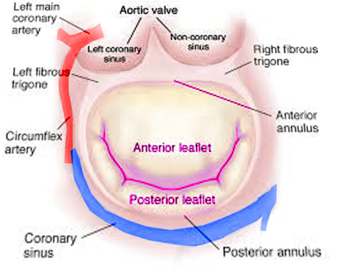
Lawrie GM. Structure, function, and dynamics of the mitral annulus: importance in mitral valve repair for myxamatous mitral valve disease. Methodist DeBakey cardiovascular journal. 2010 Jan-Mar:6(1):8-14     [PubMed PMID: 20360652]


[22]

Gazoni LM,Fedoruk LM,Kern JA,Dent JM,Reece TB,Tribble CG,Smith PW,Lisle TC,Kron IL, A simplified approach to degenerative disease: triangular resections of the mitral valve. The Annals of thoracic surgery. 2007 May;     [PubMed PMID: 17462375]


[23]

Algarni KD,Suri RM,Schaff H, Minimally invasive mitral valve surgery: Does it make a difference? Trends in cardiovascular medicine. 2015 Jul;     [PubMed PMID: 25640311]


[24]

Colli A, Manzan E, Zucchetta F, Bizzotto E, Besola L, Bagozzi L, Bellu R, Sarais C, Pittarello D, Gerosa G. Transapical off-pump mitral valve repair with Neochord implantation: Early clinical results. International journal of cardiology. 2016 Feb 1:204():23-8. doi: 10.1016/j.ijcard.2015.11.131. Epub 2015 Nov 23     [PubMed PMID: 26655529]


[25]

Flameng W, Herijgers P, Bogaerts K. Recurrence of mitral valve regurgitation after mitral valve repair in degenerative valve disease. Circulation. 2003 Apr 1:107(12):1609-13     [PubMed PMID: 12668494]


[26]

Chikwe J, Goldstone AB, Passage J, Anyanwu AC, Seeburger J, Castillo JG, Filsoufi F, Mohr FW, Adams DH. A propensity score-adjusted retrospective comparison of early and mid-term results of mitral valve repair versus replacement in octogenarians. European heart journal. 2011 Mar:32(5):618-26. doi: 10.1093/eurheartj/ehq331. Epub 2010 Sep 16     [PubMed PMID: 20846993]

Level 2 (mid-level) evidence

[27]

Vassileva CM, Ghazanfari N, Spertus J, McNeely C, Markwell S, Hazelrigg S. Heart failure readmission after mitral valve repair and replacement: five-year follow-up in the Medicare population. The Annals of thoracic surgery. 2014 Nov:98(5):1544-50. doi: 10.1016/j.athoracsur.2014.07.040. Epub 2014 Sep 22     [PubMed PMID: 25249161]


[28]

Silaschi M, Chaubey S, Aldalati O, Khan H, Uzzaman MM, Singh M, Baghai M, Deshpande R, Wendler O. Is Mitral Valve Repair Superior to Mitral Valve Replacement in Elderly Patients? Comparison of Short- and Long-Term Outcomes in a Propensity-Matched Cohort. Journal of the American Heart Association. 2016 Jul 28:5(8):. doi: 10.1161/JAHA.116.003605. Epub 2016 Jul 28     [PubMed PMID: 27468927]


[29]

Coutinho GF, Garcia AL, Correia PM, Branco C, Antunes MJ. Negative impact of atrial fibrillation and pulmonary hypertension after mitral valve surgery in asymptomatic patients with severe mitral regurgitation: a 20-year follow-up. European journal of cardio-thoracic surgery : official journal of the European Association for Cardio-thoracic Surgery. 2015 Oct:48(4):548-55; discussion 555-6. doi: 10.1093/ejcts/ezu511. Epub 2015 Jan 5     [PubMed PMID: 25564214]


[30]

Kernis SJ, Nkomo VT, Messika-Zeitoun D, Gersh BJ, Sundt TM 3rd, Ballman KV, Scott CG, Schaff HV, Enriquez-Sarano M. Atrial fibrillation after surgical correction of mitral regurgitation in sinus rhythm: incidence, outcome, and determinants. Circulation. 2004 Oct 19:110(16):2320-5     [PubMed PMID: 15477410]


[31]

Badhwar V, Rankin JS, Damiano RJ Jr, Gillinov AM, Bakaeen FG, Edgerton JR, Philpott JM, McCarthy PM, Bolling SF, Roberts HG, Thourani VH, Suri RM, Shemin RJ, Firestone S, Ad N. The Society of Thoracic Surgeons 2017 Clinical Practice Guidelines for the Surgical Treatment of Atrial Fibrillation. The Annals of thoracic surgery. 2017 Jan:103(1):329-341. doi: 10.1016/j.athoracsur.2016.10.076. Epub     [PubMed PMID: 28007240]

Level 1 (high-level) evidence

[32]

Gillinov M, Soltesz EG. Atrial fibrillation in the patient undergoing mitral valve surgery: A once-in-a-lifetime opportunity. The Journal of thoracic and cardiovascular surgery. 2018 Mar:155(3):995-996. doi: 10.1016/j.jtcvs.2017.09.125. Epub 2017 Oct 9     [PubMed PMID: 29089092]


[33]

Gallerstein PE, Berger M, Rubenstein S, Berdoff RL, Goldberg E. Systolic anterior motion of the mitral valve and outflow obstruction after mitral valve reconstruction. Chest. 1983 May:83(5):819-20     [PubMed PMID: 6839827]


[34]

David TE, Ivanov J, Armstrong S, Christie D, Rakowski H. A comparison of outcomes of mitral valve repair for degenerative disease with posterior, anterior, and bileaflet prolapse. The Journal of thoracic and cardiovascular surgery. 2005 Nov:130(5):1242-9     [PubMed PMID: 16256774]


[35]

Jouan J. Mitral valve repair over five decades. Annals of cardiothoracic surgery. 2015 Jul:4(4):322-34. doi: 10.3978/j.issn.2225-319X.2015.01.07. Epub     [PubMed PMID: 26309841]


[36]

Bolling SF, Li S, O'Brien SM, Brennan JM, Prager RL, Gammie JS. Predictors of mitral valve repair: clinical and surgeon factors. The Annals of thoracic surgery. 2010 Dec:90(6):1904-11; discussion 1912. doi: 10.1016/j.athoracsur.2010.07.062. Epub     [PubMed PMID: 21095334]


[37]

Bridgewater B, Hooper T, Munsch C, Hunter S, von Oppell U, Livesey S, Keogh B, Wells F, Patrick M, Kneeshaw J, Chambers J, Masani N, Ray S. Mitral repair best practice: proposed standards. Heart (British Cardiac Society). 2006 Jul:92(7):939-44     [PubMed PMID: 16251225]


[38]

Chikwe J, Toyoda N, Anyanwu AC, Itagaki S, Egorova NN, Boateng P, El-Eshmawi A, Adams DH. Relation of Mitral Valve Surgery Volume to Repair Rate, Durability, and Survival. Journal of the American College of Cardiology. 2017 Apr 24:():. pii: S0735-1097(17)30677-0. doi: 10.1016/j.jacc.2017.02.026. Epub 2017 Apr 24     [PubMed PMID: 28476349]