Continuing Education Activity
The term Rolando fracture was described for the first time in 1910 by Silvio Rolando, an Italian surgeon. The eponym is used to describe a comminuted articular fracture of the base of the thumb metacarpal, while partial articular fractures at the volar-ulnar base of the thumb metacarpal are referred to as Bennett fractures. The former describes more comminuted intra-articular patterns through the base, generating the classically described "Y" or "T" morphologies. The latter is distinguished by its 2-part articular fracture pattern including the nondisplaced volar-ulnar fragment which is held in place by its ligamentous attachment to the trapezium, known as the anterior oblique ligament. This activity reviews the classification, evaluation, and management of Rolando fracture, and also illustrates the evaluation and management of Rolando fracture and reviews the role of the interprofessional team in improving care for patients with this condition.
Objectives:
- Describe the pathophysiology of Rolando fracture.
- Describe the typical imaging findings associated with Rolando fracture.
- Review the treatment considerations for patients with Rolando fracture.
- Explain the importance of the interprofessional team as it pertains to improving care coordination among the interprofessional team members when treating Rolando fracture.
Introduction
The term Rolando fracture was described for the first time in 1910 by Silvio Rolando, an Italian surgeon.[1] The eponym is used to describe a comminuted articular fracture of the base of the thumb metacarpal,[2] while the name given partial articular fractures at the volar-ulnar base of the thumb metacarpal is a Bennett fracture.[3] The former describes more comminuted intra-articular patterns through the base, generating the classically described "Y" or "T" morphologies.[1] The distinguishing feature of the latter is its 2-part articular fracture pattern including the nondisplaced volar-ulnar fragment which is held in place by its ligamentous attachment to the trapezium, known as the anterior oblique ligament.[4]
Rolando fracture patterns are typically represented by a transverse articular component, which extends between the diaphysis and epiphysis in addition to an associated longitudinal intra-articular fracture line that divides the epiphysis into two fragments, one volar and the other dorsal, often resulting in a central depression of the articular surface. Commonly and contemporarily speaking, all intra-articular fractures with multiple fragments of the base of the first metacarpal classify as Rolando-type fracture patterns.[5]
Etiology
Rolando fractures are the result of compressive forces acting along the axis of the metacarpal shaft when the trapezo-metacarpal articulation is in a flexed position.[5]
The locus minoris resistenziae (or point of least resistance, also known as the weakest part of the bone) is the anteromedial margin of the base of the first metacarpal which obliquely detaches, as in the Bennett-type fracture patterns. However, a true Rolando fracture characteristically presents with the concomitant fracture of the stronger, dorsolateral process that separates from the body of the bone, giving rise to the second fragment.[1] Multi-fragmentary fractures generally have the same mechanism of action, but a traumatic force of greater extent is applied.[6]
Epidemiology
Overall, intra-articular fractures of the base of the first metacarpal are infrequent; representing a variable percentage between 1.4% to 4% of all hand fractures.[7][8] Rolando fractures make up 15% to 20% of all thumb metacarpal base fractures.[9]
In the pediatric population, 22% of all tubular bone hand fractures occurred somewhere along the thumb ray, and in the elderly (over 65 years of age), 20% of hand fractures occurred in the thumb. Finally, only 12% of fractures in patients aged 17 to 40 years of age occurred in the thumb ray.[10] Up to 80% of all thumb fractures involve its metacarpal base.
History and Physical
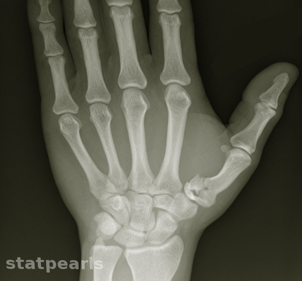
Clinical examination is required for all patients presenting with acute or chronic thumb injuries. Clinical examination alone is not enough for the differentiation between Rolando and Bennet fractures, nor does it allow identification of comminuted fracture patterns. Thus, radiographic imaging is necessary for the comprehensive evaluation of these injuries.
Rolando fractures maintain an intact volar carpal ligament, which prevents displacement of the volar fragment, whereas the dorsal fragment gets displaced by the abductor pollicis longus (APL). The thumb metacarpal shaft suffers displacement by the adductor and extensor pollicis longus (EPL).[11][12] The volar-ulnar fragment remains held in place by its attachment to the trapezium via the anterior oblique ligament, formerly described as the beak ligament.[10]
Evaluation
Diagnosis requires radiography in the two orthogonal projections, but since the thumb lies on a different plane from the rest of the hand,[10] the use of specific views can help to identify and classify the lesion.[6] In particular, the projection of Robert and that of Bett, help to evaluate the articular congruence and the degree of displacement of the fragments.
- Robert view (true AP of the thumb)
- The forearm is in maximum pronation with the dorsum of the thumb lying against the cassette
- True lateral of the thumb
- Hand pronated 30 degrees; radiographic beam angled 15 degrees distally
- Oblique view
In particular, the Robert view allows us to obtain an accurate anteroposterior view; it requires that the back aspect of the thumb rests on the radiographic plate, with the hand in overpronation. The Bett view, instead, allows an actual lateral image of the tarsometatarsal joint. It can be obtained by placing the palmar aspect of the hand on the radiographic cassette, pronated with an inclination between 15 and 35 degrees with the beam inclined at 15 degrees distal to proximal.[10]
CT scans obtained with collimation, and slice thickness between 0.5 and 1.0 mm, complemented by multiplanar and 3D reconstructions can provide valuable information for surgical planning.[13] The associated ligaments and tendons injuries, as well as osseous abnormalities, can be examined with MRI. For this purpose, MRI should be performed using small fields of view (FOVs) and small extremity coils to optimize the signal-to-noise ratios.[13]
Treatment / Management
This fracture is much more complex to treat than Bennett fracture patterns due to its intrinsic instability.[10][14] The choice of the most appropriate treatment method is influenced mainly by the number of fragments and the degree of displacement. In general, the following treatment algorithm is recommended[15]:
- Closed reduction and thumb spica splinting/immobilization
- Indicated for extra-articular fractures or minimally displaced 2-part articular fractures (i.e., Bennett fractures) with less than 1mm displacement
- Closed reduction and percutaneous pinning (CRPP)
- Percutaneous reduction pinning with K-wire fixation is necessary to set either:
- Extra-articular fractures with more than 30 degrees of angulation following closed reduction
- Rolando fracture patterns with minimal (i.e., less than 1 mm) of displacement or when fracture patterns are
- Comminuted fracture patterns not amenable to screw fixation
- Open reduction internal fixation (ORIF)
- Indicated when greater than 1 mm displacement in intra-articular fractures (Bennett or Rolando fracture patterns) and comminuted fracture fragments involving the metacarpal base when the fragments are large enough and amenable to screw fixation
- Distraction and external fixation
- Indicated for the following:
- Rolando fracture with >1mm displacement and significant soft tissue injury not amenable to ORIF
- Bennett, Rolando, or severely comminuted fractures with fragments too small for ORIF
Surgery can be open or arthroscopic, but in both cases, it requires an articular approach.[16] Open reduction is possible through a Wagner incision, radial to the thenar skin creases.[17] Fixation may be obtained by means of a plate and screw, tension banding and K-wires.[10] The surgical approach allows ligaments to be freed, eliminating any capsular interpositions, and the fracture reduction process to be visually monitored to obtain a temporary fixation with Kirshner wire and the application of screws or mini T-plates to fix the diaphysis and epiphysis together. If the strength of the construct allows it, early and even immediate mobilization is encouraged, prescribing a removable commissural splint for the first month.[5]
Differential Diagnosis
- Bennett fracture patterns
- Soft tissue or other osseous injuries to the thumb ray
- Includes thumb UCL injuries (include gamekeeper and skier thumb)
Prognosis
Data on the long-term outcome of Rolando fractures in the scientific literature are limited. In the series by Langhoff et al., out of 17 Rolando fractures, 82% required surgery. Among fractures treated surgically, 11 were treated by open reduction while three by percutaneous K-wire fixation. The quality of the radiologically evaluated reduction was excellent in 45.4% of patients treated with open surgery, and in none of the cases treated percutaneously. However, in a follow-up study on 16 of these patients at a median distance of 5.8 years, the quality of the obtained reduction was not related to the presence of symptoms or the onset of osteoarthritis, both accounting for 37%.[18]
Complications
Stiffness and osteoarthritis are the main long term consequences of Rolando’s fracture. Multi-fragmentary forms are considered the most severe forms and those most at risk of joint stiffness and osteoarthritis.[5]
Enhancing Healthcare Team Outcomes
• Rolando fractures are intra-articular fractures of the base of the first metacarpal with the detachment of several fragments - typically three. • Rolando fractures account for 21% of fractures of the base of the first metacarpal.• The pathogenetic mechanism is the same as that of Bennet fractures, but the damaging force is of greater magnitude.• Diagnostic imaging uses specific radiological projections and thin layer CT.• In fractures with a few large fragments, the treatment is preferentially surgical.• Ligamentotaxis with percutaneous traction may be preferred in markedly comminuted fractures but may lead to a poor quality restoration of the articular surface, and may suffer exposure to infectious complications.• Multi-fragmentary forms are more at risk of articular stiffness and osteoarthrosis.
An interprofessional team of orthopedic surgeons, orthopedic nurses, and specialist orthopedic physical therapists will result in the best outcomes. [Level 5]