Continuing Education Activity
A papilloma is a benign non-cancerous epithelial proliferation caused by the Human Papillomavirus. Most papillomas are caused by low-risk HPV forms, and self-limiting human papillomavirus (HPV) is still the leading cause of cervical cancers, causing 270,000 deaths annually worldwide, of which 85% occur in developing countries. This activity reviews the evaluation and management of papillomas and highlights the role of the interprofessional team in managing patients with this condition.
Objectives:
- Outline the typical presentation of a patient with papillomas.
- Review the risk factors for developing papillomas.
- Outline the management considerations for patients with papillomas.
Introduction
A papilloma is a benign (non-cancerous) tumor arising from an epithelial surface and usually known to grow in an outward direction. Commonly, a papilloma refers to squamous cell papillomas which appear as frond-like tumors that can develop almost anywhere on the body with squamous epithelium:
- Skin: as warts and cutaneous papillomas
- Lip
- Oral cavity
- Eyelid
- Tongue
- Pharynx
- Larynx
- Esophagus
- Cervix
- Genital tract: known as genital warts
Most of these lesions are caused by Human Papillomavirus (HPV). They are contagious upon contact with the exception of cutaneous papilloma, which is also called an acrochordon or more commonly known as a skin tag. Most of these lesions are usually self-limiting in immunocompetent individuals.[1]
Etiology
The majority of papillomas are caused by human papillomavirus (HPV). There are over 170 subtypes of the virus. Usually, types 6, 7, and 11 are mostly associated with papillomas and are called low-risk types as they don't usually cause precancerous lesions and rarely progress into cancer. HPV has been found to cause papillomas to arise almost anywhere in the body with stratified squamous epithelium, like the skin, conjunctiva, oropharynx, larynx and upper trachea as well as the anogenital tracts.[2]
Studies conducted have found HPV6/11 in 96% to 100% of all Genital wart lesions.[3][4] Infection is very contagious through direct and sexual contact. Genital HPV is spread by sustained direct skin-to-skin contact. Vaginal, anal, or oral sex are the most common ways of spread. Several studies conducted have shown that most skin tags or cutaneous papillomas of the head and neck to be non-contagious, unlike warts despite containing low-risk forms of HPV DNA, especially of type 6/11.[5][6][7] However, a study conducted by Pezeshkpoor et al. had found no significant association between skin tags and HPV.[8] This is why we term non-viral growths as acrochordons.
There is evidence of vertical transmission of human papillomavirus from mothers to their infants.[9] This may lead to the development of several papillomas in the larynx and upper trachea called Recurrent Respiratory Papillomatosis. It is a serious condition, as the papillomas may eventually enlarge causing obstruction of the airway.
It is worth noting that some papillomas are non-viral in origin as in the inverted papilloma of the urinary tract which has strongly been associated with smoking.[10] Nasal papillomas may be caused by local irritation and trauma to the mucosa, and some cutaneous papillomas have been associated with skin irritation. An example of this was cutaneous papillomas that had occurred in rats, mice, and hamsters following the local application of powerful carcinogens where they are believed to arise from the stratified squamous epithelium.[11] In addition, there are types of papillomas in which the mechanism responsible for their occurrence isn't fully understood, as in intraductal (breast duct) papillomas and choroid plexus papillomas.
Epidemiology
The global prevalence of HPV infection, the main cause of papillomas, is around 11% to 12%. However, reliable surveillance figures are difficult to obtain regarding the prevalence of warty lesions. For non-genital warts, two large population-based studies found prevalence rates of 0.84% in the US and 12.9% in Russia, with prevalence being highest in children and young adults.[12] For genital warts, the annual incidence was found to be between 0.1 to 0.2% of the population in developed countries. The highest prevalence was found amongst teens and young adults.
Acrochorda or skin tags have been reported to have a prevalence of 46% in the general population with a higher prevalence among older age groups, unlike warty papillomas. They are more common in the obese and tend to grow in areas of skin-to-skin contact. They have an equal distribution among males and females.
Pathophysiology
Papillomas arise from the skin or some mucosal surfaces depending on the different types of Human Papillomavirus involved and their affinity to different sites. For example, conjunctival papilloma is caused by HPV infection type 6, 11, 16, 33, and 45, which is somewhat dissimilar to genital warts caused by types 2, 3, 6, 11, 16, 18, and 30-32 and cutaneous papillomas (types 1-4 and 26-29).
Infection is established in the basal cell layers of the epithelium, but this involves the expression of a limited part of the viral genome. It's not until the basal cells develop and move externally through the different layers of the skin of the stratum spinosum and granulosum, that the virus begins to replicate itself, and the lesion becomes infectious.
Normally papillomavirus infects the epidermis in sites near the site of entry, but self inoculation often occurs, and the virus can infect farther sites. This phenomenon is known as the Koebner phenomenon. It has been observed that the immune system has an important role in controlling the spread of the virus, as, despite the virus infecting the intraepidermal cells that are considered "hard-to-reach" by the immune system, it was found that papillomas tend to reactivate and are more extensive in immunocompromised individuals.
Histopathology
Papillomas take origin from an epithelial surface. Complex folds of proliferating epithelium can be observed and are accompanied by a growth of supporting connective tissue and blood vessels. Typical examples are found in the skin, e.g., the common wart. Under the light microscope, these benign tumors show:
- Normally arranged epithelial cells e.g., in skin papillomas, the surface cells are squamous, and proliferation is confined to the deepest layers.
- The inner layer is a fibrovascular core with well-formed blood vessels.
- The outer layer is the epithelium, which is infolded and characterized by acanthosis, papillomatosis, and hyperkeratosis.
- The relationship of the epithelium to connective tissue is normal. They show no evidence of infiltration or invasion of the underlying connective tissues.
- In the case of conjunctival papillomas, pedunculated papillomas are usually covered by conjunctival epithelium.
History and Physical
History
- The patient usually complains of a small rough bump arising from the skin. This may either be painless or painful.
- The patient may describe habits that increase the risk factor for transmission, such as showering in communal showers, professionally handling meat products, and farm animals bare-handed or performing unsafe sex.
- The patient may report other symptoms that are more specific with the site from where the papilloma arises.
- Intraductal papillomas arising from the breast may present with bloody nipple discharge.
- Recurrent respiratory papilloma arising from the larynx and trachea may cause stridor and voice hoarseness.
- Nasal Papillomas can cause sinusitis and the loss of smell[13]
Physical Findings
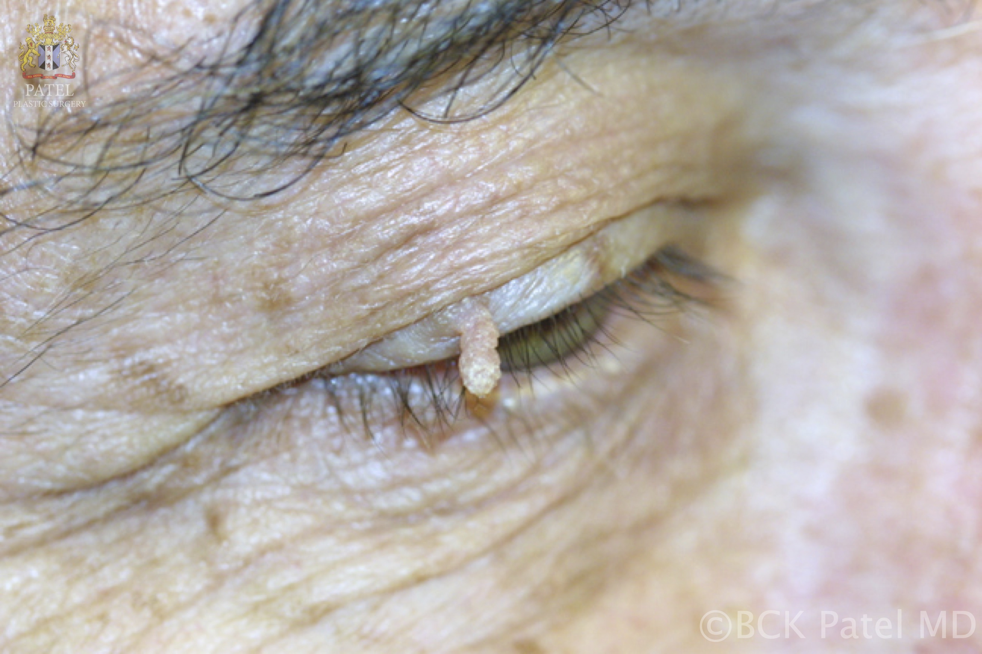
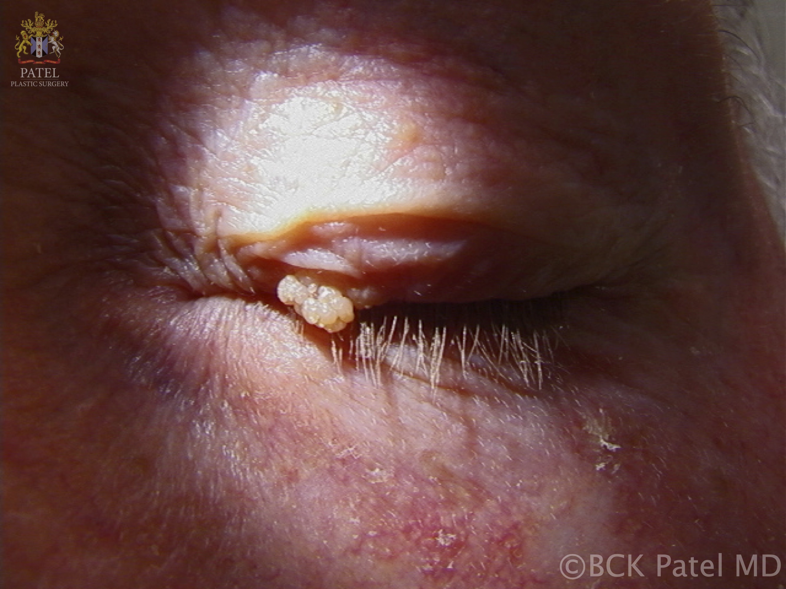
Single or multiple solid papules are observed. On the skin, it may appear as a rough solid papule, often covered with hyperkeratinized skin. Those arising on mucosal surfaces appear as a soft, pedunculated mass (supported on a stem or stalk) with numerous finger-like projections. The projections may be long and pointy or short and rounded if keratin has built-up around the lesion. Less keratinized lesions are pink or red in color and resemble a raspberry, whilst heavily keratinized lesions are white and look like the head of cauliflower.
Evaluation
Usually, most squamous cell papillomas are diagnosed upon examination and require no further investigations, especially in immunocompetent individuals where they are self-limiting and don't transform to malignant lesions. However, in those considered to have a higher risk of turning malignant, e.g: anogenital and oropharyngeal tract papillomas, they may undergo excision and a biopsy is sent for histopathological investigations if there is any question as to the diagnosis or if there is a concern for dysplasia.[14][15]
Although genital warts are usually caused by low-risk HPV subtypes, 5% to 20% of the individuals affected have been found to carry other sexually transmitted diseases. It is always important to suspect high-risk HPV infections that cause precancerous lesions in those individuals with genital warts and early screening should be initiated. Screening for cervical dysplasia/malignancy is typically accomplished through speculum examination and Pap smear.
Treatment / Management
Sometimes painless cutaneous papillomas may be left untreated and regress with time. They seem to not increase in size over time, and the potential for malignant transformation is low in immunocompetent individuals. If treatment is indicated, it varies depending on the type, size, and location of the papilloma.
- Skin warts and genital warts are managed with topical medications or procedures, such as cryotherapy or laser surgery.
- Skin warts may also be removed by excision (whether cutting it with a scalpel or cauterizing it).
- Surgical removal is a primary treatment for papillomas of the brain, breast ducts, and respiratory tract.
- Papillomas tend to recur around the primary site of infection, and the need for retreatment may be required.
- No-touch techniques of surgical removal with cryotherapy have been found to help prevent viral dissemination and reduce the papilloma recurrence.[16]
Differential Diagnosis
There is a wide range of diseases characterized by an overgrowth of the epithelial tissue giving solid papules on the skin that have to be differentiated from papillomas.
- Acrochordon
- Adenoma
- Basal cell carcinoma
- Chancroid
- Condyloma latum
- Corns & calluses
- Cysts
- Emilia
- Herpes simplex
- Keratoacanthoma
- Molluscum contagiosum: caused by a poxvirus and forms fleshy papules.
- Psoriasis
- Moles
- Neurofibromas
- Nevus lipomatosus
- Seborrheic keratoses
Prognosis
The prognosis of papillomas caused by HPV infection is usually good in immunocompetent individuals, but recurrences may occur. Some subtypes of HPV may cause vulvar intraepithelial dysplasia, cervical dysplasia, and cervical cancer. Although these subtypes don't typically cause papillomas, they are commonly present side by side with genital papillomas.
In immunocompromised individuals, papillomas often increase and spread more rapidly, and there is a higher risk of papillomas transforming into malignancy.[17]
Complications
Rarely are papillomas able to cause serious and life-threatening complications. This mainly depends on the anatomical site of the lesion.
- Recurrent respiratory papillomatosis may grow extensively and rapidly in the larynx and upper trachea, causing upper airway obstruction and stridor.
- Choroid plexus papillomas in the brain cause increased cerebrospinal fluid production and can lead to increased intracranial pressure and hydrocephalus, which may eventually lead to brain herniation and death.
- Papillomas in themselves are rarely premalignant in immunocompetent individuals; however, malignant transformation still does occur.
- A papilloma at the lacrimal punctum may grow into the punctum and canaliculus and involve the lacrimal sac and nasolacrimal duct (inverted papilloma).
Deterrence and Patient Education
Here are important points to take care of to prevent papilloma:
- Safe sex
- Routine Pap smear screen
- Wearing flip-flops while taking communal showers
- Taking caution upon the occupational handling of meat and preferably wear disposable gloves
- Warts are usually contagious upon direct contact. Skin tags, however, are not contagious
- Vaccination: HPV vaccine is available and recommended to be taken by ages 9-12 to prevent certain precancerous lesions in males and females. The 9 valent vaccine is effective against Human Papillomavirus subtypes 6,11,16,18,31,33,45,52 and 58.
Enhancing Healthcare Team Outcomes
HPV is the most common cause of papillomas of the mucous membranes and skin. There are over 175 subtypes of HPV, and some are associated with an increased risk of malignancy.
For the most part, HPV is acquired by direct contact of other infected individuals or livestock, and one of the best ways to decrease the morbidity of this infection is through education.
Patient counseling by the pharmacist is important to educate individuals about the different treatments available for warts, their benefits, and adverse effects.
It is important to educate teens who are about to start being sexually active about safe sex by their school nurse or trained teachers.
Furthermore, it is necessary to let parents know the importance of vaccinating their children before being sexually active.
Encourage sexually active females with genital warts to undergo the pap smear to determine the presence of cervical dysplasia and high-risk HPV infections. Patients with genital warts should avoid sexual activity until their warts are treated.
Patients should avoid touching their warts to avoid self inoculation.