Continuing Education Activity
Keloids are benign raised scars that are formed after cutaneous insult. Normally, cutaneous injury results in wound closure and a flat scar. Some individuals are prone to alterations in the healing process that result in excessive tissue proliferation and excess collagen in the skin during the healing process and the formation of a keloid. The keloid can appear months years after the cutaneous insult and can continue to grow indefinitely. This activity details the evaluation and treatment of keloids and highlights the role of the interprofessional team in the management and education of patients with this condition.
Objectives:
- Explain the pathophysiology of keloid formation.
- Outline the rates of keloid recurrence after steroid injections versus after surgery alone versus after surgery followed by radiation.
- Describe the optimal radiation treatment plan for treating keloids after surgical resection.
- Summarize the importance of collaboration and communication amongst the interprofessional team to improve outcomes for patients being treated for keloid scar removal.
Introduction
Keloids are benign raised scars that form by excessive tissue proliferation and excess collagen in the skin during healing. Although benign lesions, keloids can often have a negative impact on self-esteem and quality of life.[1] Under normal circumstances, cutaneous injury results in wound closure and a flat scar. However, an imbalance at any point during the wound healing process can result in continued growth of a painful and pruritic keloid scar. The keloid can appear a few months to a few years after the cutaneous insult and continue to grow indefinitely without signs of regression.[2]
Etiology
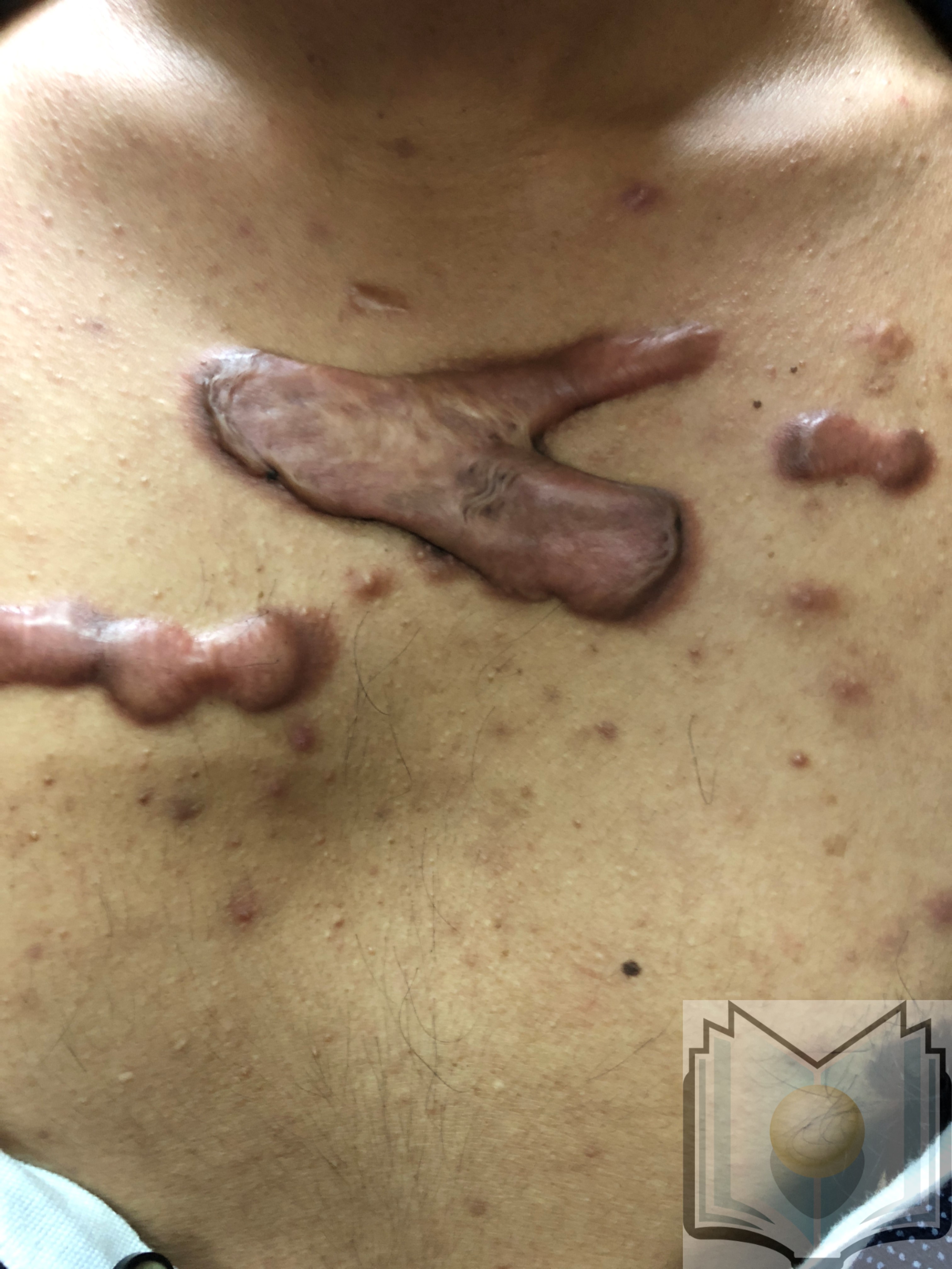
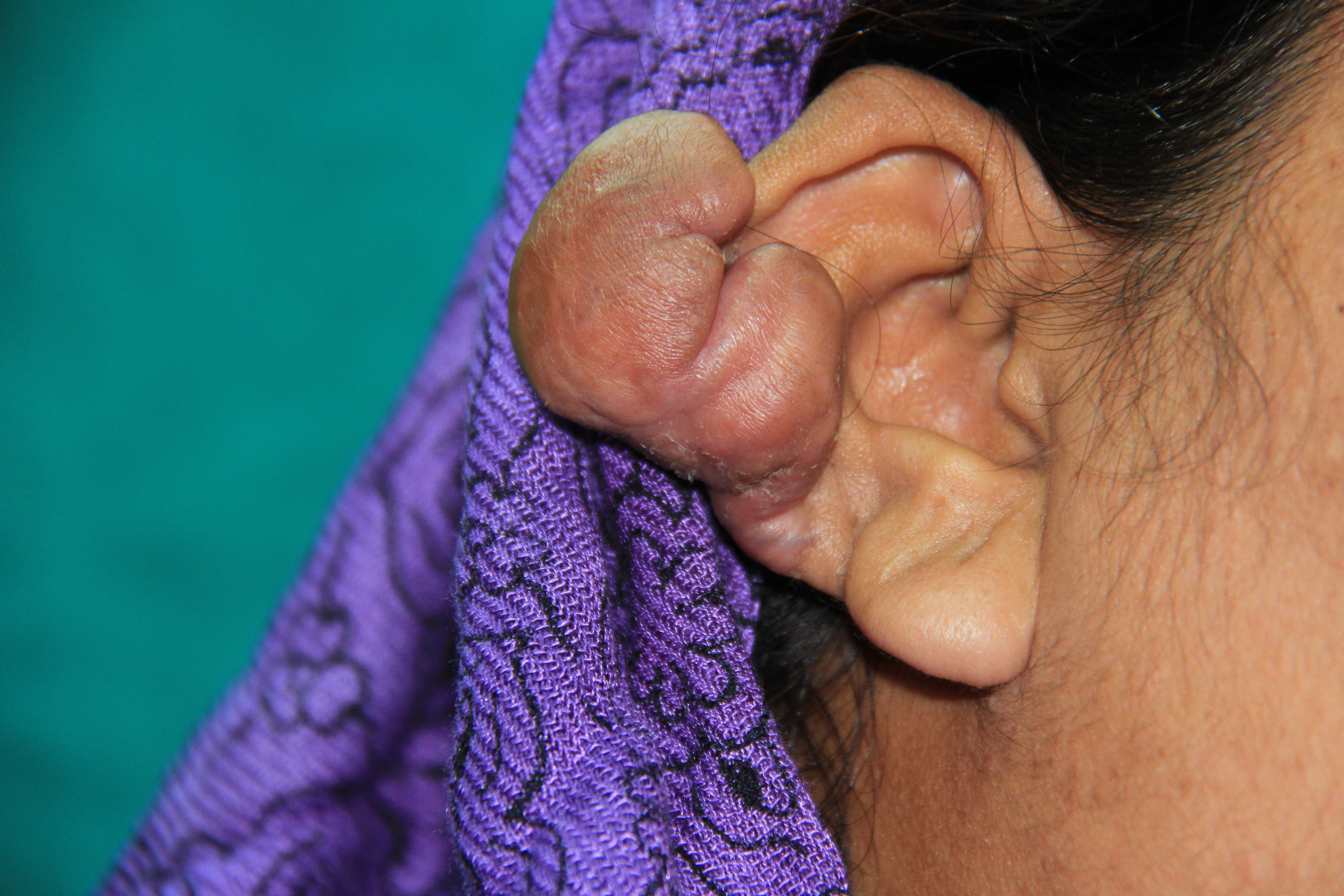
The etiology of keloid scar formation is not completely understood. Though some keloids can form spontaneously, most develop as a result of pathologic wound healing following trauma or surgery, sometimes forming years after the cutaneous injury.[2][3] Inciting events can include acne, body piercings, tattoos, insect bites, vaccinations, surgery, or mechanical force. They tend to form in regions with high skin tension, like the sternum or earlobes.
Epidemiology
The prevalence of keloids, which occur in anywhere from 30% to 90% of patients, varies depending on region and race but tends to be higher in darker skinned race individuals (non-albino African-Americans, Hispanics, and Asians) and those with a family history of keloids. Age wise, patients 10 to 30 years of age are at the highest risk, through periods of hormonal changes (puberty, pregnancy) also increase risk. Additional risk factors include having blood type A or hyper-IgE syndrome. [3][4]
Pathophysiology
The physiologic response to cutaneous injury is the formation of a scar, a process which involves inflammation, proliferation, and remodeling or maturation. Keloids form due to pathological wound healing and excess dermal fibrosis. Abnormal prolongation of the inflammatory phase of wound healing can lead to an imbalance in the destruction and deposition of extracellular matrix (ECM), which results in accumulation of collagen and other ECM matrix components. The overabundance of ECM components in the dermis and surrounding subcutaneous tissue leads to excessive scarring and keloid formation.
Numerous genetic and environmental factors are involved in the complex pathophysiology of keloids. Given that fibroblasts deposit the majority of collagen and ECM, they are thought to be the key cellular component in the fibrotic process. Other cellular players include myofibroblasts, keratinocytes, melanocytes, and mast cells. Regulatory molecules (e.g., TGF-beta, PDGF, VEGF, proteolytic enzymes) and fibrotic signaling cascades (e.g., SMAD signaling, toll-like receptor signaling, fibronectin) have also been shown to have an important role in the sequence of events leading to keloid formation. [4][5]
Histopathology
Histologically, keloids are characterized by abundant, tightly packed but disorganized collagen. The epidermal layer may appear normal, but the dermis becomes persistently infiltrated with inflammatory cells and hypo-cellular collagen bundles, made up of large, thick, and wavy type-I and type-II hyalinized fibers. Hypertrophic scars are distinguished from keloids by the presence of finer, more organized collagen bundles that run parallel to the epidermal surface. Additionally, infiltration of inflammatory cells may be present in fresh hypertrophic scars but decreases as the scar ages. [2]
History and Physical
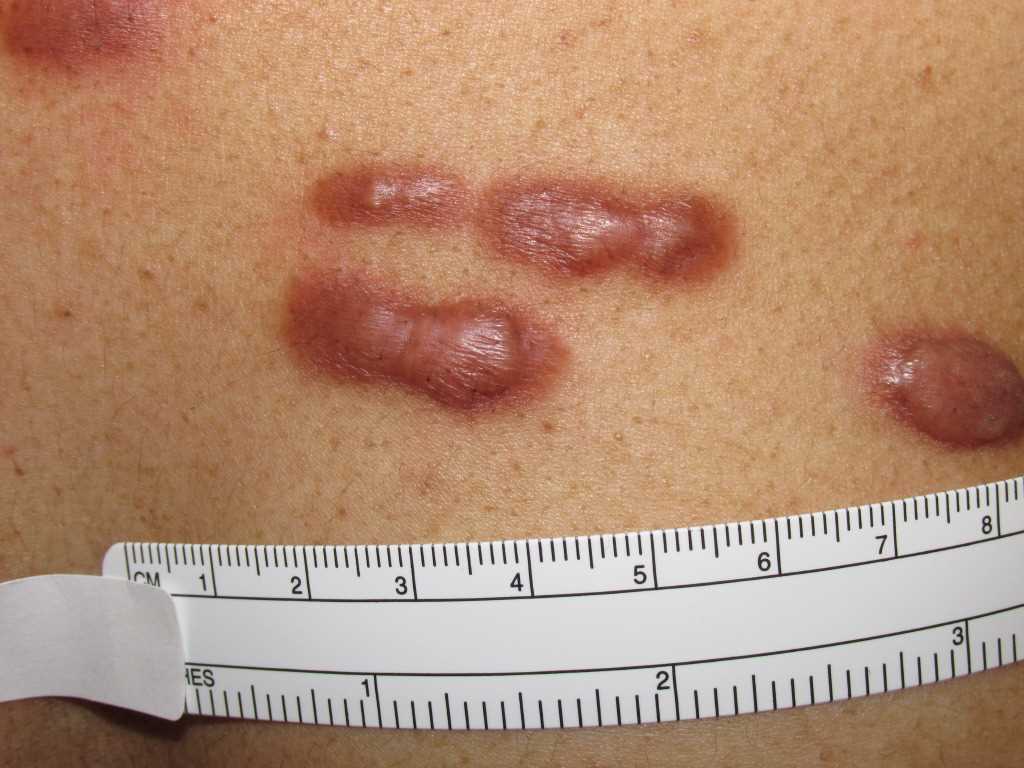
It is possible for keloids to appear spontaneously without predisposing cutaneous trauma, but most patients present with a history of surgical incisions or trauma to the skin. A mature scar should be flat and light-colored, but improper wound healing can lead to the formation of hypertrophic scars or keloids. Hypertrophic scars are elevated, erythematous, and occasionally pruritic but confined to the borders of the initial injury. Keloids are similar to hypertrophic scars in that they are elevated, erythematous, and sometimes pruritic but differ from hypertrophic scars in that they expand beyond the original borders of injury and can invade surrounding tissue. The resulting keloid causes not just a physical disturbance, but can also lead to functional impairment, cosmetic distortion, and psychological distress.
Keloid lesions can be classified into minor or major types. Minor keloids are small and focal scars with an elevation of less than 0.5 cm, while major keloids are larger and can have an elevation greater than 0.5 cm. Both types can invade surrounding tissue and continue to grow years after the initial injury. They generally do not regress on their own and can recur after surgical excision. Keloids can occur anywhere on the body, but they are most commonly seen on the neck, earlobes, chest, shoulder, upper back, sternum, knees, and ankles. [1][2][3]
Evaluation
The most common methods for evaluation of keloid scars include the use of a scoring scale (e.g., Vancouver Scar scale) for subjective evaluation and 2-dimensional photography for more objective measures. Less commonly used evaluation techniques include biochemical properties, ultrasound, laser-based techniques, and biopsy.[6]
Treatment / Management
Medical Treatment
No single medical therapy has been shown to be effective in treating keloids. Intra-lesional corticosteroids injections are a common first-line therapy for keloids to reduce scar volume, height, and related symptoms of itching and pain. Since steroid treatment has recurrence rates (9%-50%) and a high frequency of side effects (hypopigmentation, skin atrophy, telangiectasias, injection pain), intra-lesional steroid injections are usually combined with other therapies to improve outcomes. Additional treatments that do not have strong evidence for decreased recurrence or efficacy but have had varying anecdotal success include massage therapy, pressure garments, adhesive tape support/silicone gel sheeting, cryotherapy, and laser/light therapy.[3][4][7]
Surgical Treatment
Surgical excision is frequently used for the treatment of mature keloid scars but is considered ineffective as a monotherapy given the recurrence rate of 45% to 100%. To reduce the risk of recurrence, it is recommended to combine surgical excision with more conservative treatment options, such as radiotherapy, cryotherapy, or steroids. Regarding surgical technique, it is recommended to do an intramarginal fusiform excision (incomplete resection), using minimal tension and sutures to close the wound with everted borders. The use of subcutaneous/fascial tensile reduction sutures, z-plastics, and local flap transfer are thought to prevent keloid recurrence.[4][8]
Differential Diagnosis
Keloids can clinically appear similar to hypertrophic scars but have histopathological differences described above.
Radiation Oncology
Compared to surgery or radiation therapy alone, surgical excision followed by immediate postoperative radiation therapy has been shown to be the most effective treatment, with a recurrence rate of about 20%.[8] Radiation is often indicated for recurrent keloids, or for patients with a high-risk of recurrence, including marginal resections, wider spread, and unfavorable locations. Radiation starting on the same day after surgical removal of the keloid is most effective. This usually requires coordinated care between the surgeon and radiation oncologist to make sure the timing of the surgery and radiation can be optimal.
Radiation Dosing and Fractionation
There is a range of radiation prescriptions that have been used. The recommended total dose ranges from 12 to 20 Gy and historically was low dose per fractionation (3 to 4 fractions daily in 3 to 4 Gy/fx). However, newer data has suggested that using a radiation prescription that delivers a biologic effective dose (BED) of greater than 30 Gy (e.g., 13 Gy/1fx, 16Gy/2fx, 18Gy/3fx over 5 to 7 days), has better long-term control.
Radiation Treatment Planning
Simulation and Target Volume
The scar plus a 1cm radial margins, 0.5 to 1 cm depth, is targeted for radiation. Depending on the site of the lesion and concern for radiation dose to surrounding structures, either a clinical or CT simulation can be used. The patient should be placed in a reproducible position. Lead shields or absorbers can be positioned near the field to block critical structures.
For example, if a patient gets radiation to the earlobe, they would be simulated in the supine position with the head turned so that the affected ear is facing up. Then an aquaplast mask can be made to immobilize the head and replicate the rotation of the head. The mask can be cut out near the ear to allow visualization of the treatment area. The scar may be wired, and the 1 cm margin can also be wired to help with creating an electron cut out and with treatment planning. A lead shield with wax cover can be placed behind the lobe to block radiation dose to the neck and brain.
Radiation Modality
Different forms of radiation can be used to deliver dose to a superficial target, including electrons (less than or equal to 6 MeV), x-rays (70 to 150 kV), and iridium-192 brachytherapy with implants or surface applicators. The modality most commonly used to treat keloids is electrons. Electrons are generated and delivered by a standard radiation machine called a linear accelerator, the same machine that delivers photons that are used in a majority of radiation treatments. Electrons have a shallow dose penetration compared to photons and are ideal for treating superficial lesions while minimizing dose to normal underlying structures, making it ideal for skin lesions. Often a 1 cm bolus is placed over the lesion increase the dose at the skin while reducing the depth of penetration.[9]
Superficial x-ray therapy uses photon beams to deliver energy to a maximum depth of 5 mm. Similar to EBRT, superficial x-ray therapy can treat superficial lesions without damage to the underlying structure. This method is also less costly and easier to use but can result in uneven delivery of dosage since radiation drops off at the periphery.[9]
Brachytherapy is also another modality to treat keloids that have been very successful. However, it is not as widely used because all radiation centers do not have the physical radioactive source (Iridium-192) needed for brachytherapy. Usually, a surface applicator or mold would need to be created so that the catheters that would hold the Ir-192 could be placed on the scar to allow for dose penetration to the skin at risk for re-developing a keloid. Brachytherapy can deliver more precise radiation. However, the radiation dose delivery is very heterogeneous, and it could lead to increased skin toxicity. In a meta-analysis systemic review of post-excisional radiation modalities by Mankowski et al., brachytherapy was demonstrated to have the lowest recurrence rate (15%) compared to EBRT (23%) and x-ray therapy (23%).[10]
Prognosis
Numerous studies have demonstrated that a surgery followed by radiation therapy is most effective for the treatment of keloids. A literature review of over 70 studies showed that radiotherapy after surgical excision had significantly lower recurrence rates (22% +/- 4%) compared to radiation as a single modality of treatment (recurrence rate of 37% +/- 12%). The same meta-analysis revealed that among the radiation therapy modalities used for keloid treatment, post-excisional brachytherapy had the lowest recurrence rate at 15%, compared to 23% recurrence for post-excisional electron beam therapy and post-excisional x-ray therapy.
In addition to treatment modality, the rate of keloid recurrence also varies by anatomic location. Compared to keloids located on the ear, head, and neck, or extremities, keloids located on the chest and truck had the highest rate of recurrence at 34%. Keloids localized on the ear were found to have the lowest rate of recurrence (12%). This variation in recurrence rates based on anatomical location is thought to be due in part to the difference in the tensile strength of skin in each of those areas.[10]
Complications
The most common complications of radiation for keloid treatment, with occurrences around 30%, are erythema and pigment related changes (hypopigmentation, hyperpigmentation). Acute complications, which can arise during radiation or seven to ten days after treatment, are related to the total dose of radiation given. These can manifest as erythema, edema, desquamation, ulceration, or necrosis. Late complications can occur weeks to months after treatment and are related to the dose of radiation given per session. These include pigmentary changes (hypopigmentation or hyperpigmentation), alopecia, atrophy, and telangiectasis. Although seen in less than 1% of cases, infection and wound dehiscence are also possible complications of keloid treatment. Use of an emollient and steroid ointment after radiation treatment can help reduce the risk of side effects.
Pearls and Other Issues
- Keloids are a benign overgrowth of scar tissue that can lead to pain and disfigurement
- Surgery followed by radiation therapy is most effective for the treatment of recurrent keloids
- Start radiation on post-op day 1 and use a radiation prescription with BED greater than 30 Gy (i.e., 6 Gy times 3) for optimal local control
Enhancing Healthcare Team Outcomes
The management of keloids is difficult and thus the condition is best managed by an interprofessional team that includes a dermatologist, plastic surgeon, radiation expert, primary care provider and the specialist nurse. There is no effective treatment for keloids and the only reason for treatment is cosmesis. If the patient does not have problems with aesthetics, keloids should be left alone. There are dozens of treatments for keloids and none is better than the other and all have the potential to worsen the original lesion. Recently radiation has been touted as a treatment for keloids. While the treatment works, it is not reliable or consistent and it is not possible to predict in which patient it will work. The primary care providers should educate the patient on different treatment options and their pros and cons. The best remedy for keloids is to do nothing.