Continuing Education Activity
Bowenoid papulosis is an uncommon sexually transmitted condition that occurs in both males and females. It is characterized by multiple well-demarcated red-brown to violaceous papules in the genital area. This activity describes the etiology, epidemiology, clinical presentation, evaluation, and management of Bowenoid papulosis and shows the importance of the interprofessional team in educating patients on safe sex.
Objectives:
Determine the etiology of Bowenoid papulosis.
Identify the clinical presentation of Bowenoid papulosis.
Assess the management and follow-up monitoring for patients with Bowenoid papulosis.
Communicate how an interprofessional team can collaborate to improve the diagnosis evaluation, management, monitoring, and education of patients about safe sex to prevent Bowenoid papulosis.
Introduction
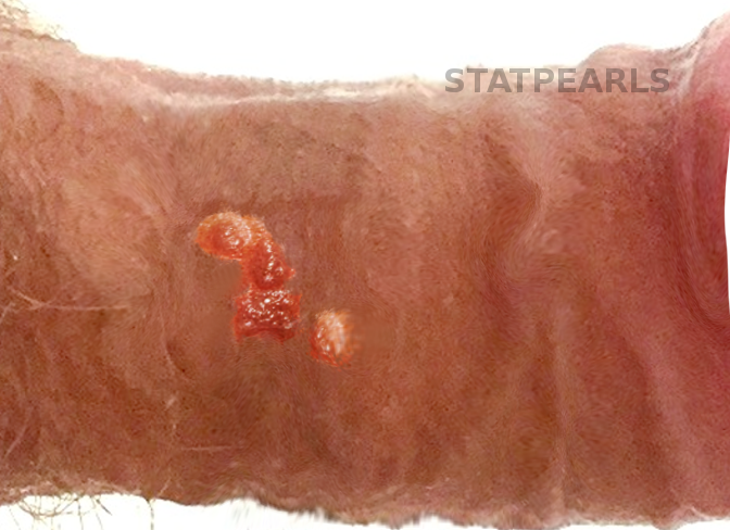
Bowenoid papulosis is an uncommon sexually transmitted condition. It was first described in 1977 by Kopf and Bart as penile papules. However, it occurs in both sexes. It tends to affect young, sexually active people. This condition was also termed “vulvar intraepithelial neoplasia (VIN)” in the vulva and termed penile intraepithelial neoplasia (PIN) in the penis.[1] The classification of this disease was confusing and included 3 clinical entities: bowenoid papulosis, Bowens disease, and erythroplasia of Queyrat. Now it is recommended that these three entities not be used to describe lesions in the anogenital area.[2] However, dermatologists still recognize Bowenoid papulosis as a distinct clinical variant. In fact, Bowenoid papulosis is induced virally by human papillomavirus (HPV) and presents as solitary or multiple skin-colored papules in the anogenital area. It can last from 2 weeks to several years. Clinically, bowenoid papulosis is assimilated to genital warts, while histologically, it has a close resemblance to squamous cell carcinoma in situ (Bowens disease). Treatment is generally conservative. Bowenoid papulosis lesions are generally considered benign with a spontaneous regress leaving no sequelae in immunocompetent persons, although a small number may transform into invasive squamous cell carcinoma (see Image. Bowenoid Papulosis).
Etiology
Bowenoid papulosis is a sexually transmitted condition associated with HPV infection. Most lesions are associated with oncogenic HPV types, mainly the HPV 16 genotype, but occasionally, HPV 18, 31, 33, 34, 35, 39, 42, 48, 51, 52, 53, and 54 are detected.[2] Bowenoid papulosis may also occur in immunocompromised individuals, such as organ transplant recipients. Smoking has recognition as a recurrence factor.
Epidemiology
Bowenoid papulosis occurs commonly in sexually active individuals and predominantly in their third to the mid-fifth decade with a mean age of 31 years. However, bowenoid papulosis can appear at any age range from 3 to 80 years. Both sexes are affected, although recent data showed an increased number of women. There are an estimated 5 cases per 100000 women.[3] The exact prevalence is unknown because bowenoid papulosis lesions are related clinically to genital warts. There is no racial predilection for bowenoid papulosis.
Pathophysiology
Detection of papillomavirus common antigen in cases of bowenoid papulosis supports the hypothesis that bowenoid papulosis results from HPV. In fact, E6 and E7 viral oncoproteins of oncogenic HPV types contribute to oncogenesis by inducing over-expression of p16 protein and human telomerase reverse transcription (hTERT).
Histopathology
Histologically, bowenoid papulosis is characterized by acanthosis with full-thickness epidermal atypia, known as Bowenoid dysplasia. Multiple metaphase mitoses are usually visible above the basal layer and scattered dyskeratotic and multinucleated keratinocytes with pleiomorphism. Histopathologic findings may also show parakeratosis and hypergranulosis. The integrity of the basement membrane is preserved. The dermis contains a superficial infiltrate of lymphocytes with perivascular accentuation. Induction of the focal epidermal hyperplasia and dysplasia is viral. Moreover, immunohistochemistry for the p16 protein reveals strong in bowenoid papulosis. Staining with an antibody to p16 protein has high specificity and sensitivity to detect this disease.
History and Physical
Bowenoid papulosis is a rare transmitted condition and typically occurs in sexually active people. It is clinically characterized by multiple well-demarcated red-brown to violaceous papules, usually less than 1 cm in size. The surface of the lesion can be flat, smooth, papillomatous, or verrucous. Some papules may coalesce into large plaques. Sometimes, bowenoid papulosis presents as warty white plaques. The distribution of lesions is commonly discrete; sometimes bowenoid papulosis can have an annular or linear exhibition. In men, bowenoid papulosis lesions primarily involve the penile shaft but may also involve the foreskin, glans, scrotum as well as the anus. Whereas in women, the lesions are usually bilateral and affect the labia major, labia minor, clitoris, inside the vagina, inguinal folds, and perianal area. The lesions are generally darker in women than in men. They are usually asymptomatic; occasionally, patients may complain of pruritus and soreness of the affected area. Extragenital bowenoid papulosis is a very rare condition, and it may involve the face, fingers, or neck, with or without concomitant genital lesions.[4]
Evaluation
Because of its potential malignant transformation, the diagnosis is usually by a skin biopsy. HPV subtyping may also be a recommended next step. Microscopic findings show typical features of Bowens disease with only a few differentiating features. The distinction rests in the circumscribed plaque-like pattern, the multiplicity of lesions, the age of the patient, and less dyskeratosis and atypia and more dilated vessels in the dermis in pathology.[4] Hence the diagnosis of bowenoid papulosis is based on clinical grounds and histopathological correlation. Furthermore, a skin biopsy is recommended in case of recalcitrant lesions to standard therapies to rule out malignancy.
An extensive assessment of HPV infection is mandatory, including examination of the oral, genital, and anal areas. Besides, anoscopy should be performed in the case of receptive anal sex. All this must be performed for the patient as well as the partner.
Treatment / Management
The treatment aims to prevent malignancy transformation and to preserve normal tissue and function. Since the disease commonly occurs in young people and frequently remits spontaneously, the management of bowenoid papulosis is generally conservative. Without treatment, bowenoid papulosis lesions may regress in an average of 8 months. Treatment modalities include locally ablative or destructive therapies such as carbon dioxide (CO2) laser vaporization, cryotherapy, electrocoagulation, 5-aminolevulinic acid-mediated photodynamic therapy (ALA-PDT), excisional surgery, and 5 fluorouracil (5FU). Moreover, topical imiquimod cream 5% once a day on an alternate day for 1 month has proven good results on limited lesions of bowenoid papulosis with viral clearance in some cases. However, relapse often occurs with all treatment modalities.[3][5][6] Furthermore, there are prophylactic vaccines to prevent infection with oncologic HPV subtypes. Moreover, prevention of recurrence correlates with cessation of cigarette smoking.
Differential Diagnosis
The differential diagnoses for bowenoid papulosis include the following:
- Genital warts
- Psoriasis
- Lichen planus
- Condylomata acuminate
- Seborrheic keratosis
- Pigmented Bowens disease
- Melanocytic Nevus
- cutaneous squamous cell carcinoma
- Warty dyskeratoma
Prognosis
Bowenoid papulosis has a variable course. Lesions may regress spontaneously or persist for several years with older persons or immunocompromised patients. It may rarely transform into Bowens disease or invasive squamous cell carcinoma.[7]
Complications
Transformation into invasive squamous cell carcinoma is rare and occurs in less than 1 % of cases, especially in immunocompromised individuals. Females with bowenoid papulosis lesions and sexual partners of male patients are at high risk of cervical or vulvar carcinomas because of the infection with a potentially oncogenic HPV.
Deterrence and Patient Education
Bowenoid papulosis rarely evolves into invasive squamous cell carcinoma; however, it is essential to educate patients regarding the risk of potential malignant transformation. The patient should receive thorough education regarding HPV infections which spread via sexual contact. Therefore, the avoidance of direct sexual contact should be strongly advocated to decrease transmission of the disease.[8] Additionally, female partners of men should receive a close follow-up.
Enhancing Healthcare Team Outcomes
The management of bowenoid papulosis is best with an interprofessional approach, including a team of clinicians. Bowenoid papulosis may develop malignant characteristics; therefore, a long-term evaluation is recommended by a dermatologist every 3 to 6 months. Thus, the gynecologist should perform a careful cervical and anal cytologic screening of female patients as well as of sexual partners of the male patient. The urologist should examine patients with urethral involvement. Patients with perianal involvement should be examined by a GI specialist. Furthermore, smoking cessation is strongly advised.