Continuing Education Activity
Ureterolithiasis, commonly known as kidney stones, is a prevalent and burdensome urological condition affecting millions worldwide. Characterized by the formation of mineral deposits within the urinary tract, ureterolithiasis presents with excruciating flank pain and various associated symptoms, posing significant challenges to patients and healthcare providers alike. Despite its often acute and debilitating nature, ureterolithiasis typically follows a benign course, with many stones passing spontaneously. However, the potential for severe complications necessitates prompt and effective management strategies.
This activity for healthcare workers is designed to enhance learners’ proficiency in evaluating and managing ureterolithiasis. A comprehensive overview of ureterolithiasis, encompassing its epidemiology, clinical presentation, diagnostic modalities, treatment options, and preventive measures, is provided. Participants develop a deep understanding of the pathophysiology of this disease and relate the process to its manifestations. Learners improve skills in clinical assessment, differential diagnosis, and applying evidence-based evaluation and treatment strategies, preparing them to collaborate within an interprofessional team caring for patients with ureterolothiasis.
Objectives:
Identify the signs and symptoms indicative of ureterolithiasis.
Implement a clinically guided diagnostic plan for a patient with possible ureteral calculi.
Apply an individualized management plan for an individual diagnosed with ureterolithiasis.
Collaborate effectively among interprofessional team members to improve outcomes and treatment efficacy for patients with ureterolithiasis.
Introduction
Ureterolithiasis Overview
Ureterolithiasis is a worldwide disease affecting millions of people at a considerable cost and placing a significant burden on the global healthcare system. This disorder is also increasing in incidence and prevalence.[1] Ureterolithiasis is associated with other systemic conditions, specifically cardiovascular disease, diabetes mellitus, metabolic syndrome, and obesity.[2]
The condition often manifests with exceedingly painful flank pain radiating toward the groin. The pain occurs suddenly without warning. Episodes often recur after resolution. Unlike patients with an acute abdomen who wish to remain still, patients with ureterolithiasis typically want to move constantly, which is characteristic of colicky pain. Nausea and vomiting are commonly associated with acute ureterolithiasis. Lower urinary tract symptoms may occur when stones approach the bladder.
This unique clinical presentation, usually accompanied by hematuria (85%), makes it a relatively easy presumptive diagnosis to make in the emergency department. However, a definitive diagnosis generally requires imaging, preferably without contrast. Standard treatment involves appropriate analgesia, antiemetics, intravenous (IV) fluids, antibiotics when indicated, and medical expulsive therapy (α-blockers), which facilitate spontaneous stone passage in patients not requiring immediate surgical intervention.
Stone passage is usually determined by the stone's size, shape, and location, and the patient's ureteral anatomy. While most stones 5 mm and smaller pass spontaneously, stones with a diameter >7 mm and calculi that have not moved in 4 to 6 weeks may need surgical intervention. The 2 procedures most commonly performed to remove ureteral stones are ureteroscopy, usually with laser lithotripsy and stone basketing, and extracorporeal shockwave lithotripsy, which breaks stones into tiny fragments that can pass easily.
Ureterolithiasis associated with an infected kidney is potentially dangerous, causing obstructive pyelonephritis and urosepsis. Such situations require urgent renal pelvis surgical drainage. Definitive surgery of the ureteral stone is postponed until the infection is controlled and the patient has clinically recovered. Medical pyelonephritis cannot be clinically distinguished from the more dangerous obstructive pyelonephritis without appropriate imaging.
Hospitalization and urological surgical intervention are required in some cases.[3][4][5] Urosepsis, renal abscess, infected stones, chronic kidney disease (CKD), obstruction, extravasation, ureteral scarring, avulsion, and stenosis are all possible complications of ureterolithiasis.
Afterward, kidney stone prevention testing with a 24-hour urine collection is suggested for all high-risk patients, including recurrent stone formers, patients with renal failure, solitary kidneys, and cystine stones, children with stones, immunocompromised individuals, and people with high surgical or anesthesia risk. Such testing is optional for all other stone formers and should be discussed with these patients. Successful stone prevention requires a willingness to commit to long-term compliance with therapy, as ureterolithiasis can recur.
Renal System Overview
The urinary system consists of the kidneys, ureters, bladder, and urethra. Kidneys filter blood to produce urine, which then travels through the ureters to the bladder for storage until elimination through the urethra during urination. Urine formation involves blood filtration in the kidneys to remove waste products and excess substances, followed by reabsorption of essential solutes and secretion of the rest. The kidneys also regulate urine concentration by adjusting water reabsorption, thus maintaining water and electrolyte balance in the body.
Kidney stones develop when certain substances in urine become highly concentrated and crystallize, forming solid masses. Factors contributing to stone formation include dehydration, dietary factors, metabolic disorders, and genetic predisposition. Once formed, kidney stones can travel down the ureters and become lodged at various points along the urinary tract, leading to obstruction and symptoms.
The ureters contain anatomical constrictions—the ureteropelvic junction (UPJ), pelvic brim, and ureterovesical junction (UVJ)—which are common sites for stone impaction. Nerves innervating the ureters include sympathetic (T10-L2), parasympathetic (S2-S4), and visceral sensory fibers from the renal plexus (T10-L1). Sympathetic nerves regulate blood flow and smooth muscle tone, while parasympathetic fibers control peristalsis. Sensory nerves transmit pain signals in response to stimuli such as distension or obstruction. Kidney stones lodged in the ureter can irritate sensory nerves, causing severe colicky pain (renal colic), the intensity and location of which depend on the stone's location and the degree of obstruction.
Etiology
Renal calculi should be retrieved whenever possible and sent for chemical analysis.[6][7] Guidelines recommend offering 24-hour urine testing to all patients with kidney stones to identify risk factors for new stone formation, enabling proper preventive therapy.[8][9][10][11][12][13] The most common risk factors for renal calculus formation are discussed below.[14][15]
Chronic Infection
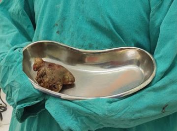
Chronic urinary tract infections can lead to the formation of a specific type of kidney stone called "struvite" or "infection stones," composed of magnesium, calcium, and ammonium phosphate. Struvite is the material that produces staghorn stones (see Image. Staghorn Renal Calculus). Urea-splitting organisms, typically Proteus or Klebsiella spp but not Escherichia coli, produce urease, which breaks down urea in the urine. Urea splitting increases renal ammonia concentrations and raises urinary pH, ultimately promoting struvite stone formation and growth.
Treatment includes infection control with total surgical removal of all possibly infected stone material.[16] A specific urease inhibitor, acetohydroxamic acid, may be useful in selected cases. See StatPearls' companion reference, "Struvite and Triple Phosphate Renal Calculi," for more information.
Cystinuria
Cystine stones account for only 1% to 2% of all kidney stones but have a high recurrence rate, are resistant to shockwave lithotripsy, and require aggressive prophylactic therapy. Only about 250 mg of cystine is soluble per liter at a neutral pH (pH 7), which increases to 500 mg per liter at a pH of 7.5. Hypercystinuria is an inheritable, autosomal recessive trait that can cause cystinuria and cystine calculus formation.
Cystine is relatively radiolucent but will appear on computed tomography (CT) scans. Treatment of cystine stones is usually surgical with ureteroscopy and laser lithotripsy. Preventive therapy involves aggressive hydration, sufficient to produce 3,000 mL of urine daily, and urinary alkalinization to maintain a urinary pH of 7.5. A thiol-based chelating agent may be used to increase the solubility of cystine if these measures are insufficient. See StatPearls' companion reference, "Cystinuria," for more information.
Hypercalciuria
Hypercalciuria, characterized by a urinary calcium level of 250 mg or more per 24 hours, is the most common chemical etiology of nephrolithiasis. Calcium-containing stones form approximately 80% of all renal calculi. Increased urinary calcium levels may be due to increased intestinal absorption of calcium, higher circulating serum calcium, reduced renal calcium reabsorption (renal calcium leak), hypervitaminosis D, hyperparathyroidism, high dietary animal protein ingestion, or systemic acidosis. Hypercalciuria increases the urinary saturation of calcium salts like oxalate and phosphate, causing crystal and calculus formation.
Treatment involves minimizing excessive daily oral calcium intake, correcting phosphate deficiencies that increase Vitamin D activity, oral phosphate supplementation, and using thiazides to decrease calcium excretion in the nephron. See StatPearls' companion reference, "Hypercalciuria," for more information.
Hyperoxaluria
Oxalate naturally occurs in plants, bound tightly to calcium in the plant's tissue fluid. Vegetable ingestion results in intestinal oxalate absorption and urinary excretion. Green leafy vegetables like spinach, rhubarb, and collard greens are particularly high in oxalate. Oxalate has no nutritional or beneficial role in human physiology. Thus, this chemical is excreted in the urine, where it can form crystals and stones with calcium.
Oxalate is considered the strongest chemical promoter of urinary stones. Normal urinary oxalate is about 40 mg daily, but optimal 24-hour urine levels generally reach 25 mg/day or less.[17] See StatPearls' companion reference, "Hyperoxaluria," for more information.
Hyperuricosuria and Aciduria
Uric acid stones account for 5% to 10% of all renal calculi. High urinary uric acid levels can promote both calcium oxalate and uric acid stone formation. Hyperuricosuria contributes to calcium stone formation. Hyperuricosuria can be secondary to a high animal protein diet or a genetic defect causing increased uric acid excretion.[18] Most pure uric acid stones are caused by high total urinary acid levels (aciduria), not hyperuricosuria. Aciduria results from metabolic acidosis, excessive animal meat ingestion, or renal tubular acidosis.
Therapy should aim for a pH of approximately 6.5 or more. Allopurinol or febuxostat (Uloric) can reduce uric acid production. Potassium citrate counters aciduria. Sodium bicarbonate may be used to increase urinary pH but carries a high sodium load. Pure uric acid stones can sometimes be dissolved by prolonged urinary alkalinization.[19] See StatPearls' companion references, "Hyperuricosuria" and "Uric Acid Nephrolithiasis," for more information.
Hypocitraturia
Citrate is the urinary equivalent of serum bicarbonate. This substance increases urinary pH, inhibiting crystal aggregation and stone formation by forming soluble calcium and magnesium complexes. Inadequate urinary citrate levels can contribute to nephrolithiasis. A urinary citrate level of 320 mg or less per day indicates hypocitraturia. Optimizing urinary citrate levels is useful in treatment. The optimal urinary citrate level is approximately 300 mg or more per liter. However, actual citrate excretion is less important than reaching the optimal urinary pH level of 6.5 when this substance is used to correct hyperuricosuria and aciduria.
Potassium citrate supplements are recommended to raise urinary citrate levels, treat systemic acidosis, and optimize urine pH in cases of uric acid stone formation and aciduria. See StatPearls' companion reference, "Hypocitraturia and Renal Calculi," for more information.
Inadequate Urinary Volume
Patients with low urine volumes, ie, <1 L per day, have increased urinary solute concentration. Urine osmolarity >600 mOsm/Kg indicates high urinary solute concentration. Urolithiasis results from high urine osmolarity, supersaturation, and urinary stasis.
The average daily urinary fluid volume in normal individuals without stones is about 1300 mL. The optimal daily urine volume for stone formers is 2500 mL, with a minimum acceptable level of at least 2000 mL. Urinary specific gravity may also be used, where the goal is to maintain a specific gravity of <1.010.[27] This amounts to about 2 additional 8-ounce glasses of water for most patients daily. Inadequate urinary fluid excretion is the most common single identifiable cause of urolithiasis.
Epidemiology
Up to 12% of adults in the United States may experience ureterolithiasis, a painfully common condition affecting 1 in every 11 individuals. Studies estimate renal calculi's lifetime prevalence in the country to be about 11% in men and 6% to 7% in women, and the overall incidence is increasing. Urolithiasis incidence in men is roughly double that in women, but the rate of incidence increase in female individuals is markedly higher.
Worldwide, the Global Burden of Disease study has indicated that urolithiasis' global incidence has increased overall from 1990 to 2019 and continues to rise. The highest rate of increase is in the Caribbean, followed by Central Asia and Africa, particularly Congo, Eswatini, Gabon, and Grenada. A difference in prevalence has also been observed among racial groups, with Whites having the highest incidence and Blacks having the lowest. This difference is likely due to ethnically based geographical and cultural preferences, dietary trends, and socioeconomic factors rather than genetic influences.
Nephrolithiasis is most commonly diagnosed in patients in their 40s, with the average age of presentation for symptomatic calculi estimated at approximately 45 years for men and 41 years for women. Over 70% of urinary stones occur in individuals between 20 and 50 years of age.[17] The greatest and fastest increase in the incidence of urolithiasis is among women and children, particularly adolescents.[18]
Geographic location and climate influence urinary stone prevalence. The "stone belt" in warmer latitudes has higher rates of nephrolithiasis for both genders.[19] The yearly incidence of symptomatic kidney stone disease is estimated to be 0.5% of the population in Europe and North America. Half of the cases recur within the next 10 years unless preventive measures are implemented.[20][21] Recurrence rates for symptomatic stones are estimated at 11% within 2 years, 26% within 5 years, 50% within 10 years, and 60% to 80% lifetime unless long-term prophylactic treatments are used.[22][23]
Various medical conditions increase urolithiasis risk and symptomatic kidney stone recurrences. These conditions include the following:
- Aciduria
- Bariatric (Roux-en-Y) surgery
- Bowel and intestinal resections
- Calcium phosphate urolithiasis
- Calyceal diverticulum
- Cardiovascular disease
- Chronic diarrhea
- Cystic fibrosis
- Cystinuria or history of previous cystine stones
- Diabetes
- Distal renal tubular acidosis
- Enteric hyperoxaluria
- Family history of cystinuria or nephrolithiasis
- High oxalate diet
- Horseshoe kidney
- Hyperparathyroidism
- Hypertension
- Intestinal malabsorption
- Irritable bowel syndrome
- Kidney or kidney stone surgery previously
- Lesch-Nyhan syndrome
- Low urinary volume (especially if < 1000 mL daily)
- Malabsorption
- Medullary sponge kidney
- Metabolic acidosis
- Metabolic syndrome
- Nephrocalcinosis
- Obesity
- Pediatric urolithiasis
- Personal history of urolithiasis
- Primary hyperoxaluria
- Renal failure
- Sarcoidosis
- Short bowel syndrome
- Solitary kidney
- Struvite stones
- Ureteral strictures
- Ureterocele
- Ureteropelvic junction obstruction
- Uric acid urolithiasis
- Vesicoureteral reflux
Among nonmedical factors, the male gender and White race are associated with developing renal calculi.[24][25][26][27][28][29]
Pathophysiology
Renal stones start to form in the presence of the conditions stated previously. Most ureteral calculi can pass on their own. However, calculi with a diameter of 7 mm and greater often only pass with urologic intervention, even rarely if the diameter is >10 mm. Ureteral calculi 5 mm and smaller tend to pass spontaneously. However, individual anatomic variations can make it painful for some patients to pass stones as little as 2 to 3 mm, while others do not feel pain when passing larger calculi. Neither pain severity nor stone size can reliably predict stone expulsion or prognosis.[30] Stones >10 mm in size generally remain in the renal pelvis and are unlikely to cause pain or discomfort unless they become infected, pass into the ureter, block the ureteropelvic junction, or otherwise cause some obstruction.
Most urinary calculi are composed of calcium salts, the most common being calcium oxalate (70% to 75%), followed by calcium phosphate (about 10%), uric acid (about 10%), struvite (7% to 8%), and cystine (1% to 2%).[31][32][33][34] Ureteral calculi can cause obstruction anywhere in the urinary tract but are most likely to become impacted, symptomatic, and obstructive where the ureters narrow or make an acute angle.[35] As mentioned, these regions are the UPJ, pelvic brim, and UVJ. The UPJ is where the ureter joins with the renal pelvis. At the iliac bifurcation, the ureter makes a sharp posterior turn at the pelvic brim to enter the bony pelvis. The UVJ, where the ureter enters the bladder at an oblique angle and is surrounded by detrusor musculature, is the ureter's naureter'ssegment.
Acute renal colic occurs when calculi leave the renal pelvis and travel down the ureter, where they can cause intermittent, severe colicky pain and hydroureteronephrosis. Urine can reflux into the kidney and expand the renal capsule, creating more pain. Pain severity is often influenced by the degree of obstruction, not stone size.
Pain from acute ureterolithiasis is caused by a combination of proximal ureter dilation, ureteral spasms, regional inflammatory responses, and enlargement of the affected kidney, stretching its capsule. Pain and stretch receptor stimulation in the ureter, kidney, renal pelvis, and capsule are responsible for the bulk of renal colic pain. Acute renal colic caused by ureterolithiasis typically subsides within a few hours to a day. An equilibrium state is reached at that point, preventing further proximal ureter and kidney stretching and alleviating symptoms. Failure to achieve this equilibrium warrants consideration of surgical intervention.
Proximal ureteral pressure increases, the renal pelvis distends, the kidney enlarges, the renal capsule stretches, and ureteral peristalsis increases immediately after a ureteral stone obstructs urinary flow. The renal arterial blood flow increases for the first 1.5 hours after a complete ureteral obstruction and decreases afterward. Renal arterial blood flow returns to normal after 5 hours but slowly diminishes. Renal blood flow in the affected kidney is about 50% of baseline after 3 days, decreasing to just 12% of its usual level by 8 weeks. Hydronephrosis persists at this point, but ureteral peristalsis is almost nonexistent.
Maximum intraluminal pressure from a complete ureteral obstruction often occurs between 2 and 5 hours after the blockage. At this point, patients develop severe, excruciating pain, frequently accompanied by nausea and vomiting. Renal edema and capsular stretching further contribute to acute renal colic pain.
Pyelolymphatic and pyelovenous backflow have a limited capacity to compensate for high proximal ureteral pressure. Acute ureteral obstruction decreases the glomerular filtration rate of that renal unit, while urinary excretion in the contralateral kidney increases. Total ureteral obstruction can cause calyceal rupture with urinoma formation. Obstruction also eventually leads to loss of kidney function in the affected renal unit, with possible irreversible damage starting in just 1 to 2 weeks.
Lower urinary tract symptoms like dysuria, frequency, hesitancy, urgency, and voiding difficulty may develop as the ureteral calculus moves down the ureter towards the bladder and intramural ureter.[36][37][38] Nausea and vomiting are seen in at least half of those patients presenting with acute renal colic from a ureteral calculus. These symptoms arise from the gastrointestinal and genitourinary tracts' common embryological pathways. The brain interprets signals from the affected ureter and kidney as a gastrointestinal disorder and responds with nausea and vomiting. This effect is often exacerbated by the intake of analgesic medications with gastrointestinal adverse effects, such as nonsteroidal anti-inflammatory drugs (NSAIDs) and opioids.[39][40][41] These symptoms are not influenced by renal colic pain intensity but by the ureters' and kidneys' unique innervation and embryology.[42][43][44]
History and Physical
History
Ureterolithiasis typically presents with acute renal colic. Patients often report sudden, extremely severe, colicky, unilateral flank pain radiating anteriorly and inferiorly toward the groin, medial lower abdomen, or testicle. The pain is often described as excruciating, peaking at approximately 1 to 2 hours after it starts. Patients typically cannot lay still and may be seen constantly moving or pacing around the room. Pain is only made worse by periods of nausea and vomiting. By comparison, patients with an acute abdomen want to remain completely still.[45] The pain plateaus over time. Patients report waking up in the middle of the night due to pain and may remember the exact moment when the symptoms began.[46] Lower urinary tract symptoms such as frequency, urgency, hesitancy, gross or microscopic hematuria, and dysuria may be present if the obstructing ureteral stone is close to the bladder. Patients may have a personal or family history of nephrolithiasis. Notably, nonclassical symptoms of obstructing ureteral calculi include low back pain, diarrhea, and a history of recurrent urinary tract infections.
Fever and chills are suggestive of a urinary tract infection. The combination of a urinary tract infection and obstructing ureteral calculus (obstructive pyelonephritis or pyonephrosis) is potentially life-threatening and requires urgent surgical drainage.[47]
Physical Examination
The physical examination often demonstrates a patient unable to stay still. Patients may be tachycardic but afebrile and normotensive unless septic. Midabdominal examination findings may be normal or demonstrate only mild tenderness. Costovertebral angle tenderness is often present on the side of the stone. Significant abdominal tenderness should raise concerns about alternative diagnoses such as appendicitis, diverticulitis, small bowel obstruction, cholecystitis, diverticulitis, herpes zoster, or an ectopic pregnancy.
Evaluation
Key diagnostic tools for evaluating ureterolithiasis include clinical assessment, blood tests, urine analysis, and imaging studies. These methods aim to confirm the presence of stones, assess their size and location, evaluate complications, and guide appropriate management strategies.
Laboratory Studies
A basic metabolic panel should be performed to assess electrolyte levels and kidney function, which may be deranged in chronic or recurrent ureteral obstruction. The test results may also provide clues about the renal calculi's composition and the cause of nephrolithiasis. Patients with elevated serum calcium should have a serum parathyroid hormone checked. Serum uric acid levels should be performed in individuals with calcium or uric acid calculi. A complete blood count helps determine the presence of leukocytosis. Mild leukocytosis with acute ureterolithiasis is common, but counts exceeding 15,000 WBC/μL suggest infection.[48][49]
Urinalysis often shows some degree of hematuria, as 85% of patients with ureterolithiasis demonstrate gross or microscopic blood on examination.[50] Crystalluria may or may not be present but may suggest the stone's chemical composition. Urinary pH is important as calcium oxalate, cystine, and uric acid tend to form in more acidic urine, while struvite and calcium phosphate require alkaline solutions. Urinary dipsticks are not reliable. Microscopic urinalysis is recommended.
An elevated specific gravity may suggest dehydration and the need for IV fluids. The presence of pyuria or bacteriuria may suggest a urinary tract infection or possible pyonephrosis, particularly in patients with diabetes, anesthesia or surgical risk factors, and immunodeficiency. Imaging is required to help differentiate pyelonephritis, treated medically, from pyonephrosis, which requires urgent surgical intervention. Urine cultures and appropriate antibiotics should be administered.
Imaging
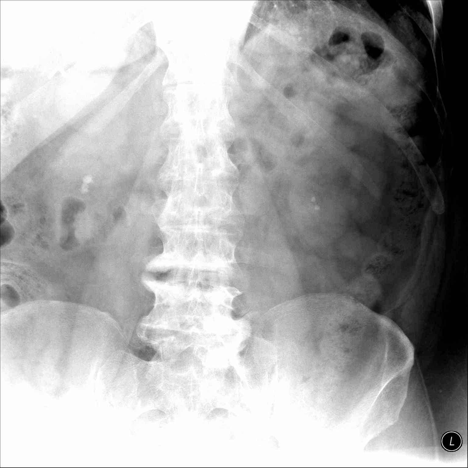
A kidney, ureter, and bladder (KUB) x-ray is a plain abdominal x-ray focusing on the kidneys, ureters, and bladder (see Image. Renal Stones on KUB Radiography). This modality can identify many calcific urinary stones except for the 10% to 20% of radiolucent calculi. KUB x-ray is inexpensive, readily available, and provides information on the ureteral stone's size, shape, and location, allowing for monitoring.[51]
A KUB x-ray is recommended before CT scanning for suspected ureterolithiasis, particularly if IV contrast is planned, to avoid obscuring calculi. About 80% of patients presenting with renal colic in the emergency department undergo CT scans.[52] A KUB x-ray becomes more helpful in monitoring stone passage after identification by CT. Without an initial KUB x-ray, a negative follow-up imaging test cannot ascertain stone passage, movement, or potential radiolucency.[53]
However, the KUB x-ray has limitations. The images cannot reliably distinguish ureteral calculi from other nearby pelvic calcifications or hydronephrosis from simple obstruction. KUB x-rays do not provide renal function information or visualize internal renal anatomy. Calculi may be obscured on x-rays by the bony pelvis, bowel gas, or abdominal organs, and stone visualization is extremely challenging in individuals with obesity and extensive pelvic calcifications. The overall sensitivity of a KUB alone for urolithiasis is only about 50%, but this increases to 87.5% for stones with a diameter >5 mm.[54] The x-ray beam widens as it travels from the focal source, causing an overestimation of stone size on KUB x-rays by roughly 10%.
KUB ultrasound can visualize larger renal calculi and determine the degree of hydronephrosis. However, this modality cannot reliably identify calculi smaller than 3 mm or determine their size or location. KUB ultrasound is preferred in pregnancy and is often the first imaging study done in the emergency department in patients with abdominal pain or suspected acute renal colic due to its low cost, availability, ease of use, and lack of radiation exposure.
The "twinkling artifact" observed on color flow Doppler ultrasound may enhance ultrasound's diagnostic specificity in identifying urinary calculi. This artifact appears as a focal area of alternating colors behind small echogenic objects, such as stones, resembling turbulent blood flow. A comet-tail artifact may accompany this finding. The twinkling artifact is particularly useful for detecting smaller urinary stones, as it improves the diagnostic sensitivity and specificity of gray-scale ultrasound from 96% and 58% to 100% and 97%, respectively. This method can detect stones as small as 2 mm with greater accuracy.
Ultrasound alone may suffice for diagnosis in some patients with a history of multiple recurrent urinary calculi, offering cost and radiation exposure reduction. However, approximately 20% of patients receive inappropriate treatment as this modality lacks specific details about obstructing stones crucial for management. A flat-plate KUB x-ray is recommended for follow-up to track stone progress and determine shape, size, location, and relative radiolucency, aiding in management decisions. Ultrasonography also often overestimates stone size, particularly for calculi 5 mm or smaller.
The advantages of KUB ultrasonography include detection of radiolucent renal calculi, hydronephrosis, bladder ureteral jets used as obstruction surrogates, and renal resistive index calculation. The European Association of Urology recommends ultrasound as the initial imaging modality, while the American Urological Association recommends unenhanced CT scans.
The renal resistive index may be calculated using Doppler ultrasonography to help identify ureteral obstruction. The formula is as follows:
Peak systolic velocity-end diastolic velocity/Peak systolic velocity
Normal values are generally 0.7 or less. Larger values suggest obstruction, particularly when unilateral, but may also be associated with medical renal disease, which typically has similarly elevated levels but bilaterally.
Combining KUB ultrasound and x-ray is a reasonably reliable, inexpensive, and efficient imaging strategy for identifying ureteral calculi in patients with acute renal colic. This combination lowers cost and reduces patient radiation exposure compared to CT while offering acceptable efficacy. The combination identifies hydronephrosis, obstruction, radioopaque, and radiolucent renal calculi and monitors their passage. Symptomatic stones often produce abnormalities on at least 1 of these imaging techniques, if not both. Combining these modalities correctly identifies obstructing stones in 90% of cases with a sensitivity of 88% and specificity of 93%.
An abdominopelvic CT scan without contrast is the gold standard for diagnosing ureterolithiasis. This modality can characterize any urinary calculus' size, number, and location, with only a few rare exceptions of medication stones. CT offers a negative predictive value of 97%, a sensitivity of 98%, and a specificity of 100%, even in obese patients. Such diagnostic accuracy and reliability are unmatched so far.
However, a KUB x-ray must be obtained before CT scanning to monitor stone passage and follow-up. As previously mentioned, IV contrast is often used during evaluation, which can obscure calculi on x-rays and prevent proper monitoring. A KUB CT scout is occasionally sufficient but often lacks detail.
Low-dose and ultra-low-dose CT protocols have been found to significantly reduce radiation exposure without appreciably affecting diagnostic sensitivity for ureterolithiasis. A standard CT scan delivers 5 to 7 mSv of ionizing radiation. In contrast, a low-dose CT scan averages <3.5 mSv, and an ultra-low-dose study generates only 1.9 mSv. By comparison, a single KUB x-ray exposure averages about 0.5 to 0.7 mSv. National urologic guidelines and the American College of Obstetricians and Gynecologists recommendations now allow the use of low-dose unenhanced CT scan imaging in some pregnant patients when ultrasonography is equivocal or inadequate.
Obstructive Pyelonephritis and Urosepsis
Obstructive pyelonephritis (pyonephrosis) is urosepsis associated with an infected kidney and an obstructing stone. This condition may initially present clinically as acute pyelonephritis. These clinical entities are hard to differentiate without imaging.
Urosepsis is a urinary infection resulting in sepsis that may present with the following:
- Acidosis
- Elevated laboratory markers for sepsis, such as lactic acid, neutrophil-to-lymphocyte ratio (NLR), and procalcitonin
- Fever
- Hypotension
- Leukocytosis (>15,000/μL)
- Pyuria
- Tachycardia
- Tachypnea
Obstructive pyelonephritis from an obstructing ureteral calculus is potentially life-threatening, warranting emergency drainage of the obstructed infected kidney. Drainage may be accomplished either by cystoscopy with double J stenting or percutaneous nephrostomy. Even with treatment, the reported overall mortality is 7.4%. Percutaneous nephrostomy is preferred in patients with a significant ureteral stone burden that is challenging to bypass cystoscopically and individuals who cannot tolerate anesthesia. See StatPearls' companion reference, "Percutaneous Nephrostomy," for more information.
Treatment delays have significant effects on morbidity and mortality, especially if more than 2 days have passed. The mortality risk increases by approximately 30% in such cases. Treatment delays appear to be related to lower patient socioeconomic status, weekend hospital presentations, and belonging to minority populations.
After drainage, definitive treatment of the obstructing stone should be postponed until the infection is well-controlled and the patient has clinically recovered fully. The waiting period is typically 1 to 2 weeks long. Antibiotic selection should be based on urine or blood culture results, but 3rd-generation cephalosporins are generally initially preferred.
The primary treatment of known or suspected pyonephrosis is renal pelvis surgical drainage. Delaying surgery to wait for clinical improvement on mere antibiotics is not justified.
Various biomarkers may be used to help identify urosepsis and track its progress but are not specific for ureterolithiasis, pyonephrosis, or urosepsis. These include C-reactive protein (CRP), interleukin-6 (IL-6), lactic acid, NLR, and procalcitonin.
CRP is a routine, cost-effective, and widely available infection and inflammation biomarker. However, CRP is not specific for bacterial infections and is slower to respond than other serum indicators. See StatPearls' companion reference, "C-Reactive Protein," for more information.
IL-6 is a pro-inflammatory cytokine with an hour-long half-life. IL-6 levels quickly rise during severe infections. A threshold level of 52.6 pg/mL or more has been suggested as a reasonable indicator of sepsis.
Lactic acid levels rise with infection. Levels >4 mmol/L (36.4 mg/dL) indicate severe tissue hypoxia and abnormal cellular metabolism and are often associated with higher endogenous catecholamine production. Lactic acid's half-life is only 20 minutes, and it may be used to monitor the progress of a severe infection.
The NLR is typically elevated in patients with sepsis. This parameter is readily available from the complete blood count and does not require special testing. While a definitive threshold level has not been confirmed, NLR ratios over 3.75 to 4 are reasonably consistent with sepsis.
Procalcitonin has proven to be a useful biomarker and tracker for bacterial infections. This parameter is generally considered superior to CRP for urosepsis due to its specificity for bacterial insults, good correlation with other indicators, and quick rise in the presence of infection. See StatPearls' companion reference, "Procalcitonin," for more information.
Risk factors for developing pyonephrosis in patients with ureteral calculi include the following:
- Diabetes
- Elevated infection biomarkers, as explained above
- Fat stranding on CT imaging
- Greater degree and severity of hydronephrosis
- Hypoalbuminemia
- Immunosuppressive state
- Larger ureteral stone size (>5 mm)
- Low platelets
- Older patients
- Pyuria
- Renal failure
Treatment / Management
Initial Management
The important aspects of the initial management of acute ureterolithiasis include analgesia, IV fluids, and antiemetics. Patients should remain nil per os (NPO), especially if hospitalized until surgery is deemed unnecessary. However, some admitted patients are given food before urology evaluates them for possible surgery, delaying necessary procedures due to recent oral intake.
Analgesia is vital in the initial management of symptomatic ureterolithiasis. The pharmacologic options include IV ketorolac, opioid, or acetaminophen. Some patients may benefit from IV lidocaine.
Ketorolac has become the standard initial analgesic for patients presenting with acute renal colic. This NSAID avoids the gastrointestinal and respiratory adverse effects of opioids while providing comparable and possibly superior pain relief, as confirmed by numerous studies.[55][56][57] The typical dose is 15 to 30 mg IV or intramuscularly (IM).[58][59][60][61] Ketorolac may also be given as a continuous infusion in selected cases.[62][63][64][65] This NSAID is contraindicated during pregnancy and in patients with renal failure (glomerular filtration rate < 30 mL/minute), a history of significant upper gastrointestinal bleeds, and an NSAID or aspirin allergy.
Injectable acetaminophen or opioids may be given instead of ketorolac or as needed for additional analgesia.[66][67] Among opioids, meperidine and pethidine should generally be avoided due to their increased tendency to cause nausea and vomiting.[68][69][70] Morphine is the preferred injectable opioid for renal colic pain due to its effectiveness, especially when combined with ketorolac.[71][72] Hydromorphone has a similar adverse effect profile to morphine, but is significantly more potent and faster-acting. However, this opioid also wears off more quickly.[73] IV lidocaine has also shown good analgesic activity in patients with acute renal colic from ureterolithiasis.[74][75][76][77][78]
Antiemetics and IV hydration are generally recommended for symptomatic patients. Ondansetron is considered the most effective antiemetic agent based on limited studies.[79][80][81] IV fluids are generally recommended for patients with symptoms of acute renal colic to prevent dehydration from emesis and lack of oral intake.
Medical Expulsive Therapy
Medical expulsive therapy is generally recommended for patients with ureterolithiasis treated on an outpatient basis. Various α-blockers reduce muscle tension in the distal ureter, where α-1 adrenergic receptors are concentrated, and facilitate spontaneous stone expulsion.[82][83] Data on their effectiveness are somewhat conflicting, but most studies and consensus guidelines indicate that medical expulsive therapy is helpful, especially in treating distal ureteral calculi.[84][85][86][87][88][89][90][91] Several randomized, multicenter trials and subgroup analyses suggest that medical expulsive therapy is most effective when used for larger (5-10 mm) distal ureteral stones.[92][93]
Of the available agents, silodosin at 0.8 mg generally gives the best results. Tamsulosin and alfuzosin are comparable, while nifedipine is less effective.[94][95][96][97] Tadalafil has also been suggested as an aid for medical expulsive therapy, but available data are conflicting.[98][99][100][101] However, combining tadalafil with tamsulosin or silodosin improves its efficacy.[102][103]
Medical expulsive therapy is relatively ineffective for larger, proximal ureteral stones. Oral corticosteroids, increased oral fluid intake, and diuretics have been used but do not have any immediate benefits in acute obstructive ureterolithiasis treatment.[104][105] Additional high-quality randomized controlled studies are needed to determine medical expulsive therapy’s effectiveness and the optimal medication, dosage, or drug combination.
Indications for Surgical Intervention
Most patients with small to moderately-sized stones and well-controlled symptoms may be treated on an outpatient basis and do not require immediate surgery.
Prior stone history, gender, age, degree of hydroureteronephrosis, initial pain intensity, presence of nausea and vomiting, degree of dehydration, and stone size or shape do not reliably predict the need for surgery in patients. However, the larger and more proximal the stone, the greater the chances of requiring a stone-related surgical procedure.[106]
Indications for surgery for patients with ureterolithiasis include the following:
- Anuria, particularly if coincident with the ureteral obstruction
- Bilateral simultaneous ureteral obstructions
- Failure of the ureteral stone to pass or progress significantly after 4 to 6 weeks
- Failure to control symptoms
- Known or suspected obstructive pyelonephritis (pyonephrosis)
- Personal preference of the patient
- Preexisting renal failure
- Progressive renal damage or loss of function
- Recurrent or continuous urinary infections
- Renal cortical thinning from prolonged obstruction
- Sepsis
- Significant medical comorbidities
- Simultaneous urinary tract infection
- Solitary kidney
- Stone size of 10 mm or larger
- Uncontrollable or long-lasting (>72 hours) severe pain
- Worsening renal failure [107][108]
Surgical intervention for ureterolithiasis typically involves either extracorporeal shockwave lithotripsy (ESWL) or ureteroscopy with stone basketing and laser lithotripsy.[109] ESWL and ureteroscopy are roughly equivalent, as the overall stone-free rates are similar for ureteral calculi between these modalities.[110] ESWL offers comparable stone-free rates to ureteroscopy, with lower cost, greater safety, reduced anesthesia needs, and fewer complications, despite the potential for requiring a second treatment.[111][112][113] Additionally, ESWL has a 0% complication rate compared to ureteroscopy's 3.2%, with no significant difference in proximal stone clearance rates.[114]
Emergency drainage of an infected renal unit for obstructive pyelonephritis and urosepsis was covered earlier. Specific renal drainage techniques are covered in our StatPearls' companion references, "Percutaneous Nephrostomy" and "Double J Placement Methods Comparative Analysis." Percutaneous nephrolithotomy is generally not performed for ureteral stones and is also covered in StatPearls' companion reference, "Percutaneous Nephrostomy."
Extracorporeal Shockwave Lithotripsy
ESWL was first FDA-approved for use in the United States in 1984. Since then, millions of renal and ureteral calculi have been successfully treated with this procedure, and it is still considered 1 of the first-line surgical ureteral stone treatments. ESWL is the least invasive urological surgical modality for urinary calculi, with efficacy comparable to ureteroscopy but a much lower complication rate. ESWL stone fragmentation involves a combination of cavitation, shear stress, squeezing, spall fracturing, superfocusing, and tensile fatigue.
A shockwave generator uses various mechanisms to produce a potent pressure wave, typically focused on a predetermined target area approximately 15 cm away. This generator operates within a liquid medium to effectively transmit the shockwave energy. The pressure waves converge on a specific target area called "F2," where the energy is concentrated and the stone is located. ESWL utilizes 3 types of pressure wave generators: electromagnetic, piezoelectric, and electrohydraulic (spark-gap). Each generator type has its own strengths and weaknesses, but overall, all 3 perform comparably well and are roughly equivalent in efficacy.
Typically, electromagnetic machines are highly durable and offer consistent shockwave energy. Piezoelectric machines may have slightly limited energy delivery to the stone and a narrow target zone. On the other hand, electrohydraulic machines feature the largest target zone and potentially the highest stone fragmentation energy delivery. However, these machines require frequent electrode replacement and exhibit greater shockwave variability as the electrode wears during treatment.
The success of ESWL can be reasonably predicted based on various stone characteristics, such as chemical composition, density, location, number, size, and the skin-to-stone distance as measured on CT. Stones with many negative predictors are likely better treated with ureteroscopy.
Chemical composition is important as some stones (brushite, cystine, and calcium oxalate monohydrate) are very hard or resistant to ESWL. Calcium oxalate dihydrate and uric acid are relatively brittle and fragment easily. Meanwhile, uric acid is radiolucent and may be difficult to target, requiring IV contrast or retrograde pyelography.
The stone’s estimated density in Hounsfield units on a noncontrast CT scan may be clinically useful. Calculi with densities <1000 Hounsfield units fragment reasonably well with ESWL therapy, but stones with densities >1000 Hounsfield units often do not respond well. A lower Hounsfield unit reading is suggestive of better outcomes with ESWL.
A skin-to-stone distance measuring 10 cm or less is a useful clinical indicator of success, whether the stones are in the kidneys or ureters. The number of stones being treated is another factor that affects the success rate. Treating multiple separate calculi without overlap (shockwaves are individually directed at each stone) typically results in a lower stone-free rate than focusing all shockwaves on a single stone.
Stone size is a predictor of ESWL success. Patients with calculi smaller than 10 mm are good to excellent candidates. Individuals with stones 10 to 20 mm are acceptable candidates, with consideration for a double J stent. People with calculi >20 mm are generally poor candidates. However, ESWL may be considered in select cases, with a double J stent strongly recommended due to the risk of steinstrasse, a condition where fragmented stone particles obstruct the ureter, impairing urine flow.
Pre-ESWL stents are unnecessary except in cases of significant infection or large stone sizes. Stents may be needed for symptom control and to visualize ureteral stones. Stents are also generally recommended when treating stones in solitary kidneys. Antibiotics are not required, but the urine must be sterile before therapy.
Tips, advice, and suggestions during ESWL therapy
Pretreatment with an NSAID just before surgery reduces the need for postprocedure analgesics. For renal stones, the use of IV fluid therapy and furosemide is beneficial. A large volume of transmission gel applied to only the lithotripter's treatment head minimizes air pockets and optimizes shockwave transmission.
ESWL treatment should start with a low-pressure reading and a slow rate if the kidneys are in the target area, slowly increasing afterward. This technique helps reduce contusions and bleeding. Guidelines recommend a frequency between 60 and 90 shocks per minute. Too fast reduces efficacy, and too slow takes too long. The recommended maximum number of shocks varies by machine but is usually around 3000. However, 4000 or more may be used selectively, particularly for ureteral stones away from the kidneys.
Prone positioning may be necessary to target a stone located over the iliac crest if the lithotripter's treatment head cannot be rotated adequately. Treating the same stone with more than 2 ESWL procedures does not appear to be beneficial. Ureteroscopy should be used instead if 2 ESWL procedures on the same stone have failed. Postoperatively, the use of α-blocker medications helps stone fragments pass and improves ESWL's overall success rate. For further details and information on ESWL, see StatPearls' companion reference, "Extracorporeal Shockwave Lithotripsy."
ESWL's contraindications include the following:
- An active or untreated urinary tract infection
- Distal ureteral obstruction
- Pregnancy
- Treatment path obstructed by an aneurysm, tumor, or other pathology that may be injured
- Uncorrected coagulation disorder
- UPJ obstruction, which applies to renal calculi
Ureteroscopy
The ureteroscope was first introduced in 1980 as a 12-French rigid instrument. Semi-rigid and flexible ureteroscopes became available in 1989. Improved scopes now offer single- and dual-channel options. Flexible ureteroscopes utilize digital imaging, providing exceptionally clear views. Single-use disposable instruments are also commercially available.
Starting an α-blocker at least a week before the ureteroscopic procedure is recommended, but the optimal drug and timing have not been determined. Studies show that preoperative α-blockers reduce the need for ureteral orifice dilation, increase postoperative stone-free rates, improve ureteral access to ureteroscopies, minimize ureteral contractions, and reduce operating time.
Unlike ESWL, ureteroscopy and laser lithotripsy may be safely performed on patients with ureterolithiasis and on anticoagulation. However, ureteroscopy is associated with a lower stone-free rate and additional postoperative hematuria.
Semi-rigid ureteroscopes may be used in the distal ureter, up to the iliac bifurcation and pelvic brim. Compared to flexible ureteroscopes, semi-rigid instruments have larger irrigation channels, more direct and easier handling of accessory stone instruments, dual basket channels, lasers, wires, and a larger field of view. Some are now available with digital imaging. The ureteroscope may be advanced directly into the ureter over a guide wire or through a ureteral access sheath. Any sheaths used must be shorter than the ureteroscope. Sheaths make repeated entries into the distal ureter less traumatic, though placing them takes time, and they may also be unnecessary in some small stones. Resistance to the scope may be remedied by using a small scope, dilating the ureter, using a ureteral access sheath, or leaving a double J stent and coming back another day.
Flexible ureteroscopes have excellent reach to the proximal ureter and kidney, providing high-quality visualization with digital imaging. These instruments can also navigate the lower-pole renal calyces to address stone fragments post-ESWL therapy. A double J stent is typically employed postoperatively unless the ureter was pre-stented or if the ureteroscopy involved minimal dilation or use of a ureteral access sheath.
Disposable flexible ureteroscopes, available commercially, may be cost-effective in certain scenarios. Reserving at least a few disposable ureteroscopes for emergency use is recommended to address critical situations where a working flexible ureteroscope is unavailable.
Tips, advice, and suggestions during ureteroscopy
A flat-plate KUB X-ray and retrograde pyelogram or ureterogram are recommended immediately before inserting the ureteroscope or ureteral access sheath to visualize internal renal anatomy, stone localization, and orientation during ureteroscopy. A range of ureteral access sheaths, guide wires, baskets, and graspers must be readily available because predicting preoperatively which ones will be required is impossible. The scope must be lubricated during longer cases to facilitate removal and reinsertion. Periodic fluoroscopic checks can help with orientation and ensure that the entire kidney and all calyces are examined.
Flexible ureteroscopes are typically used with ureteral access sheaths. The sheath’s width and length should be carefully measured so it is not longer than the scope. The sheath should be placed just distal to the stone or close to the renal pelvis at the UPJ to access the kidney. A safety guide wire is recommended to maintain access, with the wire’s distal end carefully secured.
The laser technique depends on the type of laser available. Generally, fragmentation is performed with shorter bursts, higher power settings, and low-frequency settings. Vaporization ("dusting") is produced using longer bursts, lower power settings, and high-frequency settings.
A Tuohy-Borst Y adaptor enables irrigation and accommodates a small basket or laser fiber when only a single channel is accessible. Only normal saline should be used for irrigation during ureteroscopy, as sterile water irrigation can cause osmotic tissue damage if it extravasates. For further details and information on ureteroscopy for ureterolithiasis, see StatPearls' companion reference, "Ureteroscopy."
Stent discomfort can be quite bothersome for patients. Stent use is common after ureteroscopy, especially following flexible ureteroscopy with ureteral access sheath use or substantial ureteral dilation.
Double J stents are typically employed in cases involving ureteral access sheaths, impacted stones, prolonged operating times, larger stone size or burden, older patient age, solitary kidneys, ureteral trauma or damage, and heightened intrarenal complexity or complications. Leaving a stent in such situations significantly reduces postoperative complications but can lead to stent discomfort.
Double J stents are generally left after ureteroscopic surgery. However, guidelines from the American Urological Association and the European Association of Urology now allow the omission of postoperative stents after uncomplicated ureteroscopies. Uncomplicated ureteroscopy typically entails a unilateral procedure in a healthy patient with a normal contralateral kidney without requiring ureteral access sheaths or balloon ureteral dilation for stones 10 mm or smaller and with no significant residual fragments or ureteral damage.
Stent discomfort may be treated with α-blockers, antimuscarinics, or both. Mirabegron at 50 mg has been found to be more effective for stent discomfort than antimuscarinics. Pregabalin has also proven to be effective in reducing stent-related pain.
Contraindications to ureteroscopy include active or untreated urinary tract infections, patient inability to tolerate general or spinal anesthesia, impassable ureteral strictures or angulations, and inability to achieve cystoscopic access to the bladder or pass a guide wire into the affected kidney’s side. Pregnancy is not an absolute contraindication to this procedure, unlike ESWL. However, anesthesia requirements and the risk of inducing early labor should be carefully evaluated.
Comparing ESWL and Ureteroscopy in Treating Ureteral Calculi
Deciding between ESWL and ureteroscopy generally depends on instrument availability, surgeon experience, anesthesia considerations, patient preferences, estimated stone hardness and composition, radiolucency, calculus size and location, skin-to-stone distance, and individual patient clinical factors. Shared fair and honest decision-making discussions with patients are essential when reviewing these procedures' relative advantages and disadvantages. Both offer equivalent stone-free rates for ureteral stone procedures.
ESWL advantages and disadvantages
ESWL is significantly less invasive than ureteroscopy, with a lower complication rate, inherent safety, and comparable stone-free rate, though it sometimes requires a second procedure. Minimal anesthesia, typically IV sedation, is needed for ESWL, with short and predictable surgical times. ESWL is suitable for stones anywhere in the ureters or kidneys, though this procedure is best for upper urinary tract stones. ESWL does not require pre-stenting unless necessary for other reasons, such as symptom relief or infections. The procedure may be used in pediatrics and older individuals.
ESWL does not work well on radiolucent or very hard stones, such as brushite, calcium oxalate monohydrate, cystine, and uric acid, or where the unenhanced CT stone density is more than 1000 Hounsfield units. ESWL is less effective on stones larger than 20 mm or are difficult to visualize on imaging, in morbidly obese patients, and where the skin-to-stone distance exceeds 10 cm.
The number of shocks that can be safely delivered at one time is limited to 3000 to 4000. Thus, ESWL is less effective if scattered multiple stones are treated. Lower calyceal stones may be fragmented by ESWL but often fail to pass afterward.
No more than 2 ESWL procedures are recommended for a single stone. Ureteroscopy is usually recommended when a single stone needs more than 2 procedures. ESWL is contraindicated in patients who are pregnant, on anticoagulation, or have untreated coagulation disorders. For more information on ESWL, see StatPearls' companion reference, "Extracorporeal Shockwave Lithotripsy."
Ureteroscopy advantages and disadvantages
Ureteroscopy is preferred for small distal ureteral calculi and cases where ESWL is ineffective, including stones larger than 20 mm, skin-to-stone distance exceeding 10 cm, morbidly obese patients, and radiolucent or ESWL-resistant stones like brushite, calcium oxalate monohydrate, cystine, and uric acid, or stones with unenhanced CT density exceeding 1000 Hounsfield units. Ureteroscopy achieves a significantly higher initial stone-free rate than ESWL and rarely requires a second ancillary procedure.
Like ESWL, ureteroscopy can reach any urinary tract area proximal to the bladder. However, ureteroscopy is ideal for smaller, distal ureteral stones that may be removed quickly. Unlike ESWL, ureteroscopy may be performed selectively during pregnancy and in patients taking anticoagulants.
However, ureteroscopy is quite invasive, requires a general anesthetic, and can take considerable operating room time for a complex stone, especially in the kidney. This procedure has a significantly higher risk of complications than ESWL, although the overall risk is still relatively low. Performing ureteroscopy may be difficult without a guide wire. In cases where easy ureteroscopic access is not possible, a double J stent may be left, and the procedure rescheduled for a later time, requiring a second surgery. Pre-stenting can facilitate ureteroscopy, but if not performed, postoperative double J stenting is typically required, potentially leading to significant discomfort. For more information and details on ureteroscopy, see StatPearls' companion reference, "Ureteroscopy."
A 24-hour urine analysis should be performed for kidney stone prevention in high-risk patients after the renal colic episode has been successfully and definitively addressed. Such tests are obtained to evaluate calcium, citrate, creatinine, oxalate, magnesium, phosphate, uric acid, pH, and urine volume. Serum uric acid and parathyroid hormone are also recommended in patients with high or elevated serum calcium levels.
The 24-hour urine test is particularly recommended in the following situations:
- Abnormal anatomy of the urinary tract
- Gastric bypass surgery (especially Roux-en-Y)
- High risk for surgery or anesthesia
- History of multiple urinary tract infections
- History of previous stone or ureteral surgery or reimplants
- Irritable bowel syndrome or chronic diarrhea
- Multiple stone recurrences
- Nephrocalcinosis
- Pediatric or adolescent stone disease
- Positive family history of stones
- Significant obesity
- Solitary kidney
- Stone composition primarily of cystine, calcium phosphate, or uric acid
- UPJ obstruction
Only about a third of urologists order 24-hour urine testing on their kidney stone-forming patients, and only 7.4% of all ureterolithiasis patients with a high recurrence risk have 24-hour urine testing for kidney stone prophylaxis. This test is recommended for patients at risk of recurrence but motivated to prevent relapses.
Differential Diagnosis
Acute colicky flank pain, nausea, and vomiting are symptoms not unique to ureterolithiasis. This diagnosis should be only 1 of many considerations, especially in people with a history of renal calculi and comorbidities like cardiovascular disease. Flank pain resembling acute renal colic can have various causes, including the benign and life-threatening conditions listed below.
- Abdominal aortic aneurysm (dissecting, enlarging, leaking, or rupture)
- Angiomyolipoma
- Appendicitis
- Cholecystitis
- Costochondritis
- Dermatological disorders (Herpes zoster)
- Dietl crisis
- Double J stent placement or recent removal
- Diverticulitis
- Ectopic pregnancy
- Endometriosis
- Epididymitis
- Extrinsic ureteral compression (eg, from surgical clips, staples, and cancers)
- Gastrointestinal disorders (eg, irritable bowel syndrome, Crohn disease)
- Hepatitis
- Iatrogenic causes
- Inguinal hernia
- Flank or inguinal local mass or growth
- Neurological disorders and neuropathic pain
- Nutcracker syndrome
- Mesenteric ischemia
- Musculoskeletal conditions
- Ovarian pathology (cyst rupture or torsion)
- Papillary necrosis
- Pelvic inflammatory disease
- Pelvic pain syndrome
- Pleurisy and pleural pain
- Prostatitis
- Pyelonephritis
- Referred pain from back pathologies (radiculitis)
- Renal abscess, infarction, or venous thrombosis
- Retroperitoneal fibrosis
- Retroperitoneal pathology (abscess, hematoma, or tumor)
- Subcapsular spontaneous renal hematoma (Wunderlich syndrome)
- Testicular torsion
- Ureteral spasm
- Ureteral stricture
- Ureterocele
- Ureteropelvic junction obstruction
- Ureteroscopy complications
- Urinary tract infection
Meticulous history-taking, physical examination, and appropriate diagnostic testing can distinguish ureterolithiasis from this long list of differential diagnoses.
Prognosis
The prognosis of ureterolithiasis is generally favorable. However, evidence shows that this disorder is associated with other systemic conditions such as diabetes, cardiovascular disease, and obesity. Recurrence rates are very high at an estimated 50% within 10 years. Patients with recurrent stones warrant a complete metabolic evaluation to identify the etiology. Lifestyle changes and optimal medication management can be instituted to reduce relapses. Patients with this condition, especially if recurrent, incur a significant financial burden due to occupational losses and emergency department visits.
Complications
Nephrolithiasis and ureterolithiasis do not always have a benign course. Some of these conditions' complications include infected kidney stones, sepsis, ureteral scarring, perforation, renal abscess formation, bleeding, impaired renal function, and forniceal rupture. CKD may result from a urologic procedure, obstruction, or an inherent cause of the stone formation. However, the risk of end-stage renal disease from nephrolithiasis is generally low, even in recurrent cases.
Ureterolithiasis in pregnancy presents additional complications and is associated with increased spontaneous abortions, low birth weight infants, miscarriages, preeclampsia, and gestational diabetes. Thus, ESWL is contraindicated, and ureteroscopy is used only very selectively in this patient group.
Possible complications of ESWL include: [115]
- Bruising of skin in the coupling contact area
- Failure to find the calculus
- Gastrointestinal injury (usually a hematoma that resolves on its own)
- Hematuria
- Perinephric hematoma
- Renal injury (contusion)
- Renal subcapsular hematoma
- Steinstrasse
- Ureteral obstruction from a stone fragment
Steinstrasse usually resolves spontaneously. However, repeat ESWL or ureteroscopy is recommended if this condition becomes symptomatic or lasts longer than 2 weeks.[116][117][118]
Possible complications of ureteroscopy include:
- Failure due to abnormal ureteral anatomy or inability to reach obstructing stone
- Failure to find the calculus
- Fever
- "Forgotten" stents
- Hematuria
- Ileus
- Renal injury
- Renal perforation
- Segmental ureteral necrosis
- Stent discomfort
- Stone migration
- Ureteral perforation
- Ureteral spasm
- Ureteral stricture
- Urinary extravasation
- Urinary tract infection [119][120]
Patients must be counseled to seek medical help immediately if symptomatic.
Deterrence and Patient Education
Primary prevention of ureterolithiasis involves lifestyle modifications and dietary shifts to minimize the risk of calculus formation. Preventive measures include increasing fluid intake, minimizing consumption of foods high in oxalates, sodium, and animal proteins, and eating more fruits and whole grains. Engaging in regular exercise and avoiding medications that increase the risk of calculus formation are likewise important preventive measures.
Meanwhile, in patients who present with renal or ureteral calculi for the first time, education on the high rate of recurrence is imperative. Increasing calcium intake must be considered in patients at risk of recurrent stone formation. Higher dietary calcium consumption is associated with a lower relative risk of calcium oxalate stone formation, as it enhances intestinal oxalate binding, reducing its absorption and subsequent urinary excretion.
Additional outpatient testing, such as 24-hour urine analysis, is needed to identify the cause of calculus formation, especially in recurrent and high-risk cases. While such evaluations are possible, the efficacy of prophylactic measures depends significantly on the patient's willingness to comply with treatment suggestions long-term. Unfortunately, most high-risk kidney stone formers do not get such testing, and many are not even informed that they can obtain the analysis for their benefit.
Patients should be advised to use a strainer or sifter to retrieve any stone material for chemical analysis. Patients treated as outpatients should be advised to look out for warning symptoms, such as fever, chills, lightheadedness, lethargy, or difficulty urinating, that warrant further treatment.
The value and use of medical expulsive therapy should also be discussed. Routine antibiotic use is not recommended unless urinary tract infection symptoms or unexplained fever are noted or the patient is at high risk for sepsis. Mild leukocytosis is common in ureterolithiasis, even without any infection. Appropriate outpatient follow-up with the patient's primary care provider or a urologist should be recommended.
Pearls and Other Issues
Ureterolithiasis occurs due to the formation of solid masses in the urinary tract, typically comprised of concentrated substances like calcium, oxalate, and uric acid, leading to obstruction and subsequent pain. Patients with acute renal colic and ureterolithiasis should not be given anything by mouth until the necessity for immediate surgery is ruled out. Urology should be notified about all high-risk ureterolithiasis patients who present to the emergency department and those requiring admission. Diagnostic testing includes imaging studies like KUB x-rays, CT scans, and ultrasound to accurately visualize stone location and size.
ESWL is preferred for small renal calculi and offers a less invasive option with minimal anesthesia. Meanwhile, ureteroscopy is more effective for larger stones. Patient factors, stone characteristics, and procedural preferences must be considered when deciding between ESWL and ureteroscopy for stone management.
Kidney stone prevention testing with a 24-hour urine collection is suggested for all high-risk patients, including individuals with recurrent calculus formation, renal failure, solitary kidneys, cystine stones, immunodeficiency, and high surgical or anesthesia risk. This test is also appropriate for pediatric patients with nephrolithiasis but optional for all individuals with a previous history of renal calculus formation. Successful stone prevention treatment requires motivation and a willingness to commit to long-term therapeutic compliance, as the risk of this condition's recurrence is high.
Enhancing Healthcare Team Outcomes
The optimal treatment of ureterolithiasis requires an interprofessional approach. Physicians, advanced care practitioners, nurses, pharmacists, and other healthcare professionals each play crucial roles in this process. Many patients present to the emergency department or for outpatient care. Thus, emergency medicine and primary care physicians are the first clinicians to evaluate and provide initial treatment for the condition. Patients are later referred to urologists, who administer definitive renal or ureteral calculi treatment.
Nurses, nutritionists, and pharmacists are also crucial to management. Nurses monitor vital signs, administer medications, and offer support to patients with ureterolithiasis. Nutritionists recommend dietary modifications to reduce the risk of stone formation. Pharmacists ensure appropriate prescriptions for pain relief, prevention of recurrent stones, and management of any underlying conditions contributing to stone formation.
Collaborative planning is essential to develop effective strategies for managing ureterolithiasis cases. This involves establishing protocols for pain management, fluid resuscitation, antibiotic therapy, and stone removal techniques tailored to individual patient needs and preferences. Coordinated care involves integrating services across disciplines to optimize patient outcomes. This may involve scheduling appointments, arranging consultations with specialists, facilitating transitions of care, and providing patient education to promote adherence to treatment plans and preventive measures.
All team professionals should actively educate patients on the importance of dietary modifications, increasing fluid intake, the role of 24-hour urine testing, regular follow-up evaluations, and correctly taking any prescribed medications. Together, the interprofessional healthcare team can ensure patient-centered care, optimal outcomes, safety, and efficient team performance.