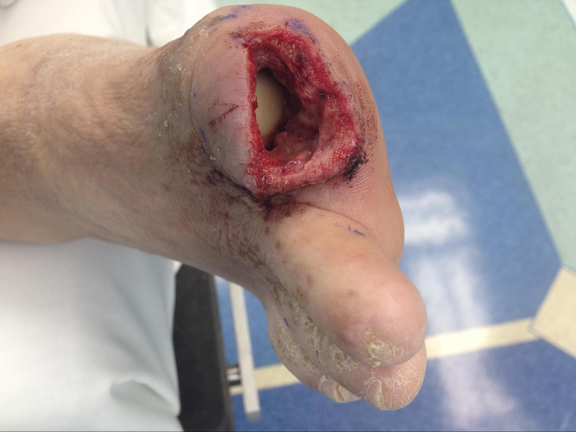
Continuing Education Activity
Digital amputation is often associated with traumatic injuries but is also seen within the elective surgery setting, such as in the case of cancer resection and management of chronic conditions such as Dupuytren's contractures or peripheral vascular disease. It is also seen as a consequence of severe sepsis, although this often results in auto-amputation. In the traumatic setting, the primary objective of management is to salvage the amputated finger to restore function, especially if the dominant hand is affected. However, this is not always possible, as there are many factors to consider. These factors include the time from injury, mechanism of injury, and degree of contamination in a traumatic finger amputation. This activity reviews the presentation, evaluation, and management of traumatic finger amputation and stresses the role of an interprofessional team approach to the care of affected patients.
Objectives:
- Summarize the goal of treatment of finger amputation.
- Describe the common causes of traumatic finger amputation.
- Outline the factors that must be considered in determining whether traumatic finger amputation is necessary.
- Explain the modalities to improve care coordination among interprofessional team members in order to improve outcomes for patients requiring traumatic finger amputation.
Introduction
Digital amputation is often associated with traumatic injuries but is also seen within the elective surgery setting, such as cancer resection and management of chronic conditions such as Dupuytrens contractures or peripheral vascular disease. It is also seen as a consequence of severe sepsis, although this often results in auto-amputation.
In the traumatic setting, the primary objective of management is to salvage the amputated finger to restore function, especially if the dominant hand is affected. However, this is not always possible, as there are many factors to consider. These factors include the time from injury, mechanism of injury, and degree of contamination [1]. In the elective setting, determinants for the level of amputation include various factors, such as oncological clearance, symptom relief, and function preservation; however, for the purpose of this paper, the primary focus will be on a traumatic finger amputation.
The basic goal in the treatment of digit amputations (especially upper limb) includes
- Preservation of functional length
- Durable coverage
- Preservation of useful sensibility
- Prevention of symptomatic neuromas
- Prevention of adjacent joint contractures
- Early return to work
- Early prosthetic fitting
Etiology
In traumatic injuries, one should assess the patient in accordance with the advanced trauma life support approach (airway, breathing, circulation, etc.) for a systematic assessment of the patient and to rule out any life-threatening injuries before referring or transferring patients for further specialist management of their finger injuries. Determining the mechanism of injury is crucial, as it could affect decisions regarding management and outcome for the patient. Sharp injuries may provide a clear amputation level, whereas blunt trauma may correlate with crush injuries at the level of the amputation, and avulsion injuries can cause distant trauma away from the level of visible injury.
Epidemiology
Amputation injuries represent approximately 1% of all trauma-related injuries presenting to the emergency department, with finger and thumb amputations being the most common.[1] Fingertip and partial digit amputation are the most frequent presentations, complete digit and multiple digit amputations are less common, with most injuries occurring at home.[2] Its incidence is higher in adults (men greater than women, approximately 4 to 1) than children (approximately 3 to 1 adult to child ratio). Working with power tools is the most common cause of injury in adults.
Pathophysiology
Typically, once a finger amputation has occurred, ischemic tolerance times are 12 hours if warm and up to 24 hours if cold. For more proximal amputations, these times are halved. The amputated part should be covered in a normal saline-soaked gauze, sealed in a plastic bag and submerged in icy water with no direct contact with ice. If there is direct contact with ice, it could result in tissue damage and render the amputated part non-viable. More proximal amputations are less tolerant of ischemia due to the presence of muscle tissue, which can undergo irreversible changes after 6 hours of ischemia.[3][4]
History and Physical
History:
The important aspects to be noted in history are hand dominance, occupation, time of injury, mechanism of injury, other associated injuries, comorbidities, and NPO status.
Physical Exam:
Level of amputation, structures involved, neurovascular status, function, and degree of contamination (if relevant). It is vital to assess the amputated part and ultimately determine its suitability for replanting respective to the mechanism of injury (e.g., crush, guillotine-style, avulsion).
Finger amputations classification is generally according to the level of amputation. The Sebastian and Chung classification is outlined below[5]:
- Zone 1 distal amputations
- Zone 1A - distal to lunula, through the sterile matrix
- Zone 1B - between lunula and nailbed
- Zone 1 proximal amputations
- Zone 1C - between flexor digitorum profundus insertion and neck of the middle phalanx
- Zone 1D - between the neck of the middle phalanx and insertion of the flexor digitorum superficialis
Evaluation
Laboratory:(optional depending on the clinical scenario)
CBC (complete blood count) to assess for blood loss
Coagulation studies (only if the patient is known to be on anticoagulants)
Imaging:
Plain radiograph of the affected finger/hand and amputated part; this allows assessment of bony injuries, bone quality, and guide decisions regarding bony fixation methods. Angiograms are normally not requested unless it forms part of investigations for other injuries.
Treatment / Management
Initial management - first aid, ATLS approach to the patient, preserve amputated part, tetanus vaccination, and antibiotics as per local hospital guidelines.
In the traumatic setting, the ideal candidate for replantation should be young, healthy, sharp mechanism of injury (giving a clear amputation level), minimal tissue destruction, and contamination.[3]
Indications for replantation[1][3]:
- Thumb amputation - loss of thumb represent approximately 40 to 50% loss of hand function
- Multiple finger amputations
- Amputations at or proximal to palm
- Pediatric patients with finger amputation(s) at any level
- Single finger amputation distal to the insertion of the flexor digitorum superficialis (zone I) (studies have shown that replantation distal to this insertion point had better outcomes than those proximal)
- Patient consideration - specialist requirement, e.g., occupation or pre-morbid compromised hand function
Contraindications and relative contraindications[3]:
- Single-digit injury through flexor tendon zone II
- Smoking
- Severe crush
- Mangled limb
- Heavy contamination
- Segmental injuries
- Prolonged warm ischaemic time
- Medically unfit
- Improperly preserved amputated part.
- Avulsion injuries
- Other life-threatening injuries
- Mentally unstable
- Previous surgery to the affected finger
- 'Redline' or 'red ribbon' sign (seen in vessels during surgery), which predicts the level of intimal damage in the vessel
Once the patient arrives in the operating theater, the amputated part should undergo a careful assessment for suitability for replanting. All structures should be dissected and identified, especially the neurovascular bundle. If no suitable vessels are identified, then replantation should not proceed. Usually, there is an order for repair of structures:
- Bone fixation with or without bone shortening to allow repair of soft tissue
- Tendon repair - extensor and flexor tendons
- Nerve repair
- Arterial anastomosis
- Venous anastomosis (if suitable veins are present)
Bone fixation should be simple and quick to perform, but it also depends on the configuration of bony injuries. Usually, two Kirschner wires are an option, but other fixation methods may also be used (i.e., plate fixation). Occasionally, bone shortening is required before fixation to allow for soft tissue closure and repair of neurovascular structures without excessive tension.
In case the restoration is not possible, and the clinician decides to opt for amputating or forming a stump, then provide a mobile, stable, painless stump with the least interference from the remaining tendon and joint function to ensure the most useful result. Whenever possible, use volar skin for the stump coverage because it provides skin that is thicker and more sensate than dorsal skin. The options for rearrangement of volar tissue to cover the stump include:
- Fillet flaps
- Volar V-Y flaps
- Bilateral V-Y flaps
- Homodigital island flaps
In an acute setting, the "dog ears" should be left to decrease the tension and the likelihood of ischemia to the remaining flaps, and these dog ears disappear over time. Regarding the treatment of the bone in a digital amputation/stump formation, the bone under the stump end must be smooth. The remaining bone chips and devitalized bone should be removed. The articular cartilage can be preserved when the amputation occurs at the level of the interphalangeal (IP) joint. This can provide a shock pad for trauma and potentially causes less pain under than skin than the bone edges.
While addressing the nerve at the stump end, it is vital to avoid neuroma formation in this location. The nerve end should be in a position away from the stump end or an anticipated point-of-contact pressure. To decrease the likelihood of neuroma formation at the stump end, traction neurectomy of the digital nerve should be performed bilaterally for each digital amputation. Tendon insertions should be preserved where possible as the preservation of a tendon insertion enhances the active mobility and function of an amputation stump. Adhesions can result, and so early mobilization of the digit is recommended.
Differential Diagnosis
Digit amputations tend to be a straight forward presentation, but infectious processes can mimic amputation leading to loss of fingers/toes.
Prognosis
Previous studies have found these factors to have a substantial effect on functional outcome[6]:
- Level of the amputation
- Number of fingers injured
- Circumstance and mechanism surround the injury
- Time from injury
Survival rates following replantation have been reported between 80 to 90%, although these figures have come from specialist institutions. It is also worth noting that venous repair improves survival regardless of the level of amputation. This fact is important, as the venous repair is not always possible, and as a result, it could lead to venous congestion. One solution would be to make a small distal incision to allow venous drainage or the use of leeches.
Complications
Complications classify according to the time of onset[1]
- Early complications
- Arterial insufficiency
- Arterial thrombosis presents typically as a pale, cool and pulseless digit
- It is vital during the post-operative period to maximize blood flow through the anastomoses and prevent thrombosis
- Venous insufficiency
- Venous congestion typically presents as a purple digit with brisk capillary refill and swelling
- Concerns of possible anastomosis failure or thrombosis should prompt urgent return to theatre for salvage - in cases of venous congestion, leech therapy or anticoagulation may be considered to improve venous return
- Infection
- Late complications
- Cold intolerance
- Tendon adhesions
- Stiffness
- Bony malunion
- Altered sensation
Postoperative and Rehabilitation Care
Post-operative management:
- Maintain adequate hydration and circulation volume
- Analgesia
- Keep the affected limb elevated and warm
- Frequent monitoring of the replant capillary refill, color, and temperature
- Avoid dressings changes in the first 48 to 72 hours to minimize manipulation of the repair
- Consider anticoagulation
- In cases of artery-only replants, consider stab incision to the distal amputated tip and apply heparin soaked gauze to allow venous drainage or use leeches instead. This treatment can end once the finger becomes pink with normal capillary refill thus indicating adequate venous drainage
Some patients require further surgery to improve their function, such as tenolysis, bone grafting, tendon transfer, etc.[1] On average, following upper limb amputations, patients return to work within 2 to 3 months after injury.[1] Studies show that functional recovery is better in more distal injuries than proximal, both in terms of movement and power.[6]
Deterrence and Patient Education
- Good health and safety regulations - to provide a safe working environment and reduce occupation-related injuries
- Public information leaflet/public awareness campaign - same objective as above, but to ensure a safe home environment for work and recreation, e.g., BBC News article in May 2018, warning the public of DIY and gardening accidents.
Pearls and Other Issues
- Once a patient with an amputation injury arrives in hospital, a speedy but thorough assessment is essential to minimize the delay to definitive surgical management.
- Often, amputated parts are brought to the emergency department (although sometimes forgotten at the scene or referring hospital) in inappropriate storage. As a specialist center, it is crucial to inform referring units of the best way to preserve the amputated part, to label it with the patient's details, and keep it with the patient to avoid the loss.
- Early involvement of specialists where possible
- Take account of patient factors, i.e., age, occupation, comorbidities, and also patient wishes - replantation requires long and complex surgery, hospital admission, the risk of complications, long rehabilitation, and risk of an incomplete return to normal function.
- This may not be acceptable in some patients, especially in those who are self-employed and cannot take prolonged time off work.
- As such, the terminalization of the affected finger may allow early return to work and normal function for the patient - if possible, the patient needs to understand their options and the potential outcome of each.
- Good rehabilitation process - early involvement of hand therapists
Enhancing Healthcare Team Outcomes
Surgical replantation requires a multi-professional approach. In terms of surgery, specialist surgeon(s) would be required to perform the procedure efficiently and effectively. During the recovery period, skilled nursing care experienced in follow-up care of patients with amputations is vital to ensure good monitoring of the replant, and allow early detection of pending complications. Early involvement of occupational hand therapists is also vital (some say the most important part) to patient recovery and the return of normal function.
Evidence generally shows good outcomes in distal amputations, especially if venous repair is possible. However, the evidence is limited in patients who do not proceed to replant.
Even after the replantation, patients often require an extensive interprofessional rehabilitation effort of nurses, occupational therapists, and physical rehabilitation physicians to regain the function of the digit.

