Continuing Education Activity
Angular cheilitis describes an inflammatory skin process of variable etiology occurring at the labial commissure, the angle of the mouth. It is usually a symptom of another condition and leads to saliva-induced maceration of the structurally susceptible epithelium at the labial commissures. Symptoms are often mild, and the condition can go untreated for years before being brought to the attention of a medical provider. The observation of angular cheilitis should prompt an evaluation for oral candidiasis. As the elderly are especially susceptible to angular cheilitis, providers should be more vigilant in this population. This activity examines when this condition should be added to the list of differential diagnoses and how to evaluate a patient with it properly. This activity highlights the role of the interprofessional team in caring for patients with this condition.
Objectives:
- Identify the etiology of angular cheilitis.
- Review the workup of a patient with angular cheilitis.
- Outline the treatment and management options available for angular cheilitis.
- Explain interprofessional team strategies for improving care and outcomes in patients with angular cheilitis.
Introduction
Angular cheilitis (AC) is a descriptive diagnosis for an inflammatory skin process of varied etiology occurring at the labial commissure – the angle of the mouth. "Angular," or commissural, refers to a localized lip inflammation (i.e., “cheilitis,” from the Greek chilos or “lips”) that is distinguishable from the more generalized cheilitides that have different causes. The angles of the mouth are points of interface for the squamous epithelium of the face and oral mucosa. They are also a mechanically dynamic hinge for the oral aperture that endures more motion and tensile forces than the rest of the lips. Thus, the commissures are especially susceptible to certain stresses.
Diffuse cheilitides may be a function of environmental, chemical, or infectious exposures. They may also reflect an internal condition, deficiency, or derangement. They include eczematous cheilitis, contact cheilitis, drug-induced cheilitis, infective cheilitis, actinic cheilitis, glandular cheilitis, granulomatous cheilitis, exfoliative cheilitis, plasma cell cheilitis, and nutritional cheilitis. Diffuse cheilitides will not be discussed here.
Angular cheilitis is also called angular cheilosis, angular stomatitis, commissural stomatitis, rhagades, or perleche (from the French for “through licking”). Rhagades is a general term for fissuring of the skin in areas of motion, especially the labial commissures and nose.
Etiology
The following are alterations in mouth structure leading to changes in lip approximation and increased salivary pooling and maceration at the labial commissures:
- The normal loss of skin turgor due to aging, smoking, or rapid weight loss
- Loss of vertical dimension of the face due to severe tooth wear, edentulous states, and mal-fitted dentures increasing overhang of the upper lip onto the lower one (overclosure)
- Retrognathic malocclusion
- Deepened furrowing of the skin dependent on the commissures (marionette lines)
- Conditions associated with enlarged lips such as oro-facial granulomatosis (OFG). Up to 20% of OFG patients suffer from AC, but Candida is usually not isolated from lesions
- Down syndrome: 25% of patients have AC due to macroglossia leading to tongue protrusion and drooling[1]
Atopic Dermatitis
Allergic or irritant contact dermatitis causes up to 22% of cases of AC and 25% to 34% of generalized cheilitis. Common causes include nickel (in individuals with orthodontic braces)[2], foods (due to flavorings and preservatives), toothpaste, mouthwash, the sunscreen component of expired lip balm, lip cosmetics (due to preservatives, sodium laurel sulfate, emollients, colophony, Cocamidopropyl betaine), acne products, and chewing gum. It may be impossible to distinguish irritant and allergic contact dermatitis without a patch test.[3]
Immune deficiency causes AC, often via the development of oral candidiasis (thrush) with extension to the labial commissures. Chronic steroid use (inhaled or oral), HIV/AIDS, thymic aplasia, a severe combined immunodeficiency syndrome (SCID), DiGeorge syndrome, hereditary myeloperoxidase deficiency, and Chediak-Higashi syndrome. Blood dyscrasias and malignancies probably also imbue some immune suppression as seen in acute leukemia and agranulocytosis.
Nutritional deficiencies are less common in developed countries but exist in susceptible populations such as the elderly, celiac disease patients, the impoverished, the mentally ill, vegans, and their solely breastfed infants not receiving vitamin supplementation. Patients who undergo bariatric surgery and ileal resection also have nutritional deficiencies and that is why they are more prone. Chronic gastritis, chronic pancreatitis, Crohn disease, and pernicious anemia are also important risk factors. Up to 25% of AC has iron or vitamin B deficiency. The following are associated with angular cheilitis:
- Vitamin B deficiencies (especially cyanocobalamin, folate, riboflavin)[4]
- Trace mineral deficiencies (zinc or iron)
- General protein malnutrition
Manifestations of Systemic Diseases - Sjogren Syndrome, Inflammatory Bowel Disease
- Sjogren syndrome (SS): AC is the most common oral lesion found in SS, followed by atrophic glossitis and oral candidiasis. This is according to a systematic review by Serrano, et al. which incorporated the data of 2426 patients with SS. The prevalence of AC ranged from 2% to 81%, with the largest population reporting frequency of 20-40%. SS is a rheumatologic disease characterized by xerostomia ("dry mouth") and hyposialia ("decreased salivation"). This results from lymphocytic infiltration and destruction of salivary glands. Surprisingly, in this patient population, there is an inverse relationship between salivary flow and the presence of candidiasis, which is the opposite of what is the case for non-Sjogren’s AC. This may be explained by proper levels of saliva allowing for mucosal lubrication, tissue healing, and local immunity. Salivary IgA inhibits the binding of candida species to mucosal surfaces and flushes Candida from the oral cavity. Dentures still predispose SS patients to AC, since dental orthotics act as a reservoir for Candida and is a risk factor for oral candidiasis.[5]
- Inflammatory bowel disease (Crohn disease more than ulcerative colitis) can contribute to AC in several ways, one of which is general malnutrition impeding wound healing.
Infection
Infection is the most common cause of AC and the organisms listed below have been isolated in over 50-80% of lesions.[6]
- Risk Factors
- Increased exposure to infecting microbes or factors that increase the microbial burden of skin flora such as poor hygiene, oral thrush, gingival disease/poor dentition
- Diabetes causing overgrowth of Candida due to increased salivary glucose levels and increased adherence to mucous membranes[7]
- Decreased local immunity due to an immunocompromised state from chronic steroid use, chemotherapy, or HIV/AIDS
- Depletion of normal oral flora from prolonged antibiotic use enabling the proliferation of Candida species
- Specific Organisms
- Candida (especially C. Albicans) is the most common cause of AC and it is a normal commensal flora of the mouth in the yeast form, with 40% to 60% of healthy individuals acting as a reservoir. This explains why Candida is found in 93% of cases of AC but is described as the sole pathogen in only about 20% to 50% of the cases. Poor oral hygiene plays a role by increasing colony burden, especially when dentures are present. Since the hyphal form of this dimorphic yeast is the pathogenic variant, potassium iodide staining can help distinguish whether Candida is an innocent bystander yeast or a culprit pathogen. Uncontrolled diabetes mellitus is a major risk factor because of relative immunodeficiency and the increased availability of glucose as a substrate for fungi. Candida is typically the primary pathogen invading macerated labial commissures, setting the stage for subsequent bacterial infection. Infantile AC is almost always associated with concomitant oral candidiasis (thrush) and needs treatment to prevent a recurrence.
- Staphylococcus aureus is the sole pathogen in approximately 20% of cases. The anterior nares harbor Staphylococcus and decolonization therapy with anti-staphylococcal ointments should be applied to the nares if Staphylococcus aureus is identified as a causative agent.
- Beta-hemolytic streptococci, isolated in 8% to 15% of cases, is less commonly a sole pathogen. The anterior nares can harbor Streptococcus and Staphylococcus.
- Polymicrobial infections cause most AC and a combination of Candida albicans and S. aureus comprises 60% to 75%.
Recurrent Mechanical, Chemical, or Thermal Injury
Repeated mechanical, chemical, and thermal insults to labial commissures or conditions make the angles of the mouth more susceptible to injury.
- Xerostomia contributes to 5% of cases of AC and the causes include:
- Radiation treatment
- Sjogren syndrome
- Medications causing xerostomia and xeroderma such as isotretinoin, acitretin, indinavir, sorafenib, anticholinergic medications, and anticancer drugs
- Hypervitaminosis A
- Environmental exposures (dry heat, cold)
- Repetitive behaviors leading to salivary overexposure and commissural maceration. Habit-induced cheilitis is sometimes considered a distinct entity from AC and termed either “factitious cheilitis” or “perleche”
- Nervous tics such as over-licking of lips
- Thumb-sucking, lollipops
- Sialorrhea, drooling, or hypersialia
- Aggressive dental flossing[8]
- Smoking
- Acute mechanical stress at the labial commissures such as post-tonsillectomy
Idiopathic AC with no Identifiable Cause
Since infection is the most common cause and maceration from saliva exposure the most common risk factor, empiric treatment with antifungal and/or antibiotic creams are reasonable but, long-term emollient therapy may be necessary in unresponsive or recurrent cases. Any case of idiopathic AC, after it has undergone adequate investigation, should raise a red flag for nutritional deficiencies or malignancy (the latter, especially in unilateral cases that fail to respond to any therapy.) A rare cause of AC is glucagonoma – a pancreatic endocrine tumor that causes a syndrome of dermatitis, diabetes, weight loss, anemia, and AC.
Epidemiology
Angular cheilitis (AC) occurs with a prevalence of 0.7% in the general American population, although it can occur more frequently in select groups. It is the most common bacterial/fungal infection of the lips. It has a bimodal distribution, occurring most frequently in children, and then again in adults (age 30 to 60). The elderly have about an 11% prevalence of AC, but there is a 3-fold incidence in denture-wearers, a prevalence of up to 28%, and is twice as frequent in men (but this risk seems to be more associated with denture use and comorbidities than chronological age.) Predisposing factors include immunodeficiency, and up to 10% of HIV-positive individuals have oral thrush, with or without concomitant AC. Patients with inflammatory bowel disease more frequently get AC, with 7.8% of Crohn patients and 5% of ulcerative colitis patients developing AC during some time in their disease course. In rare conditions such as orofacial granulomatosis, the incidence is as high as 20%.[9]
Pathophysiology
Most cases of angular cheilitis (AC) are ultimately due to physical maceration at the angular commissures due to overexposure to saliva. The digestive enzymes in saliva can act even on body tissues if allowed prolonged contact. Continued saliva exposure induces contact dermatitis and eczematous reaction at the commissures. The compromised integrity of the stratum corneum epithelium allows local commensal organisms to infect the area. Frequently, colonizing Candida albicans establishes and invades the susceptible tissue. This may then allow bacterial superinfection with staph and strep species. Thus, risk factors are those that increase saliva retention at the commissures, increase exposure to culprit microbes, cause direct tissue inflammation, or inhibit wound healing and immunity. Non-infectious causes of AC are further discussed in the etiology section.
History and Physical
History
- Include questioning about the history of dental procedures and denture use.
- Pain need not be a major complaint and, when present, is usually mild and the feeling is usually described as “dry,” “itchy,” “sore,” “irritated,” “burning”. The sensation does not extend beyond the lesion itself. If present, opening the mouth exacerbates pain. AC may be severe enough to make eating difficult and worsen malnutrition, but it is rarely a primary cause.
- Asking about symptoms of systemic disease such as diarrhea, hematochezia, abdominal pain (for Crohn disease), or dry eyes and dry mouth (for Sjogren syndrome) is appropriate.
- If thrush is present, ask about appreciable risk factors such as people suffering from diabetes, proton-pump inhibitor use, steroid use, HIV status.
Physical
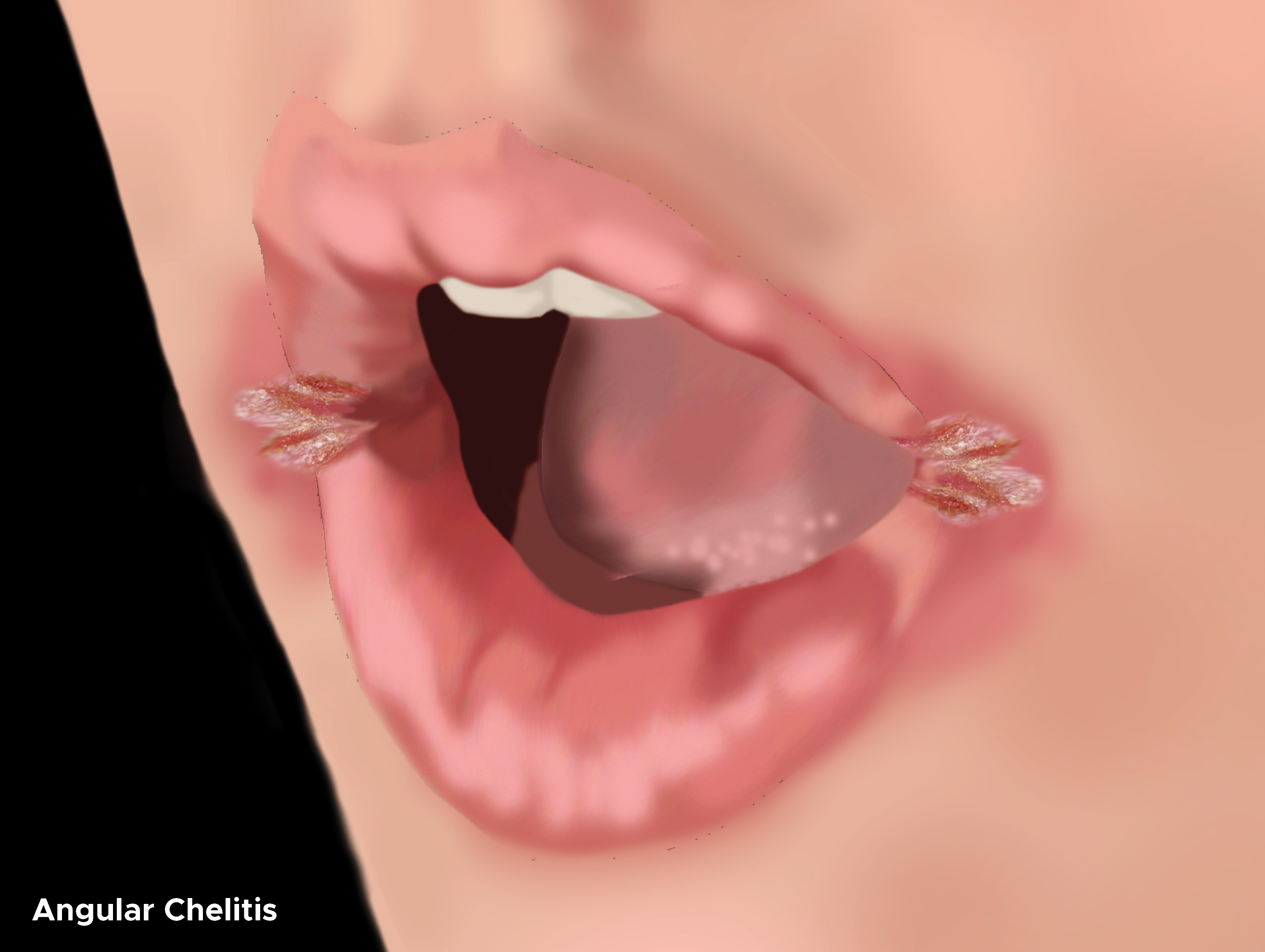
- As an inflammatory process, angular cheilitis presents with classic features of red, edematous, often painful patches of skin at the labial commissures. They are roughly triangular lesions. Mild cases may just show some pinkish erythema with adjacent lips either normal or chapped. As the condition progresses, moisture causes the superficial skin to macerate and erode, leading to small, gray-white lesions bordered by reddened mucosa. In more moderate cases, skin becomes papular, eczematous, and more fissured. These more established lesions can be bluish-white with an associated exfoliative scale surrounded by erythema. In severe cases, fissuring is deep enough to cause bleeding, but this is rare in AC. If the inflammation is enough, damaged skin can exude and crust, but this is more typical of late lesions. Bacterial AC is more likely to have honey-colored exudates, pustules, and purulent exudates. Leukoplakia can infrequently be observed.
- Longstanding AC may also be suppurative, exfoliative, and develop granulation tissue.
- AC is usually isolated to the angles of the mouth with limited involvement of the adjacent face or lips. When AC extends beyond the vermillion border, it radiates from the angles as fissures (rhagades) and follows existing marionette lines, as this is the natural streambed for salivary flow.
- AC is frequently bilateral and symmetrical unless there is a risk factor for developing the lesion that disproportionately affects one commissure over the other. Asymmetric cases that do not have an obvious mechanical cause contributing to unilateral salivary exposure should raise red flags for less frequent etiologies.
- Always examine the oral cavity for oral candidiasis (thrush) and treat it, if present. Oral candidiasis may be one of the following:[10]
- Acute or chronic pseudomembranous (thrush) - white, well-defined plaques on the bucca, tongue, palate, and uvula; reveal erythematous (sometimes hemorrhagic) mucosa when scraped off
- Acute or chronic atrophic or erythematous patches and plaques - large, diffuse, well-demarcated, on the palate and tongue, associated with soreness
- Chronic hyperplastic nodules or plaques - firm, leathery, and irremovable lesions on the bucca, palate, tongue, and labial commissures; they may also extend to the skin and nails
- Candida-associated lesions
- Denture stomatitis
- Median rhomboid glossitis – erythematous patches of atrophic papillae on the central dorsum of the tongue
- Angular cheilitis – as described above
- Keratinized primary lesions superinfected with Candida
- Leukoplakia
- Lichen planus
- Lupus erythematosus
Syndromic Presentations
- Ariboflavinosis (vitamin B2 deficiency): Cheilosis, angular cheilitis, photosensitivity, magenta glossitis, stomatitis, pharyngitis, and pseudo-syphilis (seborrhea-like dermatitis of the scrotum, vulva, philtrum, or nasolabial folds). As iron absorptions may be impaired, this deficiency can also lead to normocytic anemia.
- Pellagra (hypovitaminosis B3): Classic signs are dermatitis, diarrhea, dementia, and glossitis. AC can occur.
- Vitamin B5 deficiency: Rare; AC, glossitis, seborrheic dermatitis-like rash around the eyes, nose, and mouth
- Vitamin B6 deficiency:Sideroblastic anemia, cognitive or psychiatric depression, hypertension, and hyperhomocysteinemia, neuropathy, conjunctivitis, oral (aphthous-like) ulcers called stomatitis, atrophic glossitis, angular cheilitis, and intertrigo.
- Folate/Vitamin B12 deficiency: Megaloblastic anemia (pernicious anemia) and neurological symptoms. The latter include peripheral neuropathy (paresthesia, ataxia, decreased sensation), cognitive impairment/dementia, motor deficits (absent reflexes.) Lack of vitamin B12 can also cause glossitis and angular cheilitis. Patients susceptible to hypovitaminosis B12 also usually are folate deficient.
- Iron deficiency: Microcytic anemia (fatigue, exertional dyspnea, palpitations, headache, pica), glossitis, angular cheilitis, koilonychia (spoon nails), and alopecia areata (non-scarring hair loss). If the patient is also experiencing dysphagia, a barium swallow or upper endoscopy may reveal esophageal webs, making the diagnosis of Plummer-Vinson syndrome (AKA, sideropenic dysphagia or Paterson-Brown-Kelly syndrome).
- Zinc deficiency: Constellation of AC along with alopecia, diarrhea, dermatitis, and oral ulcers (especially on the tongue and buccal surfaces). Causes include acrodermatitis enteropathica, an autosomal recessive condition that impairs zinc absorption.
Evaluation
The diagnosis of angular cheilitis (AC) is often purely clinical. Therefore, laboratory investigation is usually only performed after treatment failure. However, because an infection is the most common cause, testing for Candida or bacterial culture can be performed at diagnosis.
Investigation of underlying medical conditions that may contribute (nutritional deficiencies, immunocompromised states, systemic diseases) is at the discretion of the medical provider. If first-line antifungal/antibiotic combination yields no clinical improvement in 2 to 3 weeks, testing should include Hgb level with MCV, iron profile with ferritin, folate, and vitamin B2/B6/B12 levels, and fasting blood glucose.
Candidal AC Suspected
- Light microscopy: For confirmation, lesion scrapings can be taken and stained with periodic acid Schiff (PAS) technique. Positive results show red/purple hyphae (indicative of infection) or yeast (which can be simple colonization). Gram stain shows these bodies are dark blue. Hyphae and yeast are also visible on KOH slides.
- Germ tube test (Reynolds-Braude phenomenon) in sheep/human serum at 37 C for 2 to 4 hours
- Chlamydospore formation in CMA or rice starch agar incubated at 25 C for 2 to 3 days
- Sugar assimilation assay: Test for fermentation of glucose and maltose (but not sucrose or lactose), yielding pale pink coloration in the presence of tetrazolium reduction medium
- Fungal culture: Sabouraud dextrose agar and antibiotics, cornmeal agar, or CHROM agar
- Candida strain typing: Serotyping, isoenzyme profiles, morphotyping, resistance pattern
- Immunodiagnosis: ELISA, RIA, CIE, PHA, and LPA
Bacterial AC Suspected
- Bacterial culture with sensitivities
Oral Candidiasis Confirmed
- HIV testing
- Diabetes testing (random blood glucose, fasting blood glucose, glucose tolerance test, or HgbA1c testing)
Nutritional Deficiency AC Suspected
- Folic acid: Serum level
- Vitamin B12: Serum level; serum homocysteine and serum or urinary methylmalonic acid levels are more reliable indicators of deficiency than vitamin B12 serum levels
- Vitamin B2: Urinary riboflavin excretion or erythrocyte glutathione reductase activity assay
- Iron: Serum iron profile (iron level, iron saturation, ferritin level, TIBC
- Zinc: Serum zinc level
Allergic or Irritant Contact AC Suspected
- Patch testing to distinguish allergic contact dermatitis
Malignancy Suspected
Treatment / Management
Treatment depends on non-infectious or infectious etiology. Empiric treatment includes a focus on infection as the most common etiology. Since the most common risk factors involve saliva-induced eczema and the resultant maceration, an effort to protect the labial commissures topical barrier application (petrolatum jelly, emollients, or lip balm) is important, and often sufficient for idiopathic cases of AC.[11]
Fungicidal Medications
Fungal infections require topical fungicidal medications applied to the labial commissures, usually 3 times daily for 2 weeks.
- Nystatin 100,000 units/mL ointment topically twice per day (BID)
- Gentian violet solution topically BID to 3 times per day (TID) is effective in children if a purple discoloration is acceptable
- Ketoconazole 2% cream topically
- Clotrimazole 1% cream topically
- Miconazole 2% cream topically (with or without hydrocortisone 1%): Mixed staphylococcal and candidal infections respond best to this treatment because of its inherent gram-positive bacteriostatic activity, thus being used as first-line treatment by some providers
- Iodoquinol 1% cream topically BID to TID, usually combined with hydrocortisone 1% cream
Topical Antiseptics or Antibiotics
Bacterial infections require topical antiseptics or antibiotics. Application of the same preparation to the anterior nares (usually 4 to 5 times daily) can prevent recurrent infection when colonization is present. Treatment course is for 1 to 2 weeks.
- Mupirocin 2% ointment TID to 4 times per day (QID)
- Fusidic acid 2% cream (with or without hydrocortisone 1%) applied QID topically as an antistaphylococcal regimen
Oral (systemic) Antifungals
Nystatin is used in mild cases or thrush and those isolated to the oral cavity. Triazoles treat moderate and severe cases of oral candidiasis and any cases extending into the esophagus. When triazoles are used, they obviate the need for topical antifungals. However, they are inhibitors of hepatic cytochrome P450 system and may interact with other drugs. Fluconazole has the highest level of evidence. [12]
- Nystatin 5 mL of 100,000 units/mL suspension, swish and swallow QID for 7 to 14 day for oral candidiasis (no oral bioavailability)
- Clotrimazole 1 troche sucked on 5 times per day for 7 to 14 days for mild oropharyngeal candidiasis refractory to nystatin
- Fluconazole 200 mg orally (PO) for 1 day, then 100 mg PO daily for 7 to 14 days (can be increased to 200 mg daily for severe cases or immunosuppression). This is more effective than nystatin in immunocompromised patients.
- Itraconazole 200 mg PO daily for 2 to 4 weeks or 200 mg swish and swallow QID without food for 7 to 14 days
- Posaconazole 100 mg PO BID for 1 day, then 100 mg PO daily for 7 to 14 days but the dose can be increased to 400 mg PO BID for fluconazole/itraconazole-refractory cases
- Voriconazole: Only recommended when treatment with fluconazole and either itraconazole or posaconazole have failed
- Caspofungin 70 mg PO once, then 50 mg PO daily until 2 days after symptoms/lesions resolved
- Amphotericin 30 to 40 g per d until 2 days after symptoms/lesions resolved (40 to 50 g per day for neutropenic patients)
- Further discussion for the systemic treatment of oral candidiasis are beyond the scope of this review
Oral (Systemic) Antibiotics
These rarely warranted unless lesions are extensive or treatment failure to topical antibiotics; should warrant culture, sensitivities, and consideration of an alternative diagnosis
Topical Glucocorticoids
Topical glucocorticoids are monotherapy in strictly inflammatory processes or add-on therapy to anti-candidal or antibacterial regimens to decrease inflammation, enhance healing of erosions, and prevent relapses.
- Desonide 0.05% ointment topically
- Clotrimazole cream topically BID for 2 weeks
- Hydrocortisone 1% ointment topical BID to TID for 2 weeks (added to iodoquinol 1% cream or fusidic acid 2% cream)
Nutritional Replacement/Supplementation
Nutritional replacement/supplementation is necessary in cases of avitaminosis, mineral deficiencies, or general malnutrition. The specifics are beyond this review.
Dental
A dentist should refit ill-fitting dentures or other dental apparati to restore facial contour. As the functional reservoir of Candida, treat dentures with an antifungal and cleaned frequently. In chronically debilitated patients, a cannula incorporated into the dentures can channel salivary flow into the oropharynx.
Sometimes, malocclusion persists despite dental realignment or is not a viable option for a patient. Other times, depressions at the commissures exist and are amenable to dermal filler therapy. Injectable fillers (collagen, hyaluronic acid) or surgical implants can change mouth shape and restore commissural anatomy. This makes saliva less likely to accumulate at the fissures. A practitioner who is well-versed in the administration of fillers should apply these fillers since the purpose is beyond the normal cosmetic application.
Improved Control of Chronic Medical Conditions that Contribute to AC
- Blood sugar control in diabetes as HgbA1c directly correlates with the incidence of AC
- Anti-retroviral therapy in HIV/AIDS as immune status indirectly correlated with the incidence of AC
Elimination of Behavioral Practices that Contribute to AC
- Tobacco smoking
- Lip licking
Treatment Failures[13]
- Failure to identify/eradicate oral candidiasis
- Resistant species of Candida
- Resistant strains of Staphylococcus or Streptococcus
- Failure to remediate underlying modifiable risk factors such as hygienic issues, ill-fitting dentures, behaviors.
- Persistent, unmodifiable risk factors
- Undiagnosed nutritional deficiency
- Undiagnosed systemic inflammatory conditions (Sjogren, IBD)
- Unidentified immunosuppression
- Undiagnosed malignancy
Follow up in 2 weeks recommended.
Differential Diagnosis
- Secondary syphilis/syphilitic papule localized to the labial commissure; more likely to be unilateral
- Erosive oral lichen planus or lichenoid oral lesions
- Impetigo
- Atopic dermatitis
- Seborrheic dermatitis
- Allergic contact cheilitis
- Irritant contact cheilitis
- Early or isolated diffuse cheilitis
- Actinic cheilitis, especially is the commissures go unprotected with sun protective lip balms
- Cheilitis glandularis
- Cheilitis granulomatosa
- Exfoliative cheilitis
Toxicity and Adverse Effect Management
- Nystatin is associated with mucositis and Stevens-Johnson syndrome
- Oral fluconazole and clotrimazole troches may cause liver function test elevation and, rarely, hepatotoxicity.
- Proton pump inhibitors decreased systemic azole absorption by raising gastric pH
- Triazoles (fluconazole, itraconazole, voriconazole, posaconazole) are inhibitors of hepatic cytochrome P450. All inhibit CYP3A4 and increase levels of the following drugs. Fluconazole and Ketoconazole also inhibit CYP2C8/9. Caution should be used with the list of drugs below. Consideration should be given to substituting amphotericin or nystatin for a triazole.
- Warfarin, monitor for 2- to 3-times increase in INR
- Vinca alkaloids
- Steroids (methylprednisolone and dexamethasone)
- Statins
- Protease inhibitors
- Phosphodiesterase type-5 inhibitors
- Phenytoin decreased the dose and monitor serum levels with fluconazole and itraconazole
- Felodipine cut the dose by 50%
- Digoxin, monitor serum digoxin levels
- Cyclosporine, decrease dose by 50% and monitor serum cyclosporine levels
- Carbamazepine, only in azole doses over 200 mg per day
- Benzodiazepines, reduce the dose
- Aripiprazole
Staging
As described in the seminal article from 1986 by Ohman, et al., staging is described in four categories. While this is largely used for academic purposes only, it can help clinicians categorize severity and response to treatment:.
- "Type I: Small rhagades limited to the corner of the mouth, adjacent skin slightly involved
- Type II: Lesion with ragged border more extensive in length and depth than Type I lesion
- Type III: Lesion consisting of several rhagades radiating from the corner of the mouth into the adjacent skin
- Type IV: Lesion presenting no rhagades, but erythema of skin contagious to the vermilion border"[14][15]
Prognosis
Angular cheilitis (AC) is a highly manageable condition. AC is mostly curable and poses no inherent risk to life and rarely results in permanent disfigurement. AC improves within the first several days of successful treatment and typically resolves by 2two weeks, thus schedule a follow up then. Chronic cases can provoke atrophy or granulation formation at the angles of the mouth. In one study done over 5 years, AC had a recurrence rate of 80%. Identification and management of underlying risk factors is a necessity to prevent recurrence. When non-modifiable risk factors exist, when modifiable risk factors go unaddressed, or when the treatment course is incomplete, repeat bouts of AC are commonplace. Common reasons for recurrence are failure to identify and treat oral candidiasis, continued poor oral and denture hygiene. If relapses are frequent, prolong treatment past 2 weeks and use preventive measures with topical emollients or antifungals.
Complications
Longstanding angular cheilitis can cause tissue atrophy and permanent scarring or discoloration.
Consultations
If asymptomatic, AC may go untreated and only recognized by a dental professional who should then be able to manage the AC if due to a correctable malocclusion or ill-fitting dentures. Symptomatic cases (itching, burning, aesthetic concerns) are often brought to primary care physicians’ attention. Empiric treatment with emollients and topical antifungals are reasonable for uncomplicated cases. The suspicion of AC must prompt an evaluation for oral candidiasis (thrush). If confirmed, thrush must be treated, and its cause investigated, for example, HIV, uncontrolled diabetes, steroid use, among others. Should symptoms suggest a systemic cause (Sjogren, IBD) or lesions extend beyond what is expected for common AC, then referral to rheumatology or dermatology is reasonable. Unilateral AC, without a definable explanation, should raise suspicion for malignancy. Cases that respond but recur to not necessarily warrant referral. Severe cases and those failing to respond to conservative empiric therapy should also be referred to dermatology or an oral pathologist. The suspicion that poorly fitting dentures play a role should prompt referral to a dentist or prosthodontist.
Deterrence and Patient Education
- Xylitol or chlorhexidine acetate/xylitol gum decreased AC in patients 60 years of age and older in a clinical trial by Simons et al.[16]
- Allergen avoidance is necessary for allergic contact dermatitis-induced AC
- Rinsing of the mouth after inhaled corticoid steroid administration in COPD/asthma patients decreases the risk of oral candidiasis
- Eradicate of staph and strep with mupirocin or bacitracin application to the anterior nares in colonized individuals who suffer from bacterial AC
- Identify reservoirs of infection such as dentures and ensure proper cleaning and possibly overnight storage in hypochlorite or chlorhexidine
- Early referral to prosthodontist is imperative if dentures are a contributing risk factor
- In edentulous and immunosuppression patients, preventative therapy of barrier ointments (petrolatum, zinc oxide) or daily imidazole cream
Pearls and Other Issues
- Every case of AC should prompt an investigation for oral candidiasis. A finding of oral candidiasis should prompt an investigation for diabetes or immunosuppression including, but not limited to, steroid use.
- Do not use topical steroid monotherapy without excluding infectious AC.
- Unilateral AC may be due to local trauma, herpetic lesions, or syphilitic papule. Cases that do not respond to treatment may also be a manifestation of malignancy.
Enhancing Healthcare Team Outcomes
Angular cheilitis is a usually a symptom of some condition that leads to saliva-induced maceration of structurally susceptible epithelium at the labial commissures. Symptoms are often mild, and the condition can go untreated for years before being brought to the attention of a medical provider. The observation of angular cheilitis should prompt an evaluation for oral candidiasis. As the elderly as especially susceptible to angular cheilitis, practitioners should be more vigilant in this population. A thorough review of patient medication, behaviors, comorbidities, overall immune and nutritional status is imperative for effective treatment.
Due to the complexity of therapy and a variety of alternatives, a specialty trained pharmacist should work with the interprofessional team. The pharmacist should perform medication reconciliation and assure that there are no allergies to the initial medication choice. The pharmacist should assist in the education of the patient and family in regards to compliance.
The best outcomes are achieved with an interprofessional approach to the care of angular cheilitis. [Level V]