Continuing Education Activity
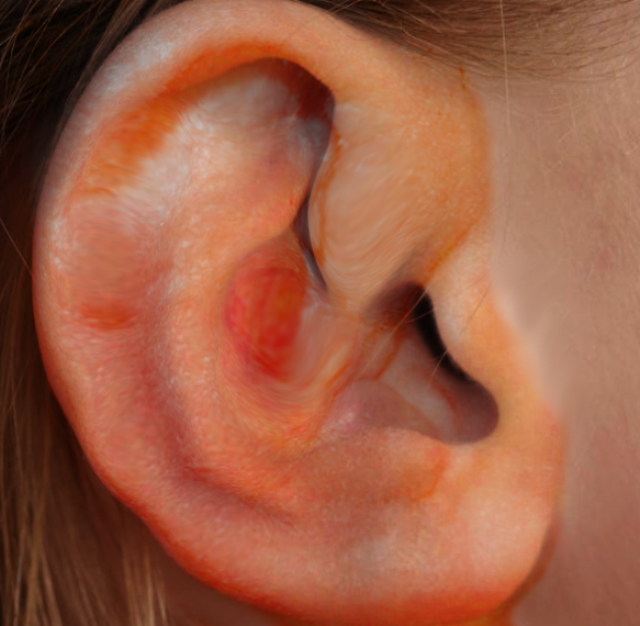
An auricular hematoma is a collection of blood underneath the perichondrium of the ear and typically occurs secondary to trauma. Auricular deformity, commonly known as "cauliflower ear" is the result of untreated or inadequately treated auricular hematoma. It is important to recognize and drain auricular hematomas since persistent hematomas can induce cartilage destruction with subsequent deformity of the ear. Treatment involves drainage and evacuation of the hematoma. This activity describes the evaluation and management of auricular hematomas and highlights the role of the interprofessional team in improving care for affected patients.
Objectives:
Identify the risk factors for developing "cauliflower ear" after an auricular hematoma.
Identify the most common adverse events associated with auricular hematomas.
Describe the management considerations for patients with auricular hematomas.
Explain the importance of improving coordination amongst the interprofessional team to enhance care for patients affected by auricular hematomas.
Introduction
An auricular hematoma is a collection of blood underneath the perichondrium of the ear and typically occurs secondary to trauma.[1] Auricular deformity, commonly known as “cauliflower ear” is the result of untreated or inadequately treated auricular hematoma. It is important to recognize and drain this collection since a persistent hematoma can induce cartilage destruction with subsequent deformity of the ear.[2] Treatment involves drainage and evacuation of the hematoma either at the bedside or in the operating room.[3] To prevent re-accumulation, it is important to place a bolster dressing post drainage procedure to close the potential space.[2] Consultation with ENT or plastic surgery is beneficial as they can provide recommendations regarding diagnosis, management, and follow up.
Etiology
Auricular hematoma is typically caused by trauma. This can be from multiple forms of trauma, such as earring placement though is more common with a larger force or direct blow to the ear such as from a motor vehicle accident. It is most commonly secondary to contact sports such as wrestling, boxing, and martial arts.[1]
Epidemiology
The exact epidemiology has not been well studied. Contact sports such as wrestling, mixed martial arts, ultimate fighting, rugby, and boxing may more readily predispose to such injuries. It could be deduced that males are at a higher risk than females; however, the exact ratio is not known. In a survey of college wrestlers, the incidence of auricular hematoma was found to be 52% for those refusing to wear headgear versus 26% who wore ear protection.[4] This places them at a higher risk of developing cauliflower ear.[5]
Pathophysiology
The auricle is composed of skin, subcutaneous tissue, musculature, and perichondrium which supplies blood to the underlying cartilage.[1] An auricular hematoma is a collection of blood between the perichondrium and underlying cartilage. The primary areas of cartilage in the ear include the tragus, helix, antihelix, triangular fossa, cymba concha, and concha cavum.[1] The blood vessels that supply the ear consist of the superficial temporal and posterior auricular artery.[6] With trauma to the ear, the perichondrium and vasculature are damaged, causing separation from the underlying cartilage and resulting in a potential space for blood to accumulate. Once blood fills this space, it causes vascular compromise of the adjacent cartilage and venous congestion that can result in histologic changes and ensuing cartilage deformity, resulting in an unsightly appearance of the external ear known as cauliflower ear.[1] A process of neocartilage development occurs that is an alteration of the normal histologic structure of the cartilage framework of the ear.
Histopathology
Histologic changes account for the altered appearance of the external ear noted after an auricular hematoma. The cartilage of the ear is usually composed of elastic cartilage. Secondary to trauma, the normal cartilage structure of the ear changes. Two weeks after the auricular hematoma develops, cartilage formation occurs on either side of the hematoma. By three weeks, the hematoma is replaced by soft tissue. By eight weeks post trauma, the soft tissue is replaced by cartilage. By fourteen weeks, bony formation, calcification, and further cartilage growth occur.[7]
History and Physical
Always start with open-ended questions and a standard history. Specific questions which are important to ask are recent trauma, pain/tenderness of ear, previous occurrences, fevers/chills, drainage from ears, change in hearing, immunosuppression, diabetes, blood-thinning medications, and hypertension. Physical exam involves a thorough evaluation of external ear. It is important to have a good understanding of the baseline anatomy of the ear to better differentiate pathology. Use of an otoscope to evaluate the external ear canal and tympanic membrane is paramount. A recent history of trauma is common, and wrestling and boxing are common risk factors. If the mechanism of trauma is large, such as a motor vehicle accident, the practitioner must rule out temporal bone trauma as well as assessing the patient for other injuries.
A proper exam includes a full head and neck exam, the details of which are beyond the scope of this article. A focused physical exam includes an evaluation of the external ear, evaluation of the tympanic membrane with an otoscope, and evaluation for any coexistent lacerations or trauma of the head and neck. It is imperative to evaluate for facial nerve weakness as the facial nerve passes through the ear and can be damaged when there is trauma to the ear. Physical exam findings consistent with auricular hematoma include contour irregularity of ear with swelling and fluctuant area overlying the ear's cartilaginous portions. Likely symptoms include pain, paresthesia, and ecchymosis.[8]
Evaluation
An auricular hematoma is typically diagnosed after a detailed history and physical. Ultrasound can be utilized to evaluate ear swelling and to rule out an auricular abscess. If significant trauma has occurred, there is concern for a foreign body or an abscess or it is determined that it is important to evaluate middle or inner ear structures, CT or MRI can be ordered. CT and MRI should not be used routinely to evaluated auricular hematomas.
If there is evidence of erythema, warmth to the area, diffuse pain on palpation of cartilage, evidence of external auditory canal swelling, or drainage, then the diagnosis of auricular hematoma is less likely. Typically, hearing is not affected by isolated auricular trauma and if the patient has subjective hearing loss, then expanding the differential diagnosis is waranted. In summary, auricular hematomas are generally a clinical diagnosis.
Treatment / Management
Once a hematoma is diagnosed the next step is determining whether treatment should occur in the operating room or at the bedside. It is important to discuss with the patient the risks, benefits, and alternatives to treatment. If the hematoma occurred in an acute setting < 48 hours, an attempt at drainage is appropriate. Keep in mind that patients can opt for no treatment which is acceptable as long as the patient knows the risks and possible poor cosmetic outcome of non treatment.
General Procedure Steps for Auricular Hematoma Drainage:
1) Gather the necessary equipment and make sure there is appropriate lighting.
2) Ensure adequate exposure and place patient in the supine position with the head of the bed elevated.
3) The patients head should be turned so that the unaffected ear is facing towards the stretcher and the affected ear is towards the ceiling.
4) Supplies should consist of an 11 blade or 15 blade scalpel and/or an 18-gauge needle with a 10 cc syringe, suction canister, tubing and suctioning instrument (Frasier), a hemostat, toothed forceps, suture supplies with scissors, bolster material, local anesthetic, and local skin cleansing material.[1]
5) Appropriate hand hygiene should be practiced and gloves should be worn during the procedure. Application of sterile gloves, gowning, headlamp use, and or Loupes to optimize vision are optional.
6) After the patient is positioned properly, the ear is cleaned with a local cleansing agent such as povidone-iodine.
7) Local anesthesia should then be injected or applied topically to the site where the incision or aspiration will be performed (e.g., lidocaine, bupivacaine, LET gel). For best results, the anesthetic can be injected in an auricular block pattern or directly into the site of the auricular hematoma. Several minutes after injection the level of local anesthesia should be assessed. This can be performed by grabbing the tissue of the planned incision with toothed forceps to determine if the area is numb. There are two methods that can potentially be used to drain the auricular hematoma. You should choose the method you will utilize prior to start of the procedure. One method is to incise and drain the hematoma using a scalpel the other is needle aspiration
Incision and drainage:
I. First complete steps 1 through 7 listed above under general procedure steps.
II. Next make a linear incision can be made on the skin overlying the swelling or hematoma.[1] The goal of the incision is to drain the fluid collection; however, making the incision in a cosmetically appealing site is ideal. Incision in areas of concavity will heal with a more aesthetically pleasing results compared to areas of convexity.
III. After the incision is made, hemostats and suction can be used to evacuate the hematoma.
IV. Once all the hematoma is removed, the site can be irrigated with normal saline.
V. A bolster dressing is applied. The bolster serves to close the dead space or potential space where the hematoma formed. When using dental rolls as a bolster, two rolls should be used. Each roll should be placed so it to runs parallel with the incision line on either side of the ear. Two vertical mattress sutures should be placed through the dental rolls to secure the bolster.[1] A permanent suture material such as nylon is appropriate. The suture is ideally on a Keith Needle; however, this is not mandatory. Adequate bolster is applied when there is no potential space for accumulation of hematoma; however, it is important to make the sutures lose enough to preserve the vascular supply of the ear.
VI. Bacitracin can be applied to the incision site post procedure.
VII. It is important to remove all instruments and dispose of sharps appropriately once the procedure is deemed complete. Proper wound care instructions, follow-up, and disposition should be explained to the patient and/or family.
Needle Aspiration:
I. First complete steps 1 through 7 listed above under general procedure steps.
II. The alternate procedure utilizes an 18-gauge needle to aspirate the hematoma. Some studies suggest that an 18-gauge needle may be acceptable for auricular hematoma evacuation when the hematoma is under 2 cm.[1] If the needle aspiration technique is used a bolster should be applied to the affected area of the ear after complete removal of the hematoma.
III. A bolster dressing is applied. The bolster serves to close the dead space or potential space where the hematoma formed. When using dental rolls as a bolster, two rolls should be used. Each roll should be placed so it to runs parallel with the incision line on either side of the ear. Two vertical mattress sutures should be placed through the dental rolls to secure the bolster.[1] A permanent suture material such as nylon is appropriate. The suture is ideally on a Keith Needle; however, this is not mandatory. Adequate bolster is applied when there is no potential space for accumulation of hematoma; however, it is important to make the sutures loose enough to preserve the vascular supply of the ear.
IV. Bacitracin can be applied to the incision site post procedure.
V. It is important to remove all instruments and dispose of sharps appropriately once the procedure is deemed complete. Proper wound care instructions, follow-up, and disposition should be explained to the patient and/or family.
Bolster Options Post Hematoma Evacuation:
There are several variances in the type of bolster used, however, the goal is the same: eliminate the potential space for fluid to accumulate.[1] Newer strategies include the use of splinting material that can be molded to the ear.[2] A recent case reports using fibrin glue to secure the perichondrium to the cartilage to reduce the risk of separation.[3] Bolster dressing can be removed after 5 to 7 days. Antibiotic use is left to the discretion of the physician. If cauliflower ear does form, excision with repair may be undertaken in the form of otoplasty; however, this will require referral to ENT or plastic surgery.[1]
Differential Diagnosis
It is important to rule out infectious, autoimmune, and traumatic sources of auricular swelling. The most common differential diagnosis should include auricular hematoma, perichondritis, auricular abscess, cellulitis, Winkler disease (relapsing perichondritis), a temporomandibular disorder resulting in external ear pain, laceration, normal anatomic variance, erysipelas, sunburn, and skin cancer.[4]
Complications
The risks are minimal; however, they do occur and must be conveyed to the patient for adequate consent to the procedure with a procedure. Always discuss the risks, benefits, and alternatives to management and provide adequate follow up to ensure patient follow-up with ENT or plastic surgery. The complications of an auricular hematoma can be cosmetic, infection, pain, paresthesia, allergy or anaphylaxis to anesthesia or local anesthetic, unsightly scar formation, and re-accumulation of blood after drainage.[4] The typical cauliflower ear deformity is usually the result of untreated or repeated auricular hematomas. The typical appearance of a cauliflower ear is a misfolded ear with several soft tissue humps abnormally located in the ear, overlying where normal cartilage would be found.
Postoperative and Rehabilitation Care
Patients with auricular hematomas can generally be managed as outpatients. Consultation and/or referral to otolaryngology or plastic surgery is recommended. Patients are instructed to take antibiotics if prescribed, pain medications as prescribed and follow-up as directed. The patient should limit physical activity for 10 to 14 days and avoid contact sports for 1 to 2 weeks. If a splint is used, this will need to be removed in 5 to 7 days. Antibiotics can be given while the splint is in place.
Consultations
Appropriate consults and referrals for auricular hematomas are Otolaryngology or plastic surgery. Otolaryngology should also be considered if there is concern for associated external, middle, or inner ear pathology.
Deterrence and Patient Education
Patient education is important in helping prevent recurrence auricular hematomas after resolution. Educating the patient on how to reduce the risks for trauma is imperative. The challenge is that patients are usually involved in contact sports and unwilling to abstain from these sports for an adequate time period.[5] In this situation, athletes can be instructed to wear appropriate head protection to limit the chance of recurrent trauma to the ear. Additionally, if there is any possible trauma to ear, in the acute setting, the use of ice to the area may beneficial in intervals of 15 to 20 minutes to reduce any potential hematoma formation.
Enhancing Healthcare Team Outcomes
An auricular hematoma can present itself in many different circumstances for evaluation by healthcare specialists. Appropriate diagnosis and/or referral to otolaryngology or plastic surgery is imperative post procedure to help improve patient cosmesis and decrease the chances of the patient requiring a more invasive procedure, such as otoplasty, in the future. It is also important to recognize the difference between auricular hematomas and other high-risk diagnoses. A thorough ear exam is imperative to complete a full evaluation to rule out damage to other structures of the ear.[4] This is important because if an external ear infection is mislabeled as an auricular hematoma in a diabetic or immunocompromised individual the consequences can be deadly.