Continuing Education Activity
Feeding tubes are tubes inserted into the gastrointestinal tract to provide a patient with enteral nutrition or for decompression. Tubes come in a variety of sizes, lengths, and materials, depending on the longevity of the tube, placement method, and location. This activity outlines the different types of feeding tubes and reviews the role of the interprofessional team in evaluating and treating patients who undergo feeding tube insertions.
Objectives:
- Identify the different types of feeding tube procedures and their particular indications.
- Describe the differences between the various feeding tube placement techniques.
- Summarize the potential complications of feeding tube placement.
- Outline interprofessional team strategies for improving care coordination and communication to advance the care of patients with feeding tubes and to improve their outcomes.
Introduction
Feeding tubes are tubes mainly inserted into the gastrointestinal (GI) tract to provide a patient with a route for enteral nutrition, though they can also be used for decompression of the GI tract. Tubes come in a variety of sizes, lengths, and materials, depending on the longevity of the tube, placement method, and location. When a patient is unable to intake any or adequate caloric nutrition by mouth, severe malnutrition may result, which will ultimately inhibit healing and contribute to critical illness. A feeding tube is an excellent option to provide the patient with enteral nutrition and can be accomplished in many ways.
Anatomy and Physiology
Prior to the placement of a percutaneous or open gastrostomy tube, it is important that the practitioner review the anatomy of the stomach and surrounding areas. The stomach is divided up into different anatomical parts. The gastroesophageal junction is the point at which the esophagus meets the stomach. The fundus of the stomach is the superior portion lying closest to the diaphragm. The pylorus is the valve that leads out of the stomach into the duodenum. Next to pylorus is the antrum, and the remainder of the body is called the body. Ideally, a feeding tube is placed in the anterior body of the stomach.
Attached to the greater curvature is the greater omentum, which forms the gastrocolic ligament and attaches to the transverse colon. The proximity to which needs to be taken into consideration, especially during the placement of a percutaneous gastrostomy tube.
Not only is normal anatomy extremely important, but it also important to consider variants of normal. A person with hepatomegaly may have a left lobe of the liver that crosses the past midline. Though preoperative imaging is not needed, if prior imaging is available, it is recommended to review it prior to performing a feeding tube procedure. In addition, it is imperative that the practitioner reviews the patient’s surgical history and perform a thorough inspection of the abdomen.
Indications
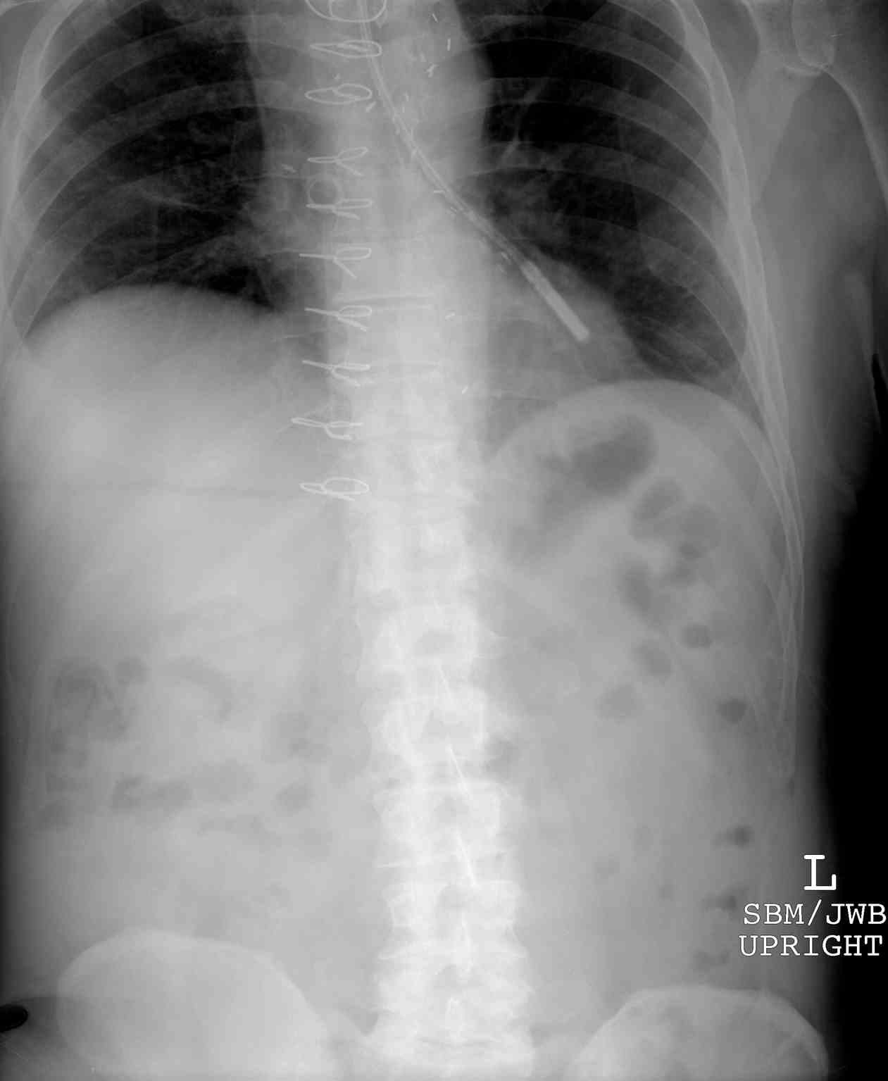
The length of time that a patient will require a feeding tube for enteral nutrition must first be addressed. If a patient will only need enteral nutrition for a short period of time, or if the length of time is not known, a temporary feeding tube may be placed. This is most commonly used to provide enteral nutrition to patients requiring intubation and mechanical ventilation and therefore, cannot swallow. This tube is most commonly an 8-12 French flexible tube inserted orally or via a nare with the tip ending in the stomach or jejunum. The location of the distal tip is most commonly confirmed with plain film x-ray.
Need for long term enteral feedings, usually more than 30 days, are often managed by gastrostomy tube placement. Dysphagia, or inability to swallow without aspiration of oral intake, is one of the most common reasons for this, as well as moderate to severe malnutrition.[1] Less commonly, a gastrostomy tube used for fixation or pexy of the stomach to the anterior abdominal wall after reduction or repair of a para esophageal hernia to prevent herniation of the stomach into the chest.
Gastrostomy tubes are shorter than the nasoenteric tubes described above but have a larger diameter of 18-20 French. Gastrostomy tubes are usually made of a more flexible material, as they are intended to be left in place for a longer duration of time.
Gastrojejunal tubes are dual lumen tubes with one lumen ending in the stomach and one post-pyloric in the jejunum. These are often indicated if a patient requires decompression of the gastric portion and feeding via the jejunal lumen. These are most commonly placed with fluoroscopic guidance but can be performed with endoscopy in the hands of a practiced provider.
Jejunal or post-pyloric feedings are also indicated anatomy has been altered, such as Billroth II or Whipple procedures, gastric outlet obstruction, gastroparesis, recurrent aspiration, severe pancreatitis, hyperemesis gravida, proximal enteric fistula, postoperative anastomotic gastroenteric stenosis.[2]
Contraindications
The unwillingness of the patient or family to provide consent is always an absolute contraindication of any elective procedure. Other contraindications include hemodynamic instability, sepsis, serious coagulation disorder that cannot be corrected, severe ascites, peritonitis, abdominal wall infection near the site of planned insertion, and peritoneal carcinomatosis.
If a patient has a gastric outlet obstruction, severe gastroparesis, or history of altered anatomy such as a previous roux-en-y or gastrectomy, the surgeon should consider postpyloric enteral access as gastrostomy would be contraindicated.[1]
Preparation
The placement of a feeding tube is usually an elective procedure. There may be a few instances where a surgeon will choose to place a feeding tube in concurrent with an emergent procedure if long term tube feeding is anticipated, but this is not widely the case. The patient should, therefore, be medically optimized, anticoagulation held if possible. The anesthesia provider should exam and evaluate the patient prior to the procedure as these patients are usually in-house, allowing any preoperative concerns to be addressed prior to the procedure. The patient should be kept NPO after midnight prior to the procedure. If the patient is receiving feedings via a nasoenteric or oroenteric tube, these tube feedings should be held as well.
Technique or Treatment
Open Stamm Gastrostomy
A supraumbilical incision is made, usually midline. The anterior parietal peritoneum of the stomach is visualized. A spot on the anterior stomach is chosen to allow for the stomach to be brought towards the anterior abdominal wall without tension. Next, the omentum is divided off of the greater curvature to reduce downward tension. 2-3 concentric purse-string suture is placed with a heavy permanent suture at the spot chosen on the stomach for gastrostomy, and a gastrostomy is created.[3] An incision is made in the anterior abdominal wall at the corresponding place for the feeding tube; the feeding tube is placed through the incision and gastrostomy.
Permanent sutures can also be placed to fix the stomach to the anterior abdominal wall. The abdomen is then closed, and the feeding tube secured to the skin.
Open Witzel Jejunostomy
Multiple variations of a jejunostomy feeding tube technique are described; however, the Witzel jejunostomy feeding tube is the most commonly accepted technique. The ligament of Treitz is identified, which indicates the transition of the duodenum to the jejunum. A segment of jejunum approximately 30 cm distal to the ligament of Treitz is then chosen for jejunostomy feeding tube creation; the loop of bowel should be mobile. The feeding tube is then passed through an incision in the anterior abdominal wall at the chosen site. A purse-string of the suture is made at the on the antimesenteric border of the jejunum, and an enterotomy is made so that the tube is passed into the lumen distally. Next, a Witzel tunnel is created with interrupted permanent suture with seromuscular sutures are placed perpendicular to the feeding tube to imbricate the bowel wall over the feeding tube.[4] The jejunum is then secured to the abdominal wall at the catheter entrance site.
Laparoscopic Feeding Tube Placement
Many variations of laparoscopic approaches have been described. Ports are placed, and pneumoperitoneum is achieved. The stomach is then lifted to the anterior abdominal wall and spot chosen for tube placement. A 2-0 silk suture is then used to form a purse string around this area. Electrocautery is then used to make a small gastrotomy in the center of the purse string. The feeding tube is then inserted through the anterior abdominal wall and into the gastrotomy, tieing the purse-string suture tight around the tube. Next, the stomach is again lifted to the abdominal wall and sutured in place to the fascia.
It is also possible to perform the placement of a jejunostomy feeding tube laparoscopically. The procedure is began similar to the above described laparoscopic gastrostomy tube; however, a mobile portion of the jejunum distal to the ligament of Treitz is chosen. A purse-string is placed in the anterior mesenteric side, an enterotomy is made, and the feeding tube is passed via an incision in the anterior abdominal wall and suture tied. The jejunum is then fixed to the abdominal wall in four quadrants. Additional fixation of the distal jejunum to the anterior abdominal wall is also recommended; this is thought to decrease the chance of jejunal torsion.
Percutaneous Gastrostomy Tube
Percutaneous gastrostomy feeding tubes can be placed radiographically using fluoroscopy, or endoscopically. There complication rate for each procedure is similar, though vary in type of complication.[5] The choice between the two procedures is, therefore, usually institution and resource-dependent.
Endoscopic Percutaneous Gastrostomy Tube Placement
The Ponsky preoral pull technique is the most common technique used to perform percutaneous endoscopic gastrostomy tube placement. The endoscopic is passed thorough the oropharynx, and the esophagus is intubated. The scope is passed through the esophagus and into the stomach. After the stomach is insufflated and transilluminated, a spot is chosen for peg placement, usually 2 cm medial to the costal margin and 2 cm below the xiphoid process. The skin is localized, and a small incision is made to allow a needle to be passed through the incision into the stomach, which is visualized on the endoscopic camera. A guidewire is then passed through the needle and grasped with an endoscopic snare, which is withdrawal out of the patient's mouth, and the needle is removed. The feeding tube is attached to the guidewire and pulled into the patient's mouth and out of the anterior abdominal wall. It is secured in place with a disc or bar-shaped bumper against the skin, and repeat endoscopy is used to confirm proper placement.[6]
Percutaneous Radiographic Gastrostomy Tube
For this procedure, a nasogastric tube is placed, and the stomach is inflated with approximately 500 ml of air. The stomach is identified with fluoroscopy, and a needle is then passed through a skin incision into the stomach. A wire is then inserted through the needle, and a series of dilators are used over the wire in a Seldinger technique before the feeding tube was then inserted.[7]
Complications
Complications after percutaneously placed gastrostomy tubes include infection, tube migration, hemorrhage, stomal leak, ileus, and bowel injury.[8] Bowel injury is oftentimes hard to diagnose as a small amount of pneumoperitoneum after the procedure is to be expected. However, when compiled with pain and tenderness should be investigated further.[9] Another feared complications of percutaneous gastrostomy tubes is a transhepatic peg tube; usually, these are discovered incidentally on post-procedure imaging, but could also present with massive bleeding or sepsis. The use of ultrasound in patients with known hepatomegaly has been suggested to help avoid this complication.[10]
When an open gastrostomy or open jejunostomy tube is placed, a rare but potential complication is a leak of gastric contents or tube feeds outside of the gastrostomy but into the abdominal cavity. This would likely be due to a technical error and can be prevented by testing the feeding tube with a saline flush while inspecting for leaks prior to abdominal closure.
If a percutaneously placed feeding tube becomes dislodged prior to the stomach fusing to the anterior abdominal wall, the stomach will fall away, and the tube would then be intraperitoneal. Peritonitis and infection can occur. Emergent intervention is usually needed to either replace the gastrostomy tube or close the gastrostomy and place a new feeding tube. Both open and laparoscopic procedures aim to prevent this complication by securing the stomach or jejunum to the anterior abdominal wall.
Clinical Significance
There are a variety of different types of feeding tubes and procedures for insertion, some more invasive than others. It is an important topic for each healthcare provider to understand so that the most appropriate procedure is chosen for each individual patient.
Enhancing Healthcare Team Outcomes
It is important that all providers, including physicians, proceduralists, nurses, speech pathologists, and nutritionists, be well versed in this topic in order to provide the best care and outcomes possible for their patients. Speech pathologists are usually recommending feeding tube placement after a thorough evaluation of the patient's ability to safely swallow. A surgeon or proceduralist (including endoscopist and an interventional radiology physician) is responsible for performing that patient best suited for that patient. The nurses play a vital role as a member of the interprofessional group as they will monitor the patient before and after the procedure, as well as administer medication and nutrition through the feeding tube as well as provide daily maintenance as needed for the feeding tube. The nutritionist will calculate the number of calories and macronutrients that the patient is in need of and will recommend the type of tube feedings and the rate.
Understanding this topic has the ability to impact many patient’s lives due to its clinical significance and the importance of ensuring each patient is able to receive nutrition, hydration, and medication.