EEG Localization Related Epilepsies

Earn CME/CE in your profession:


Continuing Education Activity

An electroencephalogram (EEG) is essential to define an epileptic syndrome, especially a focal epileptic seizure. It also aids in both localizing and characterizing the seizure, and aid in the selection of the drug for treatment. Electrodes, amplifiers, and plotting devices are used to record an EEG. These are set to various montages, that create numerous channels for region-specific electrical activity monitoring. Focal seizures are associated with specific EEG patterns, that aid in detecting and discerning the focus and subsequently the treatment for the specific epileptic syndrome. This activity reviews the application, technique used, and common EEG findings in localizing epilepsies. It also highlights the role of an interprofessional team in evaluating and treating this condition.

Objectives:

  • Describe the anticipated EEG findings associated with localization-related epilepsies.
  • Explain the equipment and technique used in recording EEG in localization-related epilepsies.
  • Summarize the clinical significance, indications, contraindication, and potential complications with EEG in localization-related epilepsies.
  • Review the importance of improving care coordination amongst interprofessional team members to improve outcomes for patients affected by localization-related epilepsies.

Introduction

Epilepsy is a condition defined by the occurrence of two or more unprovoked seizures that happen at least 24 hours apart. These are typically associated with abnormal hypersynchronous discharges in the brain, resulting in clinical manifestations.[1] An electroencephalogram (EEG) is a useful tool for recording the electrical activity from the cortex and the deeper brain structures. It is a useful tool for diagnosing and classifying various seizure types.[2] Localization-related epilepsies, also known as focal epilepsies, refer to an abnormal neuronal activity arising from a localized focus and involve a limited portion of the cortex. When there is no associated impairment in consciousness, it is called a 'focal onset aware seizure' previously known as a simple partial seizure. When it is associated with impairment in consciousness, it is called a 'focal impaired awareness seizure,' earlier known as a complex partial seizure.[3]

Anatomy and Physiology

In the cortex, when a neurotransmitter is released, the postsynaptic endplate of the adjacent dendrite is stimulated, leading to the formation of an endplate postsynaptic potential (EPSP). Consequently, an electrical dipole is established across the soma of the neuron with positive charges internally and negative charges externally. This dipole then rapidly moves along the axon of the neuron as an action potential. The EEG measures the summation of EPSPs that is a net result of both excitatory and inhibitory postsynaptic potentials from groups of synchronously firing pyramidal cells.[4][5]

The synchronization between different cerebral functions is related to the dynamic interactions of segregated brain regions.[6] In the event of a seizure, an abnormality of the brain network occurs, which causes an abnormal, large super-synchronous neuronal discharge.[7][8] The evaluation of the abnormal waveforms of these discharges and their propagation facilitates the understanding of transmission pathways and the associated seizure focus.[6]

Indications

In a patient presenting with a history of seizures, the primary evaluation includes classification of the type of seizure and subsequently, the localization of the seizure focus. In the case of partial epilepsies, the clinical presentation including the type of auras, if present, along with an EEG help in localizing the focus of the lesion, fairly accurately. Imaging modalities can aid in the localization if structural lesions are suspected.

With regards to the EEG, a trained specialist is quick to identify the source of the seizure even though it may record an abnormal waveform remote to the lesion, based on its morphology and frequency, in association with the presentation. Together, they help classify the localization of lesions into mesial temporal lobe epilepsy (MTLE), lateral temporal lobe epilepsy (LTLE), frontal lobe epilepsy (FLE), parietal lobe epilepsy (PLE), or occipital lobe epilepsy (OLE). The subjects with lesions that are limited to the temporal lobe were regarded as having either MTLE or LTLE, according to the involvement of the mesial temporal structures. The subjects with asymmetrical hippocampal sclerosis (HS) with lateralization of a smaller hippocampus were also included in this study.[9]

Contraindications

Although there is no clear contraindication, relying on an EEG is the most important diagnostic tool for the confirmation of epilepsy, it should be performed only to support a diagnosis of epilepsy in adults in whom the clinical history suggests that the seizure is likely to be epileptic in origin. 

An important consideration is the overdiagnosis of epilepsy that has been a significant hurdle in treating seizures in the last few decades. A detailed clinical history is important to determine the relevance of prescribing an EEG. If performed in cases of probable syncope a false positive result on the EEG can further lead to faulty diagnosis and unnecessary treatment. Similarly, an EEG should also not be used to exclude a diagnosis of epilepsy in patients in whom the clinical presentation supports a diagnosis of a non-epileptic event. For this reason, it is generally performed after the second epileptic seizure but in certain circumstances, as evaluated by the specialist, it can be carried out after a first seizure.[10]

An in-detail history of ongoing medication and a history of cerebrovascular disease and a history of migraine headaches and sleep deprivation is also important to avoid a misdiagnosis of seizure disorder.

Equipment

The basic pieces of equipment involved in the synthesizing of an electroencephalograph are electrodes, amplifiers, and plotting equipment. Until recently, electrolytic gel and salts were used to improve the conductivity from the scalp, through the electrodes. The advent of the 'dry electrodes' hastens the scalp preparation, by obviating the need for gels and salts. This results in a more accurate EEG recording but is yet to be used in a widespread manner. As of today, the EEG electrode caps are well-tolerated by all age groups. The amplifier and plotting equipment historically was a mechanical pen and paper recording device. This has been replaced by innovative digital EEG systems that allow for faster sampling rates and simultaneously record from an increasing number of channels. These days, a standard commercially available EEG system used in clinical practice can readily obtain data from at least 128 channels, with over 10 kHz sampling rate by all channels and a 24-bit resolution at each amplifier.[4]

Interpreting an EEG involves understanding of the electrical wave progression over the brain. The basic electrode placement follows the universal 10 to 20 system and is set to the required montages. Montages are EEG electrode settings used to specifically record the endplate polysynaptic potentials (EPSP), from a focused point of interest.[11]

These fit broadly under three headings:

  • Referential montages
  • Bipolar montages
  • Laplacian montages

Referential montages: A referential montage plots the waveforms from the suspected point of focus on the head (known as an active electrode) to a reference point elsewhere on the body, including the scalp. However, the virtue of neutrality is not guaranteed in this reference electrode.

  1. Central reference - A high signal to noise ratio (SNR) is favorable for a successful EEG recording. It is a virtue of the intensity and frequency of the stimulus, as well as the active and the reference electrode. Historically, the potential at midline electrode (‘Cz') or the average potential at all electrodes, was selected as the reference. 
  2. Average reference - The average of all potentials across the brain is taken as reference.
  3. Localized reference - The average of only the surrounding potentials of a particular electrode is taken to calculate the average.

Today, a dynamic selection method for the reference electrode is proposed, which allows all electrodes to be looked upon as active electrodes. In contrast, an electrode is statistically chosen, based on the highest estimated SNR, as the reference electrode for that specific frequency stimulus.[12]

Bipolar montages: The term 'bipolar' is derived from the mechanism of recordings in this electrode placement. There exist two electrodes, both placed along an anteroposterior or in a left-over-right position in such a way, that the difference plotted is between two set regions over the brain. Hence, this is also known as differential montages.

Laplacian montages: This a different type of montage, where the second derivative is a combined weighted average of the voltages surrounding the particular electrode of interest. It is estimated using relatively complex computation in practice, and the net result depends on the particular electrode involved in the montage. It is best applicable when the focal discharges transmit a minimal field.[13]

Various types of montage settings are used for evaluation based on the suspected lobular involvement, to accurately isolate the epileptic focus.[14] The ease of reformatting and re-montaging for the purposes of localizing the abnormalities is facilitated by the digital EEG at the user's disposal.

Technique or Treatment

A systemic approach is paramount for interpreting an EEG recording. Before starting the analysis, certain factors including the patient's age, level of physical activity, mental state, level of consciousness, the influence of different biological factors, environmental factors, and pharmacological agents that can potentially influence the morphology of the waveforms need to be taken into consideration.

There is a wide variation in the EEG waveforms. A good understanding of the normal or benign variants is necessary to differential normal or benign variants from pathologic waveforms.[15] Some of these normal variants include:

  1. Wicket spikes: These waveforms appear over the temporal (anterior or mid-temporal) region during relaxed wakefulness, drowsiness, or state of light sleep.
  2. Benign epileptiform transients of sleep (BETS): These are also called as small sharp spikes (SSS) and occur in stage 1 or stage 2 of sleep.
  3. 6 Hz "phantom" spike-and-wave complex (PhSW): PhSW can be considered a smaller version of the 3Hz spike and wave pattern. These waveforms have low amplitudes and appear in the frequency of 5 Hz to 7 Hz.
  4. Rhythmic mid-temporal theta of drowsiness (psychomotor variant) or RMTD: This pattern is usually located in the mid temporal region and appears in relaxed wakefulness and drowsiness.
  5. Positive occipital sharp transients of sleep (POSTS): These waveforms appear in the occipital regions during non-rapid eye movement (NREM) sleep and are asymmetrically distributed.
  6. Subclinical rhythmic EEG discharge in adults (SREDA): This is a very rare benign EEG pattern. It resembles ictal discharges and is, at times, interpreted as such, leading to a misdiagnosis of epilepsy.
  7. 14 Hz and 6 Hz positive spikes: 6 and 14 Hz positive spikes are typically seen in the younger age group.
  8. Presence of repetitive vertex waves, especially in children.
  9. Breach rhythm: These waveforms are seen over the regions that have a skull defect. Because of the skull defect, there is increased visibility of faster frequencies that are otherwise less appreciated on scalp EEG.

EEG recording of a seizure can begin with the appearance of abnormal discharges in bursts, known as ictal epileptiform discharges. The discharges increase in frequency to rapid continuous spikes and waves, progressing to numerous spikes with buried waves, at peak seizure activity. As the activity slows down, the waves reappear and progressively reduce in frequency, and finally stop.[16] The time of seizure activity is called the ictal period, and the time between seizures is called the interictal period. EEG activity during the interictal period also reveals abnormal discharges, and these are called interictal discharges (IEDs). Since most patients present either immediately after or before a seizure, IEDs are important for validating a clinical suspicion of seizure activity in epilepsy. 

Typically, multiple EEGs are required to record IEDs. To put things into perspective, every fourth consecutive EEG in an epileptic patient has an IED frequency between 60% to 90%. The frequency of IED in a non-epileptic patient is about 0.5% to 2.5% in healthy young men and about 12% in non-epileptic patients of all age groups with progressive cerebral disorders. Specificity is probably lower and sensitivity higher in children as compared to adults.[17]

Activation methods like hyperventilation, sleep deprivation, and photic stimulation increase the appearance of IEDs and are useful for localization purposes and in being more assertive about the diagnosis of epilepsy.

Although surface EEG recordings are less sensitive than invasive studies, they are efficient in approximating the epileptogenic zone in most of the common epilepsies. The most commonly used invasive electrodes are stereotactically implanted depth electrodes and subdural strip or grid electrodes. Invasive studies are most useful when the scalp EEG does not yield a result, or when the focus is located adjacent to the eloquent cortex.[18]

Complications

There are a few concerns regarding the waveforms in EEGs. Firstly, the choice of the reference electrode should be such that it cancels out the normal waveforms and amplifies only the pathological ones. It must be remembered that an EEG is influenced by all electrical activity occurring both remotely and locally; however, the closest electrical activity would be the most prominent influencer. The reference electrode should be placed in a location where it captures all the interfering waveforms as well. The next concern for the reference electrode is that there must be a significant potential difference to facilitate the charge movement without acceleration. This means that a reference electrode placed too close to the pathological site would essentially have the same potential as the active electrode, making the recording not useful for interpretation.[4] 

Clinical Significance

The most important waveforms for clinical evaluation include delta (0.5 Hz to 4 Hz); theta (4 Hz to 7 Hz); alpha (8 Hz to 12 Hz); sigma (12 Hz to 16 Hz) and beta (13 Hz to 30 Hz) waves. Additionally, other waveforms like infra slow oscillations (ISO) (less than 0.5 Hz) and high-frequency oscillations (HFOs) (greater than 30 Hz) have increasingly been considered clinically relevant with the discovery of digital signal processing.[19][20][21][22][23]

EEG is useful in a variety of cerebral pathologies and is complemented by a spectrum of more advanced imaging modalities. EGG has been used in the evaluation and study of epilepsy, states of altered consciousness, the parasomnias, dementias, toxic confusional states, cerebral infections, and various other encephalopathies. Abnormal waveforms in EEG reflect a wide spectrum of general pathophysiological processes, raised intracranial pressure, cerebral anoxia, edema, epileptogenesis, etc., and do not specify a particular disease most of the times.[24] EEG is fundamental in discerning an array of localized epileptiform activity in the cerebral cortex. 

There exists a dynamic interaction between specialized regions in the brain that enable processing of information. This interregional communication mediates an eclectic range of neurological processes. These intercortical communications are disrupted in patients with focal epilepsy.[25] However, clinical studies have recorded abnormal activity beyond the region of pathology.[26][27][28][29] 

In a study conducted by Foldvary et al., localized ictal onset was seen in 57% of seizures. These included mesial temporal lobe epilepsy (MTLE), lateral frontal lobe epilepsy (LFLE), and parietal lobe epilepsy. Lateralized onsets predominated in neocortical temporal lobe epilepsy, and generalized onsets were seen in mesial frontal lobe epilepsy (MFLE) and occipital lobe epilepsy.[30]

Classical EEG morphologies, based on specific lobular involvement and epileptic foci are described below.

Temporal Lobe Epilepsy (TLE)

  1. Ictal EEG finding: Ictal EEG is abnormal in about 95% of the patients with focal impaired awareness seizures, with 66% of them showing an electro-decremental pattern. A 5 Hz to 7 Hz rhythmic theta discharge in the temporal regions is specific for temporal lobe epilepsy. Depth electrodes are useful in capturing this pattern and are accurate in diagnosing ipsilateral mesial temporal structures. Scalp electrodes successfully lateralize but fail to accurately localize the focus of the seizure. Ictal EEG changes are difficult to appreciate using the scalp electrodes in frontal lobe seizures because of movement artifacts. Most EEGs would require additional neuroimaging studies using various modalities like magnetic resonance imaging (MRI), interictal fluorodeoxyglucose (FDG) photon emission tomography (PET), ictal single-photon emission computed tomography (SPECT), magnetic encephalography (MEG) or functional magnetic resonance imaging (fMRI) for further assessment of the abnormalities.[31]
  2. Interictal epileptiform discharges: Isolated IED like sharps and spikes over the temporal region and temporal intermittent rhythmic delta activity (TIRDA), in addition to clinical history, are strongly correlated with a diagnosis of temporal lobe epilepsy (TLE).[32][33] In medial temporal lobe seizures, the amplitude of mesial temporal spikes is maximal at the anterior temporal scalp electrodes, and sphenoidal electrodes when used.[34]
  3. Seizure semiology:
    • Auras: Visceral sensations and fear are associated with mesial temporal lobe epilepsy, while auditory and vertiginous auras have been associated with the neocortical temporal lobe epilepsy.
    • Neocortical temporal lobe epilepsy: At seizure onset, patients with neocortical TLE are more likely to describe any type of hallucination or illusion. Automatisms and dystonic posturing are not found in neocortical TLE.[32][35]
    • Mesial temporal epilepsy: Patients present with a behavioral arrest that is observed as a blank facial expression along with loss of awareness. This change is followed by oral, facial, or alimentary automatisms such as lip-smacking, chewing, sucking, or swallowing or is accompanied by ipsilateral automatisms such as repetitive hand movements, picking, or fidgeting behavior, and contralateral abnormal posturing of limbs. Patients commonly have a period of postictal confusion. Rarely, this progresses to secondary generalization.[36]

Frontal Lobe Epilepsy (FLE)

Frontal lobe epilepsy (FLE), after temporal lobe epilepsy, is the second most common type of the localization-related epilepsy of childhood.[37] 

  1. Ictal EEG: Scalp ictal EEG changes are difficult to appreciate with most of the frontal lobe seizures due to the movement artifacts obscuring the visibility of the underlying ictal waveforms.[38]
  2. Interictal EEG: IEDs are observed in only 60% to 80% of FLE patients and have a lower localizing value than in TLE since they can be bilateral, involving multiple lobes, or even secondarily generalized.[39] The medial frontal epilepsies rarely reveal any IED, and even if they do, they are bifrontal spike and wave discharges.[40]
  3. Seizure semiology: In view of the fact that the ictal EEG in FLE is of less value due to frequent muscle and motion artifacts, analyzing the ictal semiology and clinical history are important to differentiate FLE from psychogenic non-epileptic spells (PNES). This is condition is frequently misdiagnosed as epilepsy.[41]
    • Dorsolateral frontal epilepsy: The clinical manifestations of frontal lobe seizures are diverse. Seizures originating from the premotor cortex exhibit forced contralateral head deviation or head-turning called versive seizures. Activation of contralateral frontal eye field causes lateral deviation of the eye. Aphasic seizures are caused due to Broca area involvement.[42] Seizures involving the prefrontal cortex have more variable clinical manifestations. These are described as "hyper-motor seizures." These seizures usually have somatosensory auras followed by bizarre gestures, laughing, shouting, bicycle peddling, and thrashing of the extremities.[43]
    • Mesial frontal epilepsy: Associated features include loss of consciousness followed by conjugate eye and head deviation, behavioral arrest and immediate recovery of consciousness.[44]

Parietal Lobe Epilepsy (PLE)

  1. Ictal EEG: Ictal scalp EEG is rarely localizing in PLE. Even extensive invasive studies done for localization can be inconclusive.[45]
  2. Interictal EEG: It is common to encounter multifocal and multiregional IEDs with cortical involvement, making it difficult to localize the focus. Ristic et al. rightfully called it, 'a great imitator among focal epilepsies.'[46][47]
  3. Seizure semiology: Most patients with parietal lobe seizures have no symptoms or signs suggesting parietal lobe involvement. If specific symptoms are present, they include unilateral paresthesias and pain that occur early during the seizure. Other symptoms occur due to diffuse cortical involvement and include hallucinations and distortions of space, among other symptoms.[48]

Occipital Lobe Epilepsy (OLE)

  1. Ictal EEG: During the ictal period, an EEG recording is more likely to show diffuse bi-occipital activity spreading to the temporal regions than well-localized unifocal discharges in the occipital region. 
  2. Interictal EEG: IEDs can occur either spontaneously or following photic stimulation. In spontaneously occurring IEDs, the discharges are in the form of unilateral posterior EEG slowing rather than spike-waves. It is also rarely localized over the occipital cortex. Instead, the posterior temporal region is the most common site. In contrast, photosensitive OLE requires intermittent photic stimulation to elicit IEDs. They occur in the form of either spikes/polyspikes confined to the occipital region or generalized spikes/polyspikes diffusely spread out to the posterior cortex.[49][50]
  3. Seizure semiology: Seizure in OLE often presents with visual symptoms including hallucinations, blindness, nystagmus, and rapid blinking of the eye. Rarely, it presents as a generalized tonic-clonic seizure and impaired consciousness. This signifies a spread to neighboring cortical regions and thus makes the focus localization rather difficult.[51]

For most focal onset aware seizures, the scalp EEG commonly shows no change in simple partial seizures, because the focal ictal discharge is distant or deep, or involves too small a neuronal aggregate for a synchronized activity to register on the scalp. It becomes more evident after a larger cortical area gets involved prior to secondary generalization.

Enhancing Healthcare Team Outcomes

An epileptic syndrome is a chronic disorder that heavily depends on an interprofessional team to provide a holistic and integrated approach to provide the best possible long term seizure control.[52] The neurologist, epileptologist, the EEG technician, the nurse practitioner, the pharmacist, the primary healthcare provider, the neuro-radiologist and the neurosurgeon, all are an invaluable part of the interprofessional team taking care of patients with epilepsy. While the EEG technician, is key to accurate electrode placement and consequently recording an EEG without artifacts or without waveform cancellations, the neurologist and epileptologist are responsible for clinically correlating these recordings to determine the diagnosis and formulate the next course of action. The neurosurgeons play an important role in placing the electrodes for invasive monitoring for focal localization in complex cases and maximally resecting a pathological lesion associated with epilepsy while sparing the eloquent regions. Patients with epilepsy require education about their medication usage, side effects, and the importance of compliance. The clinical team comprising of the nurse practitioners, primary care physicians, and the neurologists/epileptologist play an important role in monitoring and education. The pharmacist is in charge of dispensing the correct dosage of the drug and preventing any possible drug interactions.

Collaborating by shared decision making and communication are key elements for a good outcome. The interprofessional care provided to the patient must use an integrated care pathway combined with an evidence-based approach to planning and evaluation of all joint activities.[53] The earlier signs and symptoms of a complication are identified, the better is the prognosis and outcome. [Level 3]


(Click Image to Enlarge)
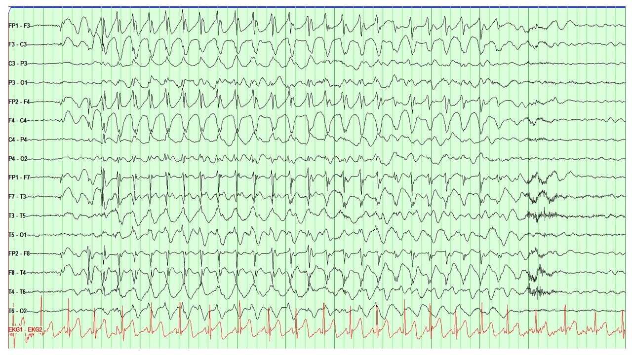
<p>EEG Showing the Characteristic 3 Hz Spike and Wave Discharges Seen in Absence Epilepsy.</p>

EEG Showing the Characteristic 3 Hz Spike and Wave Discharges Seen in Absence Epilepsy.


Contributed by AC Albuja, MD


(Click Image to Enlarge)
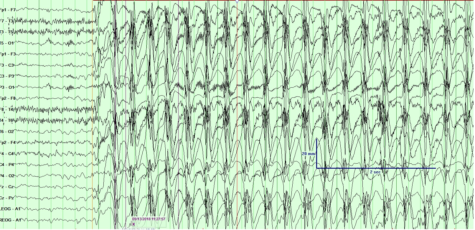
EEG - gen epileptiform
Contributed by Arayamparambil Anilkumar MD

(Click Image to Enlarge)
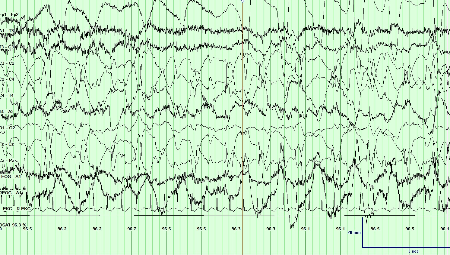
EEG Neonatal seizure
Contributed by Arayamparambil Anilkumar, MD
Details

Editor:

Appaji Rayi

Updated:

7/24/2023 11:02:51 PM

References


[1]

Scheffer IE, Berkovic S, Capovilla G, Connolly MB, French J, Guilhoto L, Hirsch E, Jain S, Mathern GW, Moshé SL, Nordli DR, Perucca E, Tomson T, Wiebe S, Zhang YH, Zuberi SM. ILAE classification of the epilepsies: Position paper of the ILAE Commission for Classification and Terminology. Epilepsia. 2017 Apr:58(4):512-521. doi: 10.1111/epi.13709. Epub 2017 Mar 8     [PubMed PMID: 28276062]


[2]

Guerreiro CA. Epilepsy: Is there hope? The Indian journal of medical research. 2016 Nov:144(5):657-660. doi: 10.4103/ijmr.IJMR_1051_16. Epub     [PubMed PMID: 28361817]


[3]

Ighodaro ET, Maini K, Arya K, Sharma S. Focal Onset Seizure. StatPearls. 2024 Jan:():     [PubMed PMID: 29763181]


[4]

Biasiucci A, Franceschiello B, Murray MM. Electroencephalography. Current biology : CB. 2019 Feb 4:29(3):R80-R85. doi: 10.1016/j.cub.2018.11.052. Epub     [PubMed PMID: 30721678]


[5]

Kirschstein T, Köhling R. What is the source of the EEG? Clinical EEG and neuroscience. 2009 Jul:40(3):146-9     [PubMed PMID: 19715175]


[6]

Mei T, Wei X, Chen Z, Tian X, Dong N, Li D, Zhou Y. Epileptic foci localization based on mapping the synchronization of dynamic brain network. BMC medical informatics and decision making. 2019 Jan 31:19(Suppl 1):19. doi: 10.1186/s12911-019-0737-8. Epub 2019 Jan 31     [PubMed PMID: 30700279]


[7]

Stam CJ. Modern network science of neurological disorders. Nature reviews. Neuroscience. 2014 Oct:15(10):683-95. doi: 10.1038/nrn3801. Epub 2014 Sep 4     [PubMed PMID: 25186238]


[8]

Ravindra VM, Sweney MT, Bollo RJ. Recent developments in the surgical management of paediatric epilepsy. Archives of disease in childhood. 2017 Aug:102(8):760-766. doi: 10.1136/archdischild-2016-311183. Epub 2017 Jan 17     [PubMed PMID: 28096104]


[9]

Ye BS, Cho YJ, Jang SH, Lee MK, Lee BI, Heo K. The localizing and lateralizing value of auras in lesional partial epilepsy patients. Yonsei medical journal. 2012 May:53(3):477-85. doi: 10.3349/ymj.2012.53.3.477. Epub     [PubMed PMID: 22476989]


[10]

National Clinical Guideline Centre (UK). The Epilepsies: The Diagnosis and Management of the Epilepsies in Adults and Children in Primary and Secondary Care: Pharmacological Update of Clinical Guideline 20. 2012 Jan:():     [PubMed PMID: 25340221]


[11]

Kutluay E, Kalamangalam GP. Montages for Noninvasive EEG Recording. Journal of clinical neurophysiology : official publication of the American Electroencephalographic Society. 2019 Sep:36(5):330-336. doi: 10.1097/WNP.0000000000000546. Epub     [PubMed PMID: 31490450]


[12]

Wu Z, Su S. A dynamic selection method for reference electrode in SSVEP-based BCI. PloS one. 2014:9(8):e104248. doi: 10.1371/journal.pone.0104248. Epub 2014 Aug 6     [PubMed PMID: 25100038]


[13]

Gordon R, Rzempoluck EJ. Introduction to Laplacian montages. American journal of electroneurodiagnostic technology. 2004 Jun:44(2):98-102     [PubMed PMID: 15328706]


[14]

Foldvary N, Caruso AC, Mascha E, Perry M, Klem G, McCarthy V, Qureshi F, Dinner D. Identifying montages that best detect electrographic seizure activity during polysomnography. Sleep. 2000 Mar 15:23(2):221-9     [PubMed PMID: 10737339]


[15]

Kang JY, Krauss GL. Normal Variants Are Commonly Overread as Interictal Epileptiform Abnormalities. Journal of clinical neurophysiology : official publication of the American Electroencephalographic Society. 2019 Jul:36(4):257-263. doi: 10.1097/WNP.0000000000000613. Epub     [PubMed PMID: 31274688]


[16]

Rosenow F, Klein KM, Hamer HM. Non-invasive EEG evaluation in epilepsy diagnosis. Expert review of neurotherapeutics. 2015 Apr:15(4):425-44. doi: 10.1586/14737175.2015.1025382. Epub 2015 Mar 16     [PubMed PMID: 25779862]


[17]

Aanestad E, Gilhus NE, Brogger J. Interictal epileptiform discharges vary across age groups. Clinical neurophysiology : official journal of the International Federation of Clinical Neurophysiology. 2020 Jan:131(1):25-33. doi: 10.1016/j.clinph.2019.09.017. Epub 2019 Nov 4     [PubMed PMID: 31751836]


[18]

Noachtar S, Rémi J. The role of EEG in epilepsy: a critical review. Epilepsy & behavior : E&B. 2009 May:15(1):22-33. doi: 10.1016/j.yebeh.2009.02.035. Epub 2009 Feb 25     [PubMed PMID: 19248841]


[19]

Vanhatalo S, Palva JM, Holmes MD, Miller JW, Voipio J, Kaila K. Infraslow oscillations modulate excitability and interictal epileptic activity in the human cortex during sleep. Proceedings of the National Academy of Sciences of the United States of America. 2004 Apr 6:101(14):5053-7     [PubMed PMID: 15044698]


[20]

CORDEAU JP. Monorhythmic frontal delta activity in the human electroencephalogram: a study of 100 cases. Electroencephalography and clinical neurophysiology. 1959 Nov:11():733-46     [PubMed PMID: 13811933]

Level 3 (low-level) evidence

[21]

Reiher J, Beaudry M, Leduc CP. Temporal intermittent rhythmic delta activity (TIRDA) in the diagnosis of complex partial epilepsy: sensitivity, specificity and predictive value. The Canadian journal of neurological sciences. Le journal canadien des sciences neurologiques. 1989 Nov:16(4):398-401     [PubMed PMID: 2804800]


[22]

Gondeck AR, Smith JR. Dynamics of human sleep sigma spindles. Electroencephalography and clinical neurophysiology. 1974 Sep:37(3):293-7     [PubMed PMID: 4136646]


[23]

Worrell G, Gotman J. High-frequency oscillations and other electrophysiological biomarkers of epilepsy: clinical studies. Biomarkers in medicine. 2011 Oct:5(5):557-66. doi: 10.2217/bmm.11.74. Epub     [PubMed PMID: 22003904]


[24]

Binnie CD, Prior PF. Electroencephalography. Journal of neurology, neurosurgery, and psychiatry. 1994 Nov:57(11):1308-19     [PubMed PMID: 7964803]


[25]

Dahal P, Ghani N, Flinker A, Dugan P, Friedman D, Doyle W, Devinsky O, Khodagholy D, Gelinas JN. Interictal epileptiform discharges shape large-scale intercortical communication. Brain : a journal of neurology. 2019 Nov 1:142(11):3502-3513. doi: 10.1093/brain/awz269. Epub     [PubMed PMID: 31501850]


[26]

Bettus G, Ranjeva JP, Wendling F, Bénar CG, Confort-Gouny S, Régis J, Chauvel P, Cozzone PJ, Lemieux L, Bartolomei F, Guye M. Interictal functional connectivity of human epileptic networks assessed by intracerebral EEG and BOLD signal fluctuations. PloS one. 2011:6(5):e20071. doi: 10.1371/journal.pone.0020071. Epub 2011 May 19     [PubMed PMID: 21625517]


[27]

Englot DJ, Konrad PE, Morgan VL. Regional and global connectivity disturbances in focal epilepsy, related neurocognitive sequelae, and potential mechanistic underpinnings. Epilepsia. 2016 Oct:57(10):1546-1557. doi: 10.1111/epi.13510. Epub 2016 Aug 24     [PubMed PMID: 27554793]


[28]

Lagarde S, Roehri N, Lambert I, Trebuchon A, McGonigal A, Carron R, Scavarda D, Milh M, Pizzo F, Colombet B, Giusiano B, Medina Villalon S, Guye M, Bénar CG, Bartolomei F. Interictal stereotactic-EEG functional connectivity in refractory focal epilepsies. Brain : a journal of neurology. 2018 Oct 1:141(10):2966-2980. doi: 10.1093/brain/awy214. Epub     [PubMed PMID: 30107499]


[29]

Tong X, An D, Xiao F, Lei D, Niu R, Li W, Ren J, Liu W, Tang Y, Zhang L, Zhou B, Gong Q, Zhou D. Real-time effects of interictal spikes on hippocampus and amygdala functional connectivity in unilateral temporal lobe epilepsy: An EEG-fMRI study. Epilepsia. 2019 Feb:60(2):246-254. doi: 10.1111/epi.14646. Epub 2019 Jan 17     [PubMed PMID: 30653664]


[30]

Foldvary N, Klem G, Hammel J, Bingaman W, Najm I, Lüders H. The localizing value of ictal EEG in focal epilepsy. Neurology. 2001 Dec 11:57(11):2022-8     [PubMed PMID: 11739820]


[31]

Pourmotabbed H, Wheless JW, Babajani-Feremi A. Lateralization of epilepsy using intra-hemispheric brain networks based on resting-state MEG data. Human brain mapping. 2020 Aug 1:41(11):2964-2979. doi: 10.1002/hbm.24990. Epub 2020 May 13     [PubMed PMID: 32400923]


[32]

Bercovici E, Kumar BS, Mirsattari SM. Neocortical temporal lobe epilepsy. Epilepsy research and treatment. 2012:2012():103160. doi: 10.1155/2012/103160. Epub 2012 Jul 16     [PubMed PMID: 22953057]


[33]

Tatum WO 4th. Mesial temporal lobe epilepsy. Journal of clinical neurophysiology : official publication of the American Electroencephalographic Society. 2012 Oct:29(5):356-65. doi: 10.1097/WNP.0b013e31826b3ab7. Epub     [PubMed PMID: 23027091]


[34]

Murro AM, Park YD, King DW, Gallagher BB, Smith JR, Yaghmai F, Toro V, Figueroa RE, Loring DW, Littleton W. Seizure localization in temporal lobe epilepsy: a comparison of scalp-sphenoidal EEG and volumetric MRI. Neurology. 1993 Dec:43(12):2531-3     [PubMed PMID: 8255452]


[35]

Maizuliana H, Usui N, Terada K, Kondo A, Inoue Y. Clinical, semiological, electroencephalographic, and neuropsychological features of "pure" neocortical temporal lobe epilepsy. Epileptic disorders : international epilepsy journal with videotape. 2020 Feb 1:22(1):55-65. doi: 10.1684/epd.2020.1132. Epub     [PubMed PMID: 32031536]


[36]

Blair RD. Temporal lobe epilepsy semiology. Epilepsy research and treatment. 2012:2012():751510. doi: 10.1155/2012/751510. Epub 2012 Mar 7     [PubMed PMID: 22957241]


[37]

Vaessen MJ, Jansen JF, Braakman HM, Hofman PA, De Louw A, Aldenkamp AP, Backes WH. Functional and structural network impairment in childhood frontal lobe epilepsy. PloS one. 2014:9(3):e90068. doi: 10.1371/journal.pone.0090068. Epub 2014 Mar 4     [PubMed PMID: 24594874]


[38]

Beleza P, Pinho J. Frontal lobe epilepsy. Journal of clinical neuroscience : official journal of the Neurosurgical Society of Australasia. 2011 May:18(5):593-600. doi: 10.1016/j.jocn.2010.08.018. Epub 2011 Feb 23     [PubMed PMID: 21349720]


[39]

Laskowitz DT, Sperling MR, French JA, O'Connor MJ. The syndrome of frontal lobe epilepsy: characteristics and surgical management. Neurology. 1995 Apr:45(4):780-7     [PubMed PMID: 7723970]


[40]

Unnwongse K, Wehner T, Foldvary-Schaefer N. Mesial frontal lobe epilepsy. Journal of clinical neurophysiology : official publication of the American Electroencephalographic Society. 2012 Oct:29(5):371-8. doi: 10.1097/WNP.0b013e31826b3c60. Epub     [PubMed PMID: 23027093]


[41]

Benbadis S. The differential diagnosis of epilepsy: a critical review. Epilepsy & behavior : E&B. 2009 May:15(1):15-21. doi: 10.1016/j.yebeh.2009.02.024. Epub 2009 Feb 21     [PubMed PMID: 19236946]


[42]

Goldberg-Stern H, Gadoth N, Cahill W, Privitera M. Language dysfunction after frontal lobe partial seizures. Neurology. 2004 May 11:62(9):1637-8     [PubMed PMID: 15136702]


[43]

Lee RW, Worrell GA. Dorsolateral frontal lobe epilepsy. Journal of clinical neurophysiology : official publication of the American Electroencephalographic Society. 2012 Oct:29(5):379-84. doi: 10.1097/WNP.0b013e31826b3c7c. Epub     [PubMed PMID: 23027094]


[44]

Kellinghaus C, Lüders HO. Frontal lobe epilepsy. Epileptic disorders : international epilepsy journal with videotape. 2004 Dec:6(4):223-39     [PubMed PMID: 15634619]


[45]

Salanova V. Parietal lobe epilepsy. Journal of clinical neurophysiology : official publication of the American Electroencephalographic Society. 2012 Oct:29(5):392-6. doi: 10.1097/WNP.0b013e31826c9ebc. Epub     [PubMed PMID: 23027096]


[46]

Ristić AJ, Alexopoulos AV, So N, Wong C, Najm IM. Parietal lobe epilepsy: the great imitator among focal epilepsies. Epileptic disorders : international epilepsy journal with videotape. 2012 Mar:14(1):22-31. doi: 10.1684/epd.2012.0484. Epub     [PubMed PMID: 22426412]


[47]

Akimura T, Fujii M, Ideguchi M, Yoshikawa K, Suzuki M. Ictal onset and spreading of seizures of parietal lobe origin. Neurologia medico-chirurgica. 2003 Nov:43(11):534-40     [PubMed PMID: 14705319]


[48]

Siegel AM, Williamson PD. Parietal lobe epilepsy. Advances in neurology. 2000:84():189-99     [PubMed PMID: 11091867]

Level 3 (low-level) evidence

[49]

Adcock JE, Panayiotopoulos CP. Occipital lobe seizures and epilepsies. Journal of clinical neurophysiology : official publication of the American Electroencephalographic Society. 2012 Oct:29(5):397-407. doi: 10.1097/WNP.0b013e31826c98fe. Epub     [PubMed PMID: 23027097]


[50]

AIRD RB, GASTAUT Y. Occipital and posterior electroencephalographic rhythms. Electroencephalography and clinical neurophysiology. 1959 Nov:11():637-56     [PubMed PMID: 13792196]


[51]

Harward SC, Chen WC, Rolston JD, Haglund MM, Englot DJ. Seizure Outcomes in Occipital Lobe and Posterior Quadrant Epilepsy Surgery: A Systematic Review and Meta-Analysis. Neurosurgery. 2018 Mar 1:82(3):350-358. doi: 10.1093/neuros/nyx158. Epub     [PubMed PMID: 28419330]

Level 1 (high-level) evidence

[52]

Epstein NE. Multidisciplinary in-hospital teams improve patient outcomes: A review. Surgical neurology international. 2014:5(Suppl 7):S295-303. doi: 10.4103/2152-7806.139612. Epub 2014 Aug 28     [PubMed PMID: 25289149]


[53]

Bosch B, Mansell H. Interprofessional collaboration in health care: Lessons to be learned from competitive sports. Canadian pharmacists journal : CPJ = Revue des pharmaciens du Canada : RPC. 2015 Jul:148(4):176-9. doi: 10.1177/1715163515588106. Epub     [PubMed PMID: 26448769]