Coronary Perfusion Pressure


Definition/Introduction

Coronary perfusion pressure (CPP) is the pressure gradient responsible for coronary and, thus, myocardial perfusion; this ensures myocardial oxygen delivery. Maintaining CPP is vital because rates of myocardial oxygen extraction are the highest of any organ at approximately 70 to 80% under resting conditions; augmentation of coronary flow by either increasing coronary perfusion pressure or inducing coronary vasodilation is, therefore, the predominant means for increasing myocardial oxygen supply.[1][2] If coronary perfusion is inadequate myocardial ischemia and ensuing infarction result, the incidence of which is approximately 790,000 per year in the United States.[1][2]

This article aims to:

  1. 1) Define CPP and describe from which pressures it is derived
  2. 2) Explain how CPP contributes to coronary blood flow
  3. 3) Explain how CPP becomes altered in cardiac disease
  4. 4) The role of reduced CPP in type 2 myocardial infarction
  5. 5) Therapeutic modification of CPP in cardiovascular disease

CPP in the left ventricle is established by the pressure gradient between the aortic diastolic blood pressure and the left ventricular end-diastolic pressure (LVEDP) [3]:

Coronary Perfusion Pressure (CPP) = Aortic Diastolic Pressure – Left Ventricular end-diastolic Pressure (LVEDP)

CPP is based on diastolic pressures because the left ventricular myocardium gets perfused during diastole rather than systole. The right and left coronary arteries both originate from the coronary sinuses at the aortic root prior to division into the right coronary and left circumflex and anterior descending arteries [4]; therefore, the pressure which drives coronary flow derives from the aortic root. These arteries extend along the epicardial surface before branching through the myocardium to form subendocardial plexuses to perfuse the myocardium. Because these vessels traverse the myocardium, myocardial contraction during systole compresses arterial branches and prevents perfusion. Therefore, coronary perfusion occurs during diastole rather than systole. LVEDP is subtracted from aortic diastolic pressure because coronary blood flow occurs from epicardial to endocardial regions.

While high left ventricular pressures are required to drive systemic circulation, the right ventricle generates lower pressures to perfuse the pulmonary circulation. Therefore, the right ventricular pressures are far lower than the pressures exerted by the left ventricle. Right ventricular perfusion occurs predominantly in systole because systolic aortic pressure exceeds systolic right ventricular pressure.[5] The right ventricle is also perfused to a lesser degree in diastole when aortic diastolic pressure exceeds right ventricular end-diastolic pressure by a smaller differential.[3][4][5]

Issues of Concern

Coronary Perfusion Pressure and Coronary Blood Flow

It is important to note that CPP is not the only determinant of coronary blood flow. While CPP provides the pressure which drives coronary perfusion, coronary autoregulation describes the process which allows coronary blood flow to match myocardial demand over a range of CPP between 60 to 180mmHg.[6] Coronary vasoconstriction and vasodilation are responsible for autoregulation; when CPP is reduced, vasodilation will improve flow, and the opposite is true when CPP becomes elevated.[1] These combined processes can be explained by Ohm’s Law[6][1]:

Flow = Difference in pressure across a vessel/resistance

Therefore, CPP represents the pressure gradient across the coronary vasculature, while resistance is mediated by autoregulation to deliver the required flow rates.[7] Multiple factors are responsible for the coronary vasoconstriction and vasodilation that occurs in autoregulation. These can categorize into neurohormonal, endocrine, metabolic, and endothelial-derived (Table 1). The combination of CPP and coronary autoregulation are crucial in ensuring adequate myocardial oxygen delivery because cardiac oxygen extraction is the highest of any organ at approximately 70 to 80% under resting conditions.[2] Clinically, systemic hypoxia and decreased coronary perfusion can cause myocardial ischemia; increasing coronary blood flow by increasing CPP and inducing coronary vasodilation are the predominant means by which myocardial oxygen delivery can increase in such circumstances. Finally, it bears mentioning that because left ventricular perfusion occurs in diastole, tachycardia decreases the relative proportion of time spent in diastole and therefore reduces myocardial perfusion.[8] Alterations in CPP and autoregulation in cardiovascular disease will be discussed before type 2 myocardial infarction, infarction resulting from decreased CPP.[7][2][8]

Table 1. Coronary autoregulation is under the control of neurohormonal, endocrine, metabolic, and endothelial-derived systems, which induce either coronary vasoconstriction or vasodilation.

Clinical Significance

Coronary Perfusion Pressure in Cardiovascular Disease

CPP becomes reduced in common cardiac conditions, including heart failure and coronary artery disease; patients with these conditions are more prone to myocardial ischemia.

The impaired ejection of blood from the left ventricle defines systolic heart failure; this increases LVEDP, and thus CPP and left ventricular perfusion are reduced.[9] LVEDP also increases in diastolic heart failure.[10] Compensatory increases in sympathetic drive initially increase myocardial contractility and blood pressure, which increases aortic diastolic pressure to maintain systemic and coronary blood flow.[11] However, increases in systolic blood pressure also increase cardiac afterload and promote cardiac remodeling. Therefore, myocardial oxygen demand increases due to hypertrophy of the myocardium and increased afterload on a background of raised LVEDP; the myocardium will be vulnerable to ischemia.[9][10][11]

Atherosclerotic plaques causing stenosis of coronary vessel lumens characterize coronary artery disease. Plaques impede flow through coronary circulation, necessitating compensatory coronary vasodilation distal to the plaque to maintain coronary flow and myocardial oxygen delivery. As stenosis progresses, the coronary flow becomes dependent on CPP. Myocardial ischemia occurs when CPP is unable to sustain coronary perfusion as autoregulation fails.[7][12] Myocardial infarction resulting from reduced coronary perfusion will be a topic of discussion below.[7][12]

Coronary Perfusion Pressure in Type 2 Myocardial Infarction

Type 1 myocardial infarction implies the rupture of a coronary atherosclerotic plaque with subsequent thrombus formation and stenosis of the arterial lumen.[13] Type 2 myocardial infarction occurs independently from coronary atherosclerotic plaque rupture instead of resulting from an imbalance in myocardial oxygen supply and demand. Decreased myocardial oxygen delivery may be caused by hypotension with reduced CPP, systemic hypoxia, or anemia. Increased myocardial oxygen demand may result from increased afterload or tachyarrhythmia. Type 2 myocardial infarction may be multifactorial; for example, tachyarrhythmia may increase oxygen demand and reduce stroke volume with subsequent hypotension and reduced CPP. According to the physiologic principles discussed above, patients with pre-existing cardiac disease, including coronary artery disease and myocardial hypertrophy, are reliant on CPP, so are less tolerant of reduced CPP and decreased myocardial oxygen delivery.[13]

Type 1 and 2 myocardial infarctions are associated with similar mortality rates [14]. However, while protocolized management of type 1 myocardial infarction has improved outcomes in recent decades, difficulties exist regarding the management of type 2 myocardial due to a lack of accepted definitions and treatment. Acute management involves the restoration of blood pressure and, thus re-establishment of CPP. This article will discuss the therapeutic modification of CPP by pharmacological and mechanical therapies will be discussed below.[14]

Therapeutic Modification of Coronary Perfusion Pressure

Two examples of therapies that modify CPP are glyceryl trinitrate and the intra-aortic balloon pump (IABP).

  • Glyceryl Trinitrate

Glyceryl trinitrate is an agent used in the acute management of type 1 myocardial infarction. Studies have shown that low-dose glyceryl trinitrate administration reduces LVEDP without reducing aortic diastolic pressure, thus increasing CPP.[15] The predominant action of Glyceryl trinitrate is central venous dilatation, which reduces cardiac preload; this reduces stroke volume according to the Frank-Starling law, and therefore, myocardial oxygen demand decreases.[16][15]

  • Intra-Aortic Balloon Pump

The IABP is the most commonly used form of mechanical support in the acutely failing heart.[17] It is placed percutaneously and sits in the descending aorta distal to the aortic arch. Inflation occurs during diastole, which increases aortic diastolic blood pressure to increase CPP and augment myocardial oxygen delivery. LVEDP and cardiac afterload are reduced, which decreases myocardial oxygen demand. IABPs, therefore, simultaneously increase myocardial oxygen supply and decreased oxygen demand.[17] 

Pearls

  • Left ventricular myocardial perfusion occurs in diastole rather than systole due to the arrangement of the coronary anatomy.
  • Coronary perfusion pressure is a significant determinant of myocardial oxygen supply; local factors regulate coronary flow across a range of coronary perfusion pressures.
  • If coronary perfusion pressure becomes acutely reduced in patients with circulatory shock, type 2 myocardial infarction may occur; this has a different etiology from the widely known type 1 myocardial infarction.

Nursing, Allied Health, and Interprofessional Team Interventions

Coronary Perfusion Pressure and Interprofessional Team Monitoring

Blood pressure is a common measurement in both hospital and community healthcare settings. Adequate blood pressure is required to drive blood flow to organs. Coronary perfusion pressure (CPP) is the term used to measure the flow through coronary arteries.[1] This section will introduce CPP and its effect on myocardial infarction, cardiopulmonary resuscitation (CPR), and hypotension.[1]

Coronary Perfusion Pressure

Blood pressure is a vital determinant of coronary blood flow. Interestingly, while the non-cardiac organs get perfused during systole (cardiac contraction) and diastole (cardiac relaxation), the high pressures generated in the heart during contraction impede coronary blood flow.[1] Therefore, coronary blood flow occurs during cardiac relaxation, and diastolic blood pressure is a major determinant of CPP. Both excessively high and low blood pressure can be hazardous to patients in hospital and community settings.[1]

Myocardial Infarction

Myocardial infarction due to inadequate blood supply to the heart is a common cause of mortality in the United States, with 790000 cases per year. Therapeutic advances and improvements in care have led to improvements in long-term survival post-myocardial infarction.[18] The involvement of interprofessional team members, including nursing staff and physical therapists, is crucial in ensuring optimal patient outcomes post-myocardial infarction. Invasive or frequent non-invasive blood pressure monitoring is undertaken in these patients to ensure blood pressure is optimally managed, as both hypotension and hypertension can be harmful in cardiac patients and should be escalated to the treating physician as either can be detrimental to patient outcomes. Hypotension reduces CPP and can worsen myocardial ischemia, while hypertension will increase myocardial oxygen demand because cardiac contraction will have a greater force against which to pump.[18]

Many patients will be treated with glyceryl trinitrate sublingual spray or intravenous infusion after myocardial infarction. At high doses, glyceryl trinitrate infusions decrease blood pressure.[19] Although a decrease in systolic blood pressure is desirable to reduce cardiac oxygen requirement, excessive decreases in diastolic blood pressure decrease CPP and worsen myocardial ischemia. Targets for blood pressure should, therefore, be established by the clinical team on commencement of therapy, and infusions titrated accordingly.[19]

Cardio-Pulmonary Resuscitation

Cardiac arrest describes a state where cardiac output ceases due to an array of potential underlying causes. CPR is necessary for patients who have had cardiac arrest to temporarily replace the oxygenation function of the lungs and the pumping function of the heart while rendering treatment to return the patient’s spontaneous circulation. It is important to maintain CPP during CPR because, due to the absence of cardiac output, these patients are unable to maintain coronary or cerebral perfusion. Studies in humans have shown that greater CPP is associated with higher rates of patient survival.[20][21] Chest compressions are a key component of effective CPR. Their use temporarily replaces the pumping function of the heart to generate CPP. Adrenaline is used in CPR protocols; its action is to increase diastolic blood pressure to increase CPP.[21] These factors illustrate the importance of protocolized, high-quality CPR delivery by the interprofessional team.[20][21] [Level 3] 

Hypotension

Hypotension is a common clinical finding in hospital inpatients and arises from a wide array of causes. Hypotension and circulatory shock, when blood pressure is unable to ensure organ perfusion, most commonly result from hypovolemia (low circulating blood volume) and sepsis (abnormal immune response to infection). When hypotension occurs to the extent that CPP is not maintained, a myocardial infarction occurs. This type of myocardial infarction results from low blood pressure rather than the presence of a thrombus in the coronary arteries. Patients who suffer from this type of myocardial infarction have a similar mortality rate compared to patients with myocardial infarction resulting from a coronary artery thrombus.[22] Protocols exist for the management of major hemorrhage while the Surviving Sepsis Campaign has produced a range of recommendations to improve recognition and management of sepsis to prevent complications, including type 2 myocardial infarction [23]. Aggressive resuscitation with intravenous fluids in sepsis and blood products in major hemorrhage is the recommended strategy to maintain target blood pressure and prevent consequences of hypotension, including myocardial infarction. Early detection through observation of vital signs and escalation is crucial in ensuring optimal patient outcomes.[22][23]

Nursing, Allied Health, and Interprofessional Team Monitoring

Frequent observation of vital signs is crucial to identify changes in the patient's condition, which may require changes in clinical management. The above examples highlight the importance of this monitoring and the physiology which underlies these conditions.


(Click Image to Enlarge)
Table 2 Coronary Perfusion Pressure
Contributed by Samuel Heward, MD

(Click Image to Enlarge)
Coronary Perfusion Table 1
Contributed by Samuel Heward, MD
Details

Editor:

Jason Widrich

Updated:

3/16/2023 11:46:34 AM

References


[1]

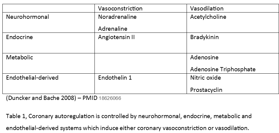
Duncker DJ, Bache RJ. Regulation of coronary blood flow during exercise. Physiological reviews. 2008 Jul:88(3):1009-86. doi: 10.1152/physrev.00045.2006. Epub     [PubMed PMID: 18626066]


[2]

Feigl EO. Coronary physiology. Physiological reviews. 1983 Jan:63(1):1-205     [PubMed PMID: 6296890]


[3]

Nguyen T, Do H, Pham T, Vu LT, Zuin M, Rigatelli G. Left ventricular dysfunction causing ischemia in patients with patent coronary arteries. Perfusion. 2018 Mar:33(2):115-122. doi: 10.1177/0267659117727826. Epub 2017 Aug 21     [PubMed PMID: 28823216]


[4]

Loukas M, Sharma A, Blaak C, Sorenson E, Mian A. The clinical anatomy of the coronary arteries. Journal of cardiovascular translational research. 2013 Apr:6(2):197-207. doi: 10.1007/s12265-013-9452-5. Epub 2013 Feb 20     [PubMed PMID: 23423864]


[5]

Brooks H, Kirk ES, Vokonas PS, Urschel CW, Sonnenblick EH. Performance of the right ventricle under stress: relation to right coronary flow. The Journal of clinical investigation. 1971 Oct:50(10):2176-83     [PubMed PMID: 5116207]


[6]

Feigl EO. Coronary autoregulation. Journal of hypertension. Supplement : official journal of the International Society of Hypertension. 1989 Sep:7(4):S55-8; discussion S59     [PubMed PMID: 2681597]


[7]

Goodwill AG, Dick GM, Kiel AM, Tune JD. Regulation of Coronary Blood Flow. Comprehensive Physiology. 2017 Mar 16:7(2):321-382. doi: 10.1002/cphy.c160016. Epub 2017 Mar 16     [PubMed PMID: 28333376]


[8]

Heusch G. Heart rate in the pathophysiology of coronary blood flow and myocardial ischaemia: benefit from selective bradycardic agents. British journal of pharmacology. 2008 Apr:153(8):1589-601. doi: 10.1038/sj.bjp.0707673. Epub 2008 Jan 28     [PubMed PMID: 18223669]


[9]

Banerjee P. Heart failure: a story of damage, fatigue and injury? Open heart. 2017:4(2):e000684. doi: 10.1136/openhrt-2017-000684. Epub 2017 Oct 15     [PubMed PMID: 29081980]


[10]

Choi S, Shin JH, Park WC, Kim SG, Shin J, Lim YH, Lee Y. Two Distinct Responses of Left Ventricular End-Diastolic Pressure to Leg-Raise Exercise in Euvolemic Patients with Exertional Dyspnea. Korean circulation journal. 2016 May:46(3):350-64. doi: 10.4070/kcj.2016.46.3.350. Epub 2016 May 27     [PubMed PMID: 27275172]


[11]

Zhang DY, Anderson AS. The sympathetic nervous system and heart failure. Cardiology clinics. 2014 Feb:32(1):33-45, vii. doi: 10.1016/j.ccl.2013.09.010. Epub     [PubMed PMID: 24286577]


[12]

Cruickshank JM. Clinical importance of coronary perfusion pressure in the hypertensive patient with left ventricular hypertrophy. Cardiology. 1992:81(4-5):283-90     [PubMed PMID: 1301256]


[13]

Gupta S, Vaidya SR, Arora S, Bahekar A, Devarapally SR. Type 2 versus type 1 myocardial infarction: a comparison of clinical characteristics and outcomes with a meta-analysis of observational studies. Cardiovascular diagnosis and therapy. 2017 Aug:7(4):348-358. doi: 10.21037/cdt.2017.03.21. Epub     [PubMed PMID: 28890871]

Level 1 (high-level) evidence

[14]

Chapman AR, Adamson PD, Mills NL. Assessment and classification of patients with myocardial injury and infarction in clinical practice. Heart (British Cardiac Society). 2017 Jan 1:103(1):10-18. doi: 10.1136/heartjnl-2016-309530. Epub 2016 Nov 2     [PubMed PMID: 27806987]


[15]

Yaginuma T, Avolio A, O'Rourke M, Nichols W, Morgan JJ, Roy P, Baron D, Branson J, Feneley M. Effect of glyceryl trinitrate on peripheral arteries alters left ventricular hydraulic load in man. Cardiovascular research. 1986 Feb:20(2):153-60     [PubMed PMID: 3085950]


[16]

Sequeira V,van der Velden J, Historical perspective on heart function: the Frank-Starling Law. Biophysical reviews. 2015 Dec;     [PubMed PMID: 28510104]

Level 3 (low-level) evidence

[17]

Parissis H, Graham V, Lampridis S, Lau M, Hooks G, Mhandu PC. IABP: history-evolution-pathophysiology-indications: what we need to know. Journal of cardiothoracic surgery. 2016 Aug 4:11(1):122. doi: 10.1186/s13019-016-0513-0. Epub 2016 Aug 4     [PubMed PMID: 27487772]


[18]

Johansson S, Rosengren A, Young K, Jennings E. Mortality and morbidity trends after the first year in survivors of acute myocardial infarction: a systematic review. BMC cardiovascular disorders. 2017 Feb 7:17(1):53. doi: 10.1186/s12872-017-0482-9. Epub 2017 Feb 7     [PubMed PMID: 28173750]

Level 1 (high-level) evidence

[19]

Marik PE, Varon J. Perioperative hypertension: a review of current and emerging therapeutic agents. Journal of clinical anesthesia. 2009 May:21(3):220-9. doi: 10.1016/j.jclinane.2008.09.003. Epub     [PubMed PMID: 19464619]


[20]

Sutton RM, Friess SH, Maltese MR, Naim MY, Bratinov G, Weiland TR, Garuccio M, Bhalala U, Nadkarni VM, Becker LB, Berg RA. Hemodynamic-directed cardiopulmonary resuscitation during in-hospital cardiac arrest. Resuscitation. 2014 Aug:85(8):983-6. doi: 10.1016/j.resuscitation.2014.04.015. Epub 2014 Apr 28     [PubMed PMID: 24783998]


[21]

Paradis NA, Martin GB, Rivers EP, Goetting MG, Appleton TJ, Feingold M, Nowak RM. Coronary perfusion pressure and the return of spontaneous circulation in human cardiopulmonary resuscitation. JAMA. 1990 Feb 23:263(8):1106-13     [PubMed PMID: 2386557]


[22]

Chapman AR,Shah ASV,Lee KK,Anand A,Francis O,Adamson P,McAllister DA,Strachan FE,Newby DE,Mills NL, Long-Term Outcomes in Patients With Type 2 Myocardial Infarction and Myocardial Injury. Circulation. 2018 Mar 20     [PubMed PMID: 29150426]


[23]

Rhodes A, Evans LE, Alhazzani W, Levy MM, Antonelli M, Ferrer R, Kumar A, Sevransky JE, Sprung CL, Nunnally ME, Rochwerg B, Rubenfeld GD, Angus DC, Annane D, Beale RJ, Bellinghan GJ, Bernard GR, Chiche JD, Coopersmith C, De Backer DP, French CJ, Fujishima S, Gerlach H, Hidalgo JL, Hollenberg SM, Jones AE, Karnad DR, Kleinpell RM, Koh Y, Lisboa TC, Machado FR, Marini JJ, Marshall JC, Mazuski JE, McIntyre LA, McLean AS, Mehta S, Moreno RP, Myburgh J, Navalesi P, Nishida O, Osborn TM, Perner A, Plunkett CM, Ranieri M, Schorr CA, Seckel MA, Seymour CW, Shieh L, Shukri KA, Simpson SQ, Singer M, Thompson BT, Townsend SR, Van der Poll T, Vincent JL, Wiersinga WJ, Zimmerman JL, Dellinger RP. Surviving Sepsis Campaign: International Guidelines for Management of Sepsis and Septic Shock: 2016. Intensive care medicine. 2017 Mar:43(3):304-377. doi: 10.1007/s00134-017-4683-6. Epub 2017 Jan 18     [PubMed PMID: 28101605]