Cerebral Ischemia

Earn CME/CE in your profession:


Continuing Education Activity

Cerebral ischemia is a common mechanism of acute brain injury that results from impaired blood flow to the brain. Cerebral ischemia represents a medical emergency; if untreated, it can result in cerebral infarctions or global hypoxic-ischemic encephalopathy, which can result in death or permanent disability. This activity reviews the etiology, epidemiology, pathophysiology, evaluation, and treatment of cerebral ischemia and highlights the role of the interprofessional team in evaluating and treating patients with this condition.

Objectives:

  • Identify the etiology of cerebral ischemia.
  • Describe the appropriate evaluation for a patient with suspected cerebral iscemia.
  • Summarize the management options available for cerebral ischemia.

Introduction

The human brain is a highly metabolically active organ, accounting for about 25% of a person's metabolic demand, despite comprising only 2.5% of a typical individual's body weight. Consequently, the brain is exquisitely sensitive to disruptions in blood flow. Complex homeostatic mechanisms act to maintain cerebral blood flow at a relatively stable rate of about 50 ml/100g of brain tissue per minute, through a process known as cerebrovascular autoregulation. When this blood flow is compromised, the result is cerebral ischemia, one of the most common mechanisms of brain dysfunction and damage. The extent of neuronal injury resulting from compromised cerebral perfusion depends upon both the extent and duration of the hypoperfusion.

Etiology

Cerebral ischemia can be global or focal. Global cerebral ischemia is a result of systemic processes, often shock. The most common cause of global brain ischemia is systemic hypotension. Transient cerebral hypoperfusion can occur when autonomic and neurohormonal mechanisms that control blood pressure and heart rate are disrupted, as in vasovagal syncope and postural tachycardia syndromes. Structural and functional heart problems, mostly arrhythmias, are the second most frequent cause of transient global brain ischemia. When the effect is transient, the condition often manifests as a presyncope or syncope. Prolonged global ischemia, on the other hand, can result in permanent neurological injury.[1]

In contrast, focal brain ischemia most commonly arises from obstruction of arterial blood flow to the brain, often as a result of thrombosis or embolism. If the ischemia persists long enough, irreversible neuronal loss occurs, resulting in an ischemic stroke. Sudden thrombosis of a previously stenotic cerebral or internal carotid artery ruptured plaque will lead to ischemia in the region supplied by the artery affected. Embolization of a clot formed in the heart or a large artery is the most common source of brain ischemia, accounting for almost 60% to 70% of brain TIA and stroke cases. Emboli of other material, such as fat, air from an iatrogenic cause, or amniotic fluid during pregnancy, are possible but much less frequent.

There are various additional rare causes of ischemia. Dissection of cervical blood vessels can rarely cause brain ischemia and may be a cause of stroke in younger patients. Vasospasm is a relatively uncommon secondary cause of brain ischemia. This can be provoked by medication, or it can be secondary to trauma, inflammation, or recent subarachnoid hemorrhage. Rarely, an infection can lead to strokes. Notably, an increased incidence of stroke has occurred with COVID-19.[2]

One simple method of classification of causes for brain ischemia and ischemic stroke is the TOAST criteria.[3] This divides ischemic stroke etiology into the categories of large artery disease (usually secondary to atherosclerosis, occlusive), cardioembolic (cardiac causes such as atrial fibrillation, valvular abnormalities, or abnormalities in cardiac wall motion), small vessel occlusive disease (affects small perforating arteries, often secondary to underlying hypertension), a stroke of other etiology (known etiology of the stroke such as vasculitis or drug use), or stroke of unknown etiology (also called a cryptogenic stroke, no cause found after medical workup).

Epidemiology

Stroke is one of the most prevalent vascular diseases in the world and persistently ranks as the 5th leading cause of death in the United States. The 2019 AHA report on heart disease and stroke statistics estimates that 20 million Americans greater than 20 years of age have had a stroke. Approximately 795,000 people have a stroke each year.[4] Prevalence increases with age, with nearly 75% of cases affecting people over the age of 65. Strokes prevalence is greater among African Americans and Hispanic people than Caucasians. The prevalence of stroke is approximately the same in both females and males, with a slight female predominance as prevalence decreased in males over the past 15 years. 

Stroke is a broad term that encompasses neurological injury resulting from any vascular cause. Ischemic stroke is the most common type, making up 87% of all strokes. They are caused by acute ischemia of an area of the brain supplied by one artery (focal ischemia) and is one major cause of disability and death in the US and the world. Syncope, the consequence of temporary ischemia to the entire brain (global ischemia), is one of the most common “symptoms” in all populations. So, brain ischemia is one of the most common causes of disability in the world.

Pathophysiology

Brain ischemia in its broad sense can be focal or multifocal, caused by a sudden closure or marked diameter reduction of the artery supplying an area of the brain, be it of previously stenotic or normal arteries (i.e. aorta, supra-aortic trunk or intracranial arteries). Brain ischemia can frequently be triggered through the lack of global brain blood supply, in more proximal causes of hemodynamic dysfunction causing sudden blood pressure fall.[5]

Ischemic brain tissue stops working in seconds and suffers necrosis in as soon as 5 minutes after complete lack of oxygen and glucose supply, compared to 20-40 minutes in other parts of the body. Some areas are particularly susceptible to ischemia, a phenomenon known as selective vulnerability. These areas include the arterial border zones, pyramidal cells in the hippocampal CA1 area, and cerebellar Purkinje cells. Arterial border zones are also known as watershed areas; one common location is the superior cerebral convexity which is the area between the territories of the anterior and middle cerebral arteries, which is highly susceptible to decreased cerebral perfusion pressure. The reason for the selective vulnerability of specific neuron types is not completely understood but is believed to be due to variations in the expression of excitatory glutamate neurotransmitter receptors on neurons.

The persistent complications of brain ischemia are due to permanent brain damage and are a final common pathway to necrosis: neuronal shrinkage and death, gliosis with astrocytosis, and loss of volume. Neuronal sensitivity to ischemia is multifactorial: the brain has a high basal metabolic rate and high cell-cell signaling activates pathways such as glutamate-induced neuronal death, dopamine-induced potentiation of ischemic injury, free radical release, zinc toxicity, and release of enzymes leading to combination apoptosis and catabolic necrosis.[6]

Histopathology

In global ischemic injury, histologic changes become apparent within 12 to 24 hours of injury. The first changes seen are shrinking or swelling of neurons. After that, "red neurons" may develop as a consequence of cytoplasmic eosinophilia, nuclear pyknosis, and other necrotic changes. Other findings of edema may also become apparent, such as empty areas within the parenchyma with widened pericellular and perivascular spaces.

Grossly, this may appear as irregular zones of discoloration with a blurring of the zones between gray and white matter. Over time, this evolves into the loss of neurons and gliosis. While most parts of the body affected by ischemia undergo coagulative necrosis, the brain is unique in that it undergoes liquefactive necrosis. This appears as viscous material containing numerous inflammatory cells such as neutrophils and cell debris under a microscope.

History and Physical

The most important part of the evaluation of any patient with neurological symptoms is the history of the presenting complaint. Cerebral ischemia results in negative symptoms related to loss of function of the ischemic tissue. Global cerebral ischemia typically presents as an alteration in consciousness. Transient alteration in consciousness, such as syncope or pre-syncope, may be precipitated by maneuvers such as standing, which may result in a drop in cerebral perfusion pressure. Often the symptoms are relieved by lowering the patient's head. Severe or persistent global cerebral ischemia may result in a patient who presents with a coma.

The symptoms of focal cerebral ischemia will depend upon the specific portions of the brain, which are ischemic. The time of onset and the time course of the presenting complaint is important to ascertain because this can have a significant impact on treatment decisions. Determining the time of symptom onset is ideal, but this may not be possible. In cases where the precise onset cannot be determined, clinical decision-making is determined by the last known time that the patient was normal.[7]

Skilled clinicians learn to recognize specific stroke syndromes, which are patterns of neurological dysfunction related to blockages in particular blood vessels. For example, lesions in the cerebral hemispheres may result in contralateral weakness and/or sensory loss. Signs of cortical dysfunction raise the concern for a large artery obstruction, such as a middle cerebral artery occlusion. Cortical dysfunction in the dominant hemisphere--typically the left--often results in aphasia. Nondominant cortical dysfunction typically presents hemineglect, though this can also be seen with dominant hemisphere infarctions. Other potential signs of cortical involvement may include visual field defects, acalculia, and hemiagnosia. Strokes related to posterior circulation pathology, e.g., the vertebrobasilar system, may present with ataxia, diplopia or ophthalmoparesis, or crossed findings. Early recognition of stroke syndromes consistent with large vessel occlusion is critical because these patients may be candidates for endovascular thrombectomy, which is discussed in more detail below. 

Because stroke outcomes depend heavily on time to treatment, the neurological community has engaged in multiple efforts to educate the public about the signs and symptoms of acute stroke. One common mnemonic is BE FAST: Balance - loss of balance, Eyes - change in vision, Facial droop, Arms- asymmetry in upper extremity strength, Speech - difficulty with speech or slurring, and Time - to alert the public that expediting time to a hospital is essential for treatment. 

In addition to eliciting determining the time of symptom onset, a focused medical history should be obtained. The presence of vascular risk factors such as hypertension, diabetes mellitus, atrial fibrillation, hyperlipidemia, and tobacco abuse should be ascertained, as this information could provide clues as to the stroke etiology. A current medication list is also important, because intravenous thrombolytics, which are often used to treat ischemic stroke, are contraindicated in patients taking anticoagulants.

The physical assessment should be focused on identifying the signs of neurological dysfunction in order to identify the location of the brain lesion. One tool commonly used by physicians to assess strokes is the NIH Stroke Scale.[8] This screening examination allows a rapid assessment of the patient's level of consciousness, oculomotor function, visual fields, motor function, sensation, cerebellar function, language, and attention or neglect. The exam findings are scored from 0 to 40, which facilitates communication between providers and allows monitoring for fluctuation of deficits over time.

Evaluation

The initial workup of symptoms suggestive of cerebral ischemia should include basic labs, including blood glucose, complete blood count, chemistry, coagulation factors, EKG, and cardiac enzymes. A stat non-contrast head CT should be obtained to rule out a hemorrhage or mass lesion.[9] Vascular imaging, such as a CT-angiogram or MR-angiogram, can be very valuable in the acute setting. Vascular imaging can help to identify the stroke etiology, especially in cases of large vessel atherosclerosis; in the case of acute large vessel occlusion, the site of the vascular obstruction may be evident.

Often in the setting of an acute ischemic stroke, a non-contrast head CT may demonstrate no discernable abnormality, especially if the patient presents early in the course of the illness. In this case, MRI or CT perfusion may be valuable in order to determine the viability of the ischemic tissue. Because the fate of ischemic neuronal tissue depends upon both the extent and duration of ischemia, patients presenting with acute focal cerebral ischemia often have a region of infarcted brain tissue (the core infarct) surrounded by a larger area of the ischemic brain which may still be viable. This potentially viable region is known as the penumbra. There has been extensive research in the last decade regarding neuroimaging modalities for determining the volume of ischemic penumbra.[10][11][12]

Although a thorough analysis of acute stroke imaging techniques is beyond the scope of this article, it is important to recognize that patients with suspected large vessel occlusions may benefit from advanced imaging to determine candidacy for endovascular interventions. If a patient presents with symptoms of large vessel occlusion, including signs of cerebrocortical ischemia, transfer to a comprehensive stroke center or thrombectomy-capable stroke center should be considered.

Treatment / Management

The management of global brain ischemia is generally targeted at correcting the underlying cause. Supportive care should be undertaken to ensure adequate cerebral blood flow; this may be facilitated by placing the patient flat or in the Trendelenburg position while other supportive measures are being taken.

In the case of acute ischemic stroke, the first priority is determining whether the patient is a candidate for acute reperfusion therapy. Intravenous thrombolytics have been in use for the treatment of acute ischemic stroke for more than 25 years. Tissue plasminogen activator (TPA) is currently the only thrombolytic agent approved by the Food and Drug Administration for the management of acute ischemic stroke, though studies into the efficacy of tenecteplase are underway.[13] TPA is approved for the treatment of acute ischemic stroke if it can be administered within 3 hours of symptom onset; however, studies indicate that it may be safe and effective if given as much as 4.5 hours after the development of symptoms.[14]

For patients with acute ischemic stroke due to occlusion of a large vessel, mechanical thrombectomy may be indicated. As discussed above, determining whether a patient is a candidate for thrombectomy relies on the time of symptom onset, the severity of the patient's deficits, the location of the large vessel occlusion, and the viability of the ischemic tissue. Patients with suspected large vessel occlusion should be transferred to a comprehensive stroke center or thrombectomy-capable center as soon as feasible. However, the decision to transfer the patient should not delay or preclude treatment with thrombolytics if the patient also meets the criteria for TPA.

Outside of the acute period, the treatment of ischemic stroke is focused on secondary prevention and promoting recovery. A thorough medical history is necessary to evaluate stroke risk factors, including diabetes mellitus, hypertension, tobacco use, hyperlipidemia, and atrial fibrillation. Management of risk factors should be optimized in order to minimize the risk of recurrent stroke.  For most stroke survivors, antiplatelet agents, such as aspirin or clopidogrel, are indicated for secondary stroke prevention. In some stroke subtypes, a short course of dual antiplatelet therapy with aspirin as well as clopidogrel may decrease the risk of early recurrence.[15]

In addition, high-intensity statin therapy has been shown to be beneficial.[16] For stroke related to atrial fibrillation, anticoagulation is indicated; factor Xa inhibitors are preferred over warfarin due to improved safety profiles.[17] Patients with symptomatic carotid stenosis, e.g., hemodynamically significant stenosis of the carotid artery ipsilateral to a stroke or transient ischemic attack, should be evaluated for carotid endarterectomy or stenting. 

For all stroke survivors, smoking cessation, maintenance of ideal body weight, and regular exercise should be encouraged. In fact, there is evidence that lifestyle modifications provide more benefit than stroke prevention medications.[18] In order to promote optimal recovery, patients with acute stroke should be cared for within interdisciplinary teams, including occupational and physical therapists and speech-language pathologists.

Differential Diagnosis

The differential diagnosis for ischemic stroke-like symptoms includes hypoglycemia, electrolyte imbalances such as hyponatremia, drug use, infections including meningitis, cardiogenic syncope, TIA, vasculitis, migraine, tumor, cerebral hemorrhage, and seizures.

Prognosis

Depending upon the duration of brain ischemia, there may be mild brain dysfunction, prolonged dysfunction with permanent damage to the brain, with or without permanent symptoms and disability. 

Patient outcomes after cerebral ischemia can range from no permanent effects (transient ischemic attack) to permanent disability to death.  The severity of impairment is linked to the patient's functional status before the ischemic event. For example, patients with mild cognitive impairment or dementia had greater relative cognitive impairment after ischemic stroke, suggestive of lower neuronal tolerance of ischemia.[19] It is also linked to patient age.[20]

Complications

While prompt reperfusion after ischemia/infarction is essential for preserving neurological function, it can precipitate tissue dysfunction and cell necrosis from the destruction of reversibly damaged cells. Cerebral ischemia-reperfusion injury can occur after thrombolysis or mechanical thrombectomy. While this restores brain flow and salvages reversibly damaged tissue, reperfusion after a longer ischemic period can cause a larger infarct than the initial occlusion. The mechanism of this injury involves leukocyte infiltration, platelet activation, complement activation, and breakdown of the blood-brain barrier leading to vasogenic ischemia.[21] 

Conversion to hemorrhagic stroke after tPA is another complication. It should be seriously considered if the patient's condition worsens after thrombolytic administrations. Symptoms can include a change in awareness or consciousness, worsening neurologic exam, increased weakness, new or worsening headache, or changes in blood pressure or pulse. If this happens, the first step should be immediate Head CT, baseline labs, and neurosurgery consult. 

Patients with acute ischemic stroke are also at risk for complications of immobility, including infections and thromboembolic complications. Due to the risk of aspiration, patients should receive a swallow screen prior to being offered food or drink by mouth. Care should be taken to minimize infection by using aspiration precautions and minimizing the use of invasive devices such as urinary catheters. Additionally, mechanical deep venous thrombosis prophylaxis (DVT) should be employed whenever possible. Pharmacological DVT prophylaxis should also be considered, though this is contraindicated within 24 hours of TPA administration and may be contraindicated for patients with a hemorrhagic conversion.

Deterrence and Patient Education

Risk factors for cerebral ischemia include heart disease, including heart conditions such as atrial fibrillation, diabetes, obesity, hypertension, atherosclerosis/hyperlipidemia, and history of strokes. Behavioral risk factors include physical inactivity, poor sleep, obesity, excessive alcohol intake, and tobacco use. Sickle cell disease and other hematologic disorders, especially hypercoagulable conditions, can also increase the risk of ischemic stroke. Minimizing risk for stroke involves treating these conditions with a healthy diet, exercise, and medication adherence. In particular, statins and blood thinners have been proven to reduce the risk of ischemic stroke.[22][23]

Enhancing Healthcare Team Outcomes

Stroke patients require care in multidisciplinary teams in order to optimize outcomes. The need for interdisciplinary care begins in the prehospital setting because of early recognition, and prompt treatment of ischemic stroke symptoms is critical.[24] Emergency medical providers are instrumental in early diagnosis, and systems of care are increasingly being organized to route patients to stroke-ready hospitals. In some areas, mobile stroke units are being employed to facilitate triage and early stroke treatment.[25] Upon arrival in the emergency department, implementation to promote rapid evaluation and facilitate thrombolytic administration have been shown to improve patient outcomes.[26]

After acute treatment decisions have been made, admission to dedicated stroke units has demonstrated improved outcomes. These units ideally incorporate trained nursing staff as well as rehabilitation providers such as physical therapists, occupational therapists, and speech-language pathologists.[16] Organized stroke units allow early detection of potential complications and facilitate rehabilitation care, which reduces mortality and has a durable effect on patient disability.[27][28][29] [Level 1]


(Click Image to Enlarge)
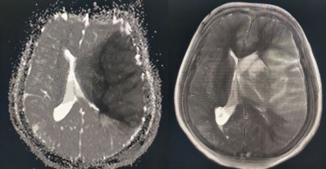
MCA infarction
MCA infarction
Contributed by Sunil Munakomi, MD

(Click Image to Enlarge)
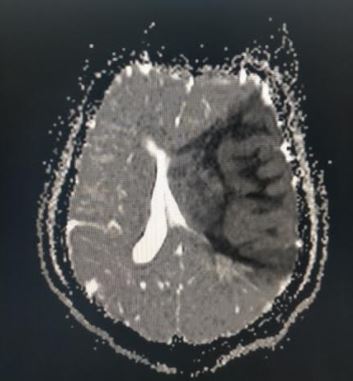
MR ADC sequence in MCA infarction
Contributed by Sunil Munakomi, MD

(Click Image to Enlarge)
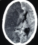
Hemispheric infarction
Contributed by Sunil Munakomi, MD

(Click Image to Enlarge)
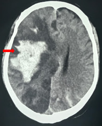
Hemorrhagic transformation within the hemispheric ischemia
Contributed by Sunil Munakomi, MD
Details

Updated:

4/3/2023 5:44:04 PM

References


[1]

Li Y, Zhang E, Yuan H. Cerebral carbon dioxide embolism after kidney cancer laparoscopic surgery with full neurological recovery: A case report. Medicine. 2020 Jul 2:99(27):e20986. doi: 10.1097/MD.0000000000020986. Epub     [PubMed PMID: 32629713]

Level 3 (low-level) evidence

[2]

Zhou Y, Li W, Wang D, Mao L, Jin H, Li Y, Hong C, Chen S, Chang J, He Q, Wang M, Hu B. Clinical time course of COVID-19, its neurological manifestation and some thoughts on its management. Stroke and vascular neurology. 2020 Jun:5(2):177-179. doi: 10.1136/svn-2020-000398. Epub 2020 May 4     [PubMed PMID: 32366614]


[3]

Adams HP Jr, Bendixen BH, Kappelle LJ, Biller J, Love BB, Gordon DL, Marsh EE 3rd. Classification of subtype of acute ischemic stroke. Definitions for use in a multicenter clinical trial. TOAST. Trial of Org 10172 in Acute Stroke Treatment. Stroke. 1993 Jan:24(1):35-41     [PubMed PMID: 7678184]

Level 1 (high-level) evidence

[4]

Benjamin EJ, Muntner P, Alonso A, Bittencourt MS, Callaway CW, Carson AP, Chamberlain AM, Chang AR, Cheng S, Das SR, Delling FN, Djousse L, Elkind MSV, Ferguson JF, Fornage M, Jordan LC, Khan SS, Kissela BM, Knutson KL, Kwan TW, Lackland DT, Lewis TT, Lichtman JH, Longenecker CT, Loop MS, Lutsey PL, Martin SS, Matsushita K, Moran AE, Mussolino ME, O'Flaherty M, Pandey A, Perak AM, Rosamond WD, Roth GA, Sampson UKA, Satou GM, Schroeder EB, Shah SH, Spartano NL, Stokes A, Tirschwell DL, Tsao CW, Turakhia MP, VanWagner LB, Wilkins JT, Wong SS, Virani SS, American Heart Association Council on Epidemiology and Prevention Statistics Committee and Stroke Statistics Subcommittee. Heart Disease and Stroke Statistics-2019 Update: A Report From the American Heart Association. Circulation. 2019 Mar 5:139(10):e56-e528. doi: 10.1161/CIR.0000000000000659. Epub     [PubMed PMID: 30700139]


[5]

Formisano L, Guida N, Mascolo L, Serani A, Laudati G, Pizzorusso V, Annunziato L. Transcriptional and epigenetic regulation of ncx1 and ncx3 in the brain. Cell calcium. 2020 May:87():102194. doi: 10.1016/j.ceca.2020.102194. Epub 2020 Mar 7     [PubMed PMID: 32172011]


[6]

Lee JM, Grabb MC, Zipfel GJ, Choi DW. Brain tissue responses to ischemia. The Journal of clinical investigation. 2000 Sep:106(6):723-31     [PubMed PMID: 10995780]


[7]

Takase KI. Cardiogenic cerebral infarction in the parietal lobe predicts the development of post-stroke epilepsy. Seizure. 2020 Aug:80():196-200. doi: 10.1016/j.seizure.2020.06.018. Epub 2020 Jun 20     [PubMed PMID: 32623354]

Level 3 (low-level) evidence

[8]

Kwah LK, Diong J. National Institutes of Health Stroke Scale (NIHSS). Journal of physiotherapy. 2014 Mar:60(1):61. doi: 10.1016/j.jphys.2013.12.012. Epub 2014 May 3     [PubMed PMID: 24856948]


[9]

Byrne D, Walsh JP, Sugrue G, Nicolaou S, Rohr A. CT Imaging of Acute Ischemic Stroke [Formula: see text]. Canadian Association of Radiologists journal = Journal l'Association canadienne des radiologistes. 2020 Aug:71(3):266-280. doi: 10.1177/0846537120902068. Epub 2020 Mar 11     [PubMed PMID: 32157894]


[10]

Albers GW, Lansberg MG, Kemp S, Tsai JP, Lavori P, Christensen S, Mlynash M, Kim S, Hamilton S, Yeatts SD, Palesch Y, Bammer R, Broderick J, Marks MP. A multicenter randomized controlled trial of endovascular therapy following imaging evaluation for ischemic stroke (DEFUSE 3). International journal of stroke : official journal of the International Stroke Society. 2017 Oct:12(8):896-905. doi: 10.1177/1747493017701147. Epub 2017 Mar 24     [PubMed PMID: 28946832]

Level 1 (high-level) evidence

[11]

Atchaneeyasakul K, Shang T, Haussen D, Ortiz G, Yavagal D. Impact of MRI Selection on Triage of Endovascular Therapy in Acute Ischemic Stroke: The MRI in Acute Management of Ischemic Stroke (MIAMIS) Registry. Interventional neurology. 2020 Jan:8(2-6):135-143. doi: 10.1159/000490580. Epub 2019 Mar 15     [PubMed PMID: 32508895]


[12]

Silva GS, Nogueira RG. Endovascular Treatment of Acute Ischemic Stroke. Continuum (Minneapolis, Minn.). 2020 Apr:26(2):310-331. doi: 10.1212/CON.0000000000000852. Epub     [PubMed PMID: 32224754]


[13]

Ramakrishnan TCR, Kumaravelu S, Narayan SK, Buddha SS, Murali C, Majeed PHA, Meenakshi-Sundaram S, Wadia RS, Sharma V, Basu I, Vijaya P, Salam KA, Barmare S, Vaid Z, Nirmal Raj KK, Wattamwar PR, Asokan K, Dhonge V, Nellikunja S, Namjoshi D, Srinivasa R, Laddhad DS, Deshpande SD, Raghunath B, Kalita J, Kumar M, Misra UK, Pradeep M, Tenecteplase in Stroke Investigators. Efficacy and Safety of Intravenous Tenecteplase Bolus in Acute Ischemic Stroke: Results of Two Open-Label, Multicenter Trials. American journal of cardiovascular drugs : drugs, devices, and other interventions. 2018 Oct:18(5):387-395. doi: 10.1007/s40256-018-0284-1. Epub     [PubMed PMID: 29948822]


[14]

Cronin CA. Intravenous tissue plasminogen activator for stroke: a review of the ECASS III results in relation to prior clinical trials. The Journal of emergency medicine. 2010 Jan:38(1):99-105. doi: 10.1016/j.jemermed.2009.08.004. Epub 2009 Sep 17     [PubMed PMID: 19765940]


[15]

Pan Y, Elm JJ, Li H, Easton JD, Wang Y, Farrant M, Meng X, Kim AS, Zhao X, Meurer WJ, Liu L, Dietrich D, Wang Y, Johnston SC. Outcomes Associated With Clopidogrel-Aspirin Use in Minor Stroke or Transient Ischemic Attack: A Pooled Analysis of Clopidogrel in High-Risk Patients With Acute Non-Disabling Cerebrovascular Events (CHANCE) and Platelet-Oriented Inhibition in New TIA and Minor Ischemic Stroke (POINT) Trials. JAMA neurology. 2019 Dec 1:76(12):1466-1473. doi: 10.1001/jamaneurol.2019.2531. Epub     [PubMed PMID: 31424481]


[16]

Powers WJ, Rabinstein AA, Ackerson T, Adeoye OM, Bambakidis NC, Becker K, Biller J, Brown M, Demaerschalk BM, Hoh B, Jauch EC, Kidwell CS, Leslie-Mazwi TM, Ovbiagele B, Scott PA, Sheth KN, Southerland AM, Summers DV, Tirschwell DL. Guidelines for the Early Management of Patients With Acute Ischemic Stroke: 2019 Update to the 2018 Guidelines for the Early Management of Acute Ischemic Stroke: A Guideline for Healthcare Professionals From the American Heart Association/American Stroke Association. Stroke. 2019 Dec:50(12):e344-e418. doi: 10.1161/STR.0000000000000211. Epub 2019 Oct 30     [PubMed PMID: 31662037]


[17]

Casula M, Fortuni F, Fabris F, Leonardi S, Gnecchi M, Sanzo A, Greco A, Rordorf R. Direct oral Xa inhibitors versus warfarin in patients with cancer and atrial fibrillation: a meta-analysis. Journal of cardiovascular medicine (Hagerstown, Md.). 2020 Aug:21(8):570-576. doi: 10.2459/JCM.0000000000001041. Epub     [PubMed PMID: 32628422]


[18]

Liljehult J, Christensen T, Molsted S, Overgaard D, Mesot Liljehult M, Møller T. Effect and efficacy of lifestyle interventions as secondary prevention. Acta neurologica Scandinavica. 2020 Oct:142(4):299-313. doi: 10.1111/ane.13308. Epub 2020 Jul 15     [PubMed PMID: 32620044]


[19]

Béjot Y, Duloquin G, Crespy V, Durier J, Garnier L, Graber M, Giroud M. Influence of Preexisting Cognitive Impairment on Clinical Severity of Ischemic Stroke: The Dijon Stroke Registry. Stroke. 2020 Jun:51(6):1667-1673. doi: 10.1161/STROKEAHA.119.028845. Epub 2020 May 13     [PubMed PMID: 32397931]


[20]

Kwah LK, Harvey LA, Diong J, Herbert RD. Models containing age and NIHSS predict recovery of ambulation and upper limb function six months after stroke: an observational study. Journal of physiotherapy. 2013 Sep:59(3):189-97. doi: 10.1016/S1836-9553(13)70183-8. Epub     [PubMed PMID: 23896334]

Level 2 (mid-level) evidence

[21]

Pan J, Konstas AA, Bateman B, Ortolano GA, Pile-Spellman J. Reperfusion injury following cerebral ischemia: pathophysiology, MR imaging, and potential therapies. Neuroradiology. 2007 Feb:49(2):93-102     [PubMed PMID: 17177065]


[22]

Katsanos AH, Lioutas VA, Charidimou A, Catanese L, Ng KKH, Perera K, de Sa Boasquevisque D, Tsivgoulis G, Smith EE, Sharma M, Selim MH, Shoamanesh A. Statin treatment and accrual of covert cerebral ischaemia on neuroimaging: a systematic review and meta-analysis of randomized trials. European journal of neurology. 2020 Jun:27(6):1023-1027. doi: 10.1111/ene.14196. Epub 2020 Mar 23     [PubMed PMID: 32133735]

Level 1 (high-level) evidence

[23]

Sussman ES, Jin M, Pendharkar AV, Pulli B, Feng A, Heit JJ, Telischak NA. Dual antiplatelet therapy after carotid artery stenting: trends and outcomes in a large national database. Journal of neurointerventional surgery. 2021 Jan:13(1):8-13. doi: 10.1136/neurintsurg-2020-016008. Epub 2020 May 15     [PubMed PMID: 32414894]


[24]

Fassbender K, Walter S, Grunwald IQ, Merzou F, Mathur S, Lesmeister M, Liu Y, Bertsch T, Grotta JC. Prehospital stroke management in the thrombectomy era. The Lancet. Neurology. 2020 Jul:19(7):601-610. doi: 10.1016/S1474-4422(20)30102-2. Epub     [PubMed PMID: 32562685]


[25]

Fatima N, Saqqur M, Hussain MS, Shuaib A. Mobile stroke unit versus standard medical care in the management of patients with acute stroke: A systematic review and meta-analysis. International journal of stroke : official journal of the International Stroke Society. 2020 Aug:15(6):595-608. doi: 10.1177/1747493020929964. Epub 2020 Jun 9     [PubMed PMID: 32515695]

Level 1 (high-level) evidence

[26]

Darehed D, Blom M, Glader EL, Niklasson J, Norrving B, Eriksson M. In-Hospital Delays in Stroke Thrombolysis: Every Minute Counts. Stroke. 2020 Aug:51(8):2536-2539. doi: 10.1161/STROKEAHA.120.029468. Epub 2020 Jun 26     [PubMed PMID: 32586222]


[27]

Glader EL, Stegmayr B, Johansson L, Hulter-Asberg K, Wester PO. Differences in long-term outcome between patients treated in stroke units and in general wards: a 2-year follow-up of stroke patients in sweden. Stroke. 2001 Sep:32(9):2124-30     [PubMed PMID: 11546906]


[28]

Krespi Y,Gurol ME,Coban O,Tuncay R,Bahar S, Stroke unit versus neurology ward--a before and after study. Journal of neurology. 2003 Nov     [PubMed PMID: 14648154]


[29]

Jauch EC, Saver JL, Adams HP Jr, Bruno A, Connors JJ, Demaerschalk BM, Khatri P, McMullan PW Jr, Qureshi AI, Rosenfield K, Scott PA, Summers DR, Wang DZ, Wintermark M, Yonas H, American Heart Association Stroke Council, Council on Cardiovascular Nursing, Council on Peripheral Vascular Disease, Council on Clinical Cardiology. Guidelines for the early management of patients with acute ischemic stroke: a guideline for healthcare professionals from the American Heart Association/American Stroke Association. Stroke. 2013 Mar:44(3):870-947. doi: 10.1161/STR.0b013e318284056a. Epub 2013 Jan 31     [PubMed PMID: 23370205]