Anatomy, Head and Neck, Pharyngeal Muscles


Introduction

The pharynx is the digestive system posterior to the nasal cavity, oral cavity, and larynx and divides into the oropharynx, nasopharynx, and laryngopharynx. A group of muscles called the pharyngeal muscles, which consist of the outer circular layer and the inner longitudinal layer, forms the lumen of the pharynx. The outer circular layer consists of the superior, middle, and inferior pharyngeal constrictor muscles. The inner longitudinal layer consists of the palatopharyngeus, salpingopharyngeus, and stylopharyngeus muscles. The pharyngeal muscles receive innervation from the vagus and glossopharyngeal nerve to work in sync to propel food from the oral cavity into the esophagus.

Structure and Function

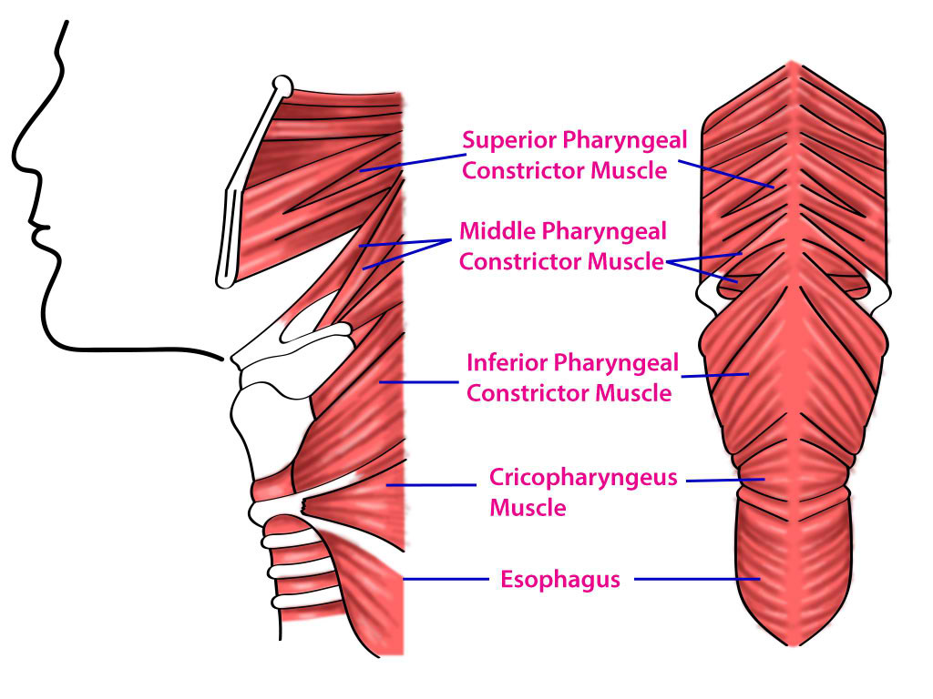
The pharynx extends from the posterior nasal and oral cavity to the cricoid cartilage before blending into the esophagus. The outer circular layer consists of the superior, middle, and inferior pharyngeal constrictor muscles. These constrictor muscles originate from bones and cartilage anteriorly and insert posteriorly to a tendinous seam called the pharyngeal raphe. The superior pharyngeal constrictor contract to narrow its lumen to assist with bolus transport as well as seal the nasopharynx to prevent food from going up — the middle and inferior pharyngeal constrictors contract to narrow its lumen to assist with bolus transport. The palatopharyngeus, salpingopharyngeus, and stylopharyngeus muscles are the inner longitudinal muscles that contract to raise the pharynx and larynx. These muscles work together to help move food bolus from the oral cavity into the esophagus.[1] 

Embryology

The muscles and nerve innervation of the pharynx derive embryologically from the third and fourth brachial arches. The third branchial arch develops into the stylopharyngeus muscle. The third arch also forms the glossopharyngeal nerve (CN IX), which is the nerve that innervates the stylopharyngeus muscle. The fourth branchial arch forms most of the pharyngeal constrictors as well as the superior laryngeal branch of the vagus nerve (CN X). Subsequently, the vagus nerve innervates most of the pharyngeal constrictor muscles.[2] 

Blood Supply and Lymphatics

Arterial supply to the pharyngeal muscle is from branches of the external carotid artery. The branches of the external carotid artery include the ascending pharyngeal, facial, lingual, and maxillary artery.[3][4] Venous drainage of the pharynx is into the pharyngeal venous plexus that eventually drains into the internal jugular vein. Lymphatic drainage of the pharynx first goes to the retropharyngeal lymph node then into the deep cervical lymph node.[5][6]

Nerves

The motor and sensory innervation of pharyngeal muscles is provided primarily by the pharyngeal plexus. The motor branch of the pharyngeal plexus comes from the vagus and glossopharyngeal nerve. The glossopharyngeal nerve provides motor to the stylopharyngeus muscle. The vagus nerve and its branches provide motor innervation to all other muscles of the pharynx. The sensory fibers of the pharyngeal muscles come from the vagus, glossopharyngeal, and the maxillary division of the trigeminal nerve.[7] 

Muscles

The outer circular layer consists of the superior, middle, and inferior constrictor muscles. The superior constrictor muscle originates from the pterygoid process, posterior end of the mylohyoid line of mandible, and the pterygomandibular ligament/raphe. This superior constrictor muscle inserts into the base of the skull at the pharyngeal tubercle and the pharyngeal raphe. The pharyngeal raphe is a midline tendinous seam where the constrictor muscles meet. When the superior constrictor muscle contracts, it constricts the upper portion of the pharynx. The middle constrictor muscle originates from the greater and lesser horn of the hyoid bone and stylohyoid ligament. The middle constrictor inserts into the median pharyngeal raphe as well as blending in with fibers of the superior and inferior constrictors. The middle constrictor muscle contracts to constrict the middle portion of the pharynx. The inferior pharyngeal constrictor muscle further divides into two muscles, the thyropharyngeus, and the cricopharyngeus muscle. The thyropharyngeus muscle originates from the thyroid cartilage and inserts into the median pharyngeal raphe. The cricopharyngeus muscle originates from the cricoid cartilage and blends into the esophageal muscle. When the inferior constrictors contract, they constrict the lower portion of the pharynx. These constrictor muscles work together and contract to push food after swallowing to propel it from the oral cavity into the esophagus.[7]  

The inner longitudinal layer consists of the palatopharyngeus, salpingopharyngeus, and stylopharyngeus muscles. The palatopharyngeus muscle originates from the posterior hard palate, the palatine aponeurosis, and inserts into the thyroid cartilage. When the palatopharyngeus muscle contracts, it elevates the pharynx superiorly.  The salpingopharyngeus muscle originates from the inferior auditory tube and inserts into the palatopharyngeus muscle. This muscle contracts to elevate the pharynx superiorly as well as opening the auditory tube when swallowing. The stylopharyngeus muscle origin is from the styloid process of the temporal bone and it inserts into the posterior thyroid cartilage as well as merging with muscle fibers of the palatopharyngeal muscle. When the stylopharyngeus muscle contracts, it elevates the pharynx and larynx. As previously mentioned, this muscle is different from the other pharyngeal muscle in that it is the only muscle innervated by the glossopharyngeal nerve.[8] These longitudinal muscles work to elevate the pharynx and larynx superiorly during swallowing.[9][10]

Physiologic Variants

There are multiple physiologic variants of the pharyngeal constrictor muscles. The superior constrictor can merge with the muscle of the middle and inferior constrictor. The inferior constrictor merges with the superior constrictor or the posterior part of the pharyngobasilar fascia.[11] Longitudinal muscle groups can have different insertion points, including into the palatine tonsil, epiglottis, and arytenoid.[1][10]

Surgical Considerations

Cricopharyngeal myotomy can be performed to improve bolus transport for a patient experiencing symptoms of block dysphagia at the cricoid level. This procedure requires cutting into the muscle of the upper esophagus and cricopharyngeus muscle.[12]

Clinical Significance

Zenker diverticulum

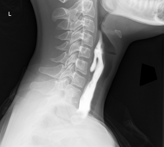
The inferior pharyngeal constrictor muscle subdivides into the thyropharyngeus and cricopharyngeus muscle. The thyropharyngeus muscle is superior to the cricopharyngeus muscle, and during normal swallowing, the thyropharyngeus contracts while the cricopharyngeus relaxes to propel the food into the esophagus. If there is incoordination of contraction and relaxation of these two muscles, intrapharyngeal pressure can rise and form a diverticulum between the weak spot of the two muscles. This structure is called Zenker diverticulum, and it can cause an accumulation of food into the pouch and can lead to dysphagia. Other complication includes gurgling, aspiration, foul breath, and neck mass.[13] 

Pharyngitis

The pharynx can become inflamed and present with pain, irritation, discomfort, and dysphagia of the throat. This condition can be the result of bacteria or viral infections. If a viral infection is suspected, treatment is often supportive and given NSAIDs for pain. If a bacterial infection is suspected, antibiotics are the therapeutic intervention. Pharyngitis caused by Streptococcus Pyogenes is promptly treated to prevent the sequela of rheumatic fever and glomerulonephritis.

Dysphagia

Dysphagia is difficulty swallowing foods or liquids, and some etiology can be due to dysmotility of the pharyngeal muscles. Stroke patients can present with dysphagia due to its effect on the pharyngeal muscle.[14][15] Dysphagia is a common issue in the elderly and is primarily of oropharyngeal or esophageal origin.[16] 

Other Issues

Cricopharyngeal achalasia

Cricopharyngeal achalasia is when the cricopharyngeal muscle fails to open adequately enough to allow the passage of bolus into the esophagus. The patient can present with symptoms of choking, regurgitation, coughing, and recurrent pneumonia. A patient diagnosed with cricopharyngeal achalasia can have it treated with cricopharyngeal myotomy or with botulinum toxin injection.[17] 

Glossopharyngeal neuralgia

Glossopharyngeal neuralgia is a disorder presenting with the repeated episodes of pain lasting from a few seconds to minutes in the tongue, throat, ear, and tonsils and is usually unilateral. The pain can worsen or triggered by swallowing, speaking, and chewing. The etiology is thought to be due to irritation of the glossopharyngeal nerve, but the source of irritation is often not found. Glossopharyngeal neuralgia is often misdiagnosed as trigeminal neuralgia since the presentation of pain can overlap and looks similar.[18]


(Click Image to Enlarge)
<p>Superior, Middle, and Inferior Pharyngeal Muscles and Cricopharyngeus Muscle</p>

Superior, Middle, and Inferior Pharyngeal Muscles and Cricopharyngeus Muscle


Contributed by T Silappathikaram


(Click Image to Enlarge)
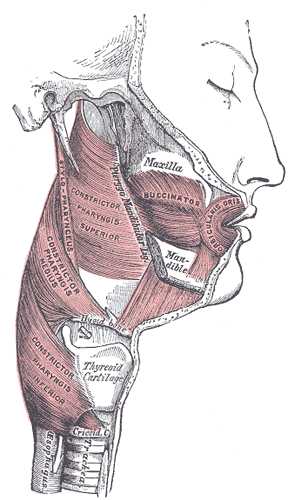
<p>Muscles of the Cheek and Pharynx

Muscles of the Cheek and Pharynx. This lateral-view image shows the buccinator, orbicularis oris, and constrictor pharyngis superior, medius, and inferior. The thyroid cartilage, trachea, upper esophagus, stylopharyngeus, maxilla, and mandible are also shown. 


Henry Vandyke Carter, Public domain, via Wikimedia Commons


(Click Image to Enlarge)
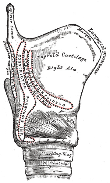
<p>The Larynx, Side view of the larynx, showing muscular attachments, Thyrohyoideus, Sternothyroideus, Constrictor pharyngis

The Larynx, Side view of the larynx, showing muscular attachments, Thyrohyoideus, Sternothyroideus, Constrictor pharyngis inferior, Stylopharyngeus


Henry Vandyke Carter, Public Domain, via Wikimedia Commons


(Click Image to Enlarge)
Zenker diverticulum Barium
Image courtesy S Bhimji MD
Details

Author

Toai Bui

Editor:

Joe M. Das

Updated:

7/24/2023 9:13:46 PM

References


[1]

Heyd C, Yellon R. Anatomy, Head and Neck, Pharynx Muscles. StatPearls. 2023 Jan:():     [PubMed PMID: 30969574]


[2]

Adams A, Mankad K, Offiah C, Childs L. Branchial cleft anomalies: a pictorial review of embryological development and spectrum of imaging findings. Insights into imaging. 2016 Feb:7(1):69-76. doi: 10.1007/s13244-015-0454-5. Epub 2015 Dec 10     [PubMed PMID: 26661849]


[3]

Hacein-Bey L, Daniels DL, Ulmer JL, Mark LP, Smith MM, Strottmann JM, Brown D, Meyer GA, Wackym PA. The ascending pharyngeal artery: branches, anastomoses, and clinical significance. AJNR. American journal of neuroradiology. 2002 Aug:23(7):1246-56     [PubMed PMID: 12169487]


[4]

Devadas D, Pillay M, Sukumaran TT. A cadaveric study on variations in branching pattern of external carotid artery. Anatomy & cell biology. 2018 Dec:51(4):225-231. doi: 10.5115/acb.2018.51.4.225. Epub 2018 Dec 29     [PubMed PMID: 30637155]


[5]

Lengelé B, Hamoir M, Scalliet P, Grégoire V. Anatomical bases for the radiological delineation of lymph node areas. Major collecting trunks, head and neck. Radiotherapy and oncology : journal of the European Society for Therapeutic Radiology and Oncology. 2007 Oct:85(1):146-55     [PubMed PMID: 17383038]


[6]

Werner JA. [The lymph vessel system of the mouth cavity and pharynx]. Laryngo- rhino- otologie. 1995 Oct:74(10):622-8     [PubMed PMID: 8672202]


[7]

Albahout KS, Lopez RA. Anatomy, Head and Neck, Pharynx. StatPearls. 2023 Jan:():     [PubMed PMID: 31334991]


[8]

Jain P, Rathee M. Anatomy, Head and Neck, Stylopharyngeus Muscles. StatPearls. 2023 Jan:():     [PubMed PMID: 31613499]


[9]

Shaw SM, Martino R. The normal swallow: muscular and neurophysiological control. Otolaryngologic clinics of North America. 2013 Dec:46(6):937-56. doi: 10.1016/j.otc.2013.09.006. Epub 2013 Oct 23     [PubMed PMID: 24262952]


[10]

Choi DY, Bae JH, Youn KH, Kim HJ, Hu KS. Anatomical considerations of the longitudinal pharyngeal muscles in relation to their function on the internal surface of pharynx. Dysphagia. 2014 Dec:29(6):722-30. doi: 10.1007/s00455-014-9568-z. Epub 2014 Aug 21     [PubMed PMID: 25142243]


[11]

Murakami K, Kuroda M, Kishi K. [Variations of the constrictor pharyngeal muscles in humans]. Kaibogaku zasshi. Journal of anatomy. 1996 Dec:71(6):638-49     [PubMed PMID: 9038006]


[12]

McKenna JA, Dedo HH. Cricopharyngeal myotomy: indications and technique. The Annals of otology, rhinology, and laryngology. 1992 Mar:101(3):216-21     [PubMed PMID: 1543330]


[13]

Nesheiwat Z, Antunes C. Zenker Diverticulum. StatPearls. 2023 Jan:():     [PubMed PMID: 29763172]


[14]

Singh S, Hamdy S. Dysphagia in stroke patients. Postgraduate medical journal. 2006 Jun:82(968):383-91     [PubMed PMID: 16754707]


[15]

Hamdy S, Aziz Q, Rothwell JC, Crone R, Hughes D, Tallis RC, Thompson DG. Explaining oropharyngeal dysphagia after unilateral hemispheric stroke. Lancet (London, England). 1997 Sep 6:350(9079):686-92     [PubMed PMID: 9291902]


[16]

Khan A, Carmona R, Traube M. Dysphagia in the elderly. Clinics in geriatric medicine. 2014 Feb:30(1):43-53. doi: 10.1016/j.cger.2013.10.009. Epub     [PubMed PMID: 24267601]


[17]

Drendel M, Carmel E, Kerimis P, Wolf M, Finkelstein Y. Cricopharyngeal achalasia in children: surgical and medical treatment. The Israel Medical Association journal : IMAJ. 2013 Aug:15(8):430-3     [PubMed PMID: 24079064]


[18]

Shah RJ, Padalia D. Glossopharyngeal Neuralgia. StatPearls. 2023 Jan:():     [PubMed PMID: 31082085]