Continuing Education Activity
Pili annulati is an inherited, rare, and benign hair shaft disorder characterized by a unique appearance of the hair with alternate light and dark bands. Pili annulati belongs to the group of hair shaft disorders without fragility. This activity illustrates the various presentations and reviews the evaluation and management of pili annulati. This activity highlights the role of the interprofessional team in collaborating to establish the diagnosis and render the best care for affected patients.
Objectives:
- Describe the etiology and pathophysiology of pili annulati.
- Explain the common physical exam findings in pili annulati
- Describe the typical trichoscopic and microscopic findings associated with pili annulati.
- Explain the importance of collaboration and communication amongst the interprofessional team to establish the diagnosis of pili annulati and to enhance patient care.
Introduction
Pili annulati (PA), also known as « ringed » or “Morse alphabet” hair, is a rare benign disorder characterized by a unique speckled and banded appearance of the hair with alternate light and dark bands.[1][2][3] This appearance results from an increased light reflex caused by a periodic occurring of abnormal air-filled cavities within the hair shafts of affected individuals.[3] Pili annulati belongs to the group of hair shaft disorders without fragility. Although there are reports of some sporadic cases, it is considered to be an inherited disorder.
Etiology
Inheritance of pili annulati is via an autosomal dominant pattern with variable expression. A locus responsible for pili annulati was mapped on the end of the telomeric region of chromosome 12q (24.32–24.33), but the responsible gene for the disease is not yet completely identified.[4] So far, a critical region of 2.9 Mb containing 36 candidates genes has been defined by recombination events.[5]
A report exists of a case of pili annulati associated with Rothmund-Thomson syndrome caused by a mutation in RECQL4.[6]
The literature also describes sporadic cases.
Epidemiology
The prevalence of pili annulati is currently unknown. It is presumed to be a rare hair shaft disorder. Since being first described in 1866, there are reports of approximately 50 cases in the literature.[7]
No racial distribution is evident for pili annulati.
Pathophysiology
Hair in pili annulati has a characteristic shiny and speckled appearance with alternating light and dark bands. The light bands that are visible with the unaided eye under a refracted light, actually correspond to dark bands when seen under light (or polarized) microscopy. By electron microscopy, these abnormal hair segments correspond to air-filled cavities within the hair shaft’s cortex.[7]
Altered light penetration of hair explains this banded appearance in pili annulati. The abnormal spaces containing air cause double diffraction and scattering of transmitted light, which results in a decrease of light transmission compared to normal areas, thus the dark appearance of abnormal areas under a light microscope. Conversely, under reflected light, the abnormal areas reflect more light and therefore appear lighter.[8]
These air-filled gaps in abnormal areas appear to result from an insufficient matrix formation or a defective assembly of structural proteins in the matrix due to an abnormal regulatory protein.[3][5][7]
Therefore, pili annulati appears to be a protein metabolic disorder. A partial dysfunction of cytoplasmic ribosomes in the differentiating cortical cells is suggested to be involved.[1]
History and Physical
Pili annulati may appear at birth or during infancy.[9] The clinical expression can be heterogeneous within the same patient at different regions of the scalp or even at different regions of the same affected hair.[6][10][11]
The clinical examination usually reveals a shiny and banded appearance of the hair with sometimes a peculiar glistening texture or even a frizzy aspect.[3] The number of white bands tends to disappear distally as the hair grows.
Pili annulati is not typically associated with increased hair fragility. Hair growth and tensile strength of hair in affected individuals are normal, but abnormal areas of the hair shafts appear to be more susceptible to weathering and present with minor surface abnormalities. In a minority of cases, an increased sensitivity at the level of the light bands with severe trichorrhexis-nodosa-like hair fracturing and breakage can be observed.[7][12]
Pili annulati is classically limited to the scalp hair, but other regions such as pubic, axillary, and beard hair, may also be affected.[3][10]
Pili annulati is easily detected in blonde hair while it can be completely obscured in black hair, as the additional pigment in dark hair tends to absorb the surrounding light and mask the banding appearance. Pili annulati become more noticeable with age as the hair becomes depigmented, increasing light transmission.[8]
There have been several reports of pili annulati associated with alopecia areata, autoimmune thyroid disorders, as well as primary immunoglobulin A deficiency. The most commonly accepted assumption is that these cases represent a coincidental concomitant manifestation, as a true pathogenetic association has not been proven.[2][3][6]
Evaluation
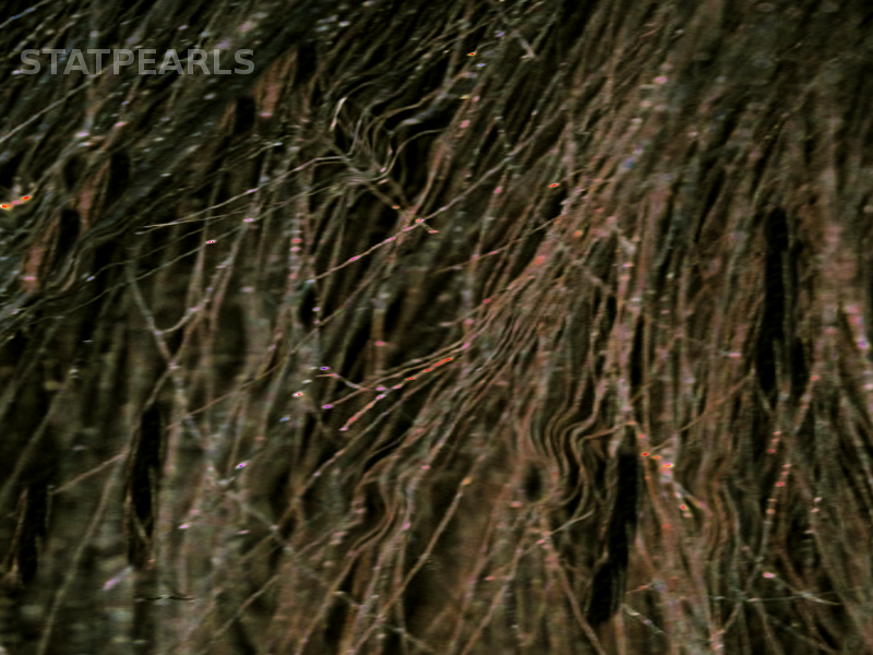
Light microscopy shows a characteristic appearance with alternating bright and dark bands in the hair shaft. The bands that appear dark in light microscopy correspond to white bands macroscopically, under reflected light, and in trichoscopy. Transmission electron microscopy of affected hairs shows a normal medulla with clusters of intermittent air-filled cavities within the cortex of the hair shafts. Scanning electron microscopy reveals a "cobblestoned" and fluted cuticle.[8][9]
Like in other hair shaft disorders, trichoscopy represents a simple and rapid method that enables the practitioners to establish the diagnosis of pili annulati without the need to pluck hairs.
In both dark and blond hairs, It demonstrates regular light-colored bands covering more than 50% of the hair shaft width, giving a “misty-like” appearance. This trichoscopic image of pili annulati may be misdiagnosed as "intermittent medulla," seen within thick hair shafts in healthy individuals. In these cases, intermittent light-colored bands cover less than 50% of the hair shaft’s thickness.[6][13]
Treatment / Management
Treatment in pili annulati is often not required as it is a benign condition in which patients rarely seek medical attention. The shiny appearance of the hair is infrequently bothersome, and some [atients even consider it attractive.
There have been a few reports of a disappearance of the "ringed" appearance of the hair after daily use of topical minoxidil.[2]
In rare forms associated with hair breakage and fragility, gentle hair care is the recommendation.
Differential Diagnosis
The primary differential diagnosis that merit consideration is pseudopili annulati. It presents with a banded clinical appearance similar to pili annulati with light and dark bands. However, this clinical aspect is an optical effect that results from a slight twisting of the hair shaft. Trichoscopy easily establishes the diagnosis as it shows twisted hairs without white bands.[3][6][8]
Prognosis
The prognosis of pili annulati is excellent as it is a benign condition that doesn’t affect the quality of life of affected patients.[2]
It is, however, essential to clarify that pili annulati becomes more obvious and manifests further with age as the pigment loss causes an increase in light transmission.[8]
Enhancing Healthcare Team Outcomes
Pili annulati is a hair shaft disorder that does not cause cosmetic issues. Therefore, affected individuals rarely seek medical attention.
Like other congenital hair shaft disorders, pili annulati may be seen at birth or during early infancy. Hence, pediatricians and pediatric nurse practitioners are likely to encounter such cases. Careful clinical examination is necessary to establish the diagnosis of pili annulati as it is an asymptomatic condition. An interprofessional and an interprofessional healthcare team approach is essential to render to best patient care, including physicians and specialty-trained nursing staff.